175
Pakistan Veterinary Journal
ISSN: 0253-8318 (PRINT), 2074-7764 (ONLINE)Accessible at: www.pvj.com.pk
Enteric Diseases of Poultry with Special Attention to
Clostridium perfringens
Hafez Mohamed Hafez*
Institute of Poultry Diseases, Faculty of Veterinary Medicine, Free University Berlin, Königsweg, 14163 Berlin, Germany *Corresponding author: [email protected]
A R T I C L E H I S T O R Y A B S T R A C T Received:
Revised: Accepted:
October 10, 2010 April 18, 2011 April 21, 2011 Key words:
Cholangiohepatitis Clostridium perfringens Enteric diseases Necrotic enteritis Poultry
The enteric health of growing poultry is imperative to success of the production. The basic role of poultry production is turning feed stuffs into meat. Any changes in this turning process, due to mechanical, chemical or biological disturbance of digestive system (enteric disorders) is mostly accompanied with high economic losses due to poor performance, increased mortality rates and increased medication costs. The severity of clinical signs and course of the disorders are influenced by several factors such as management, nutrition and the involved agent(s). Several pathogens (viruses, bacteria and parasites) are incriminated as possible cause of enteric disorders either alone (mono-causal), in synergy with other micro-organisms (multi-causal), or with non-infectious causes such as feed and /or management related factors. In addition, excessive levels of mycotoxins and biogenic amines in feed lead to enteric disorders. Also factors such as high stocking density, poor litter conditions, poor hygiene and high ammonia level and other stressful situation may reduce the resistance of the birds and increases their susceptibility to infections. Under field conditions, however, it is difficult to determine whether the true cause of enteric disorders, is of infectious or non-infectious origin. In recent years and since the ban of use of antimicrobial growth promoters in several countries the incidence of intestinal disorders especially those caused by clostridial infection was drastically increased. The present review described in general the several factors involved in enteric disorders and summarized the available literatures about Clostridium perfringens infection in poultry.
©2011 PVJ. All rights reserved To Cite This Article: Hafez HM, 2011. Enteric diseases of poultry with special attention to Clostridium perfringens. Pak Vet J, 31(3): 175-184.
INTRODUCTION
The basic role of poultry production is turning feed stuffs into meat. Broilers and meat turkeys are very efficient at both growth and feed conversion rate. Any slight alteration from the optimal condition is mostly accompanied by disruption of the growth process and all over performance. To reach the maximal potential of development, considerable demands should be placed on good intestinal health.
Enteric disorders are one of the most important groups of diseases they affect poultry and are continuing to cause high economic losses in the many areas world-wide due to increased mortality rates, decreased weight gain, increased medication costs, and increased feed conversion rates. Several pathogens (viruses, bacteria and parasites) are incriminated as possible causes of enteric disorders either alone (mono-causal), in synergy with different other microorganisms (multi-causal) or with
non-infectious causes such as feed and /or management related factors (Table 1). Under field conditions, however, it is difficult to determine weather the true cause of enteric disorders in poultry is of infectious or non-infectious origin (Hafez, 2001).
Since the first report of Moore et al. (1946), it is generally known, that supplementation of poultry feed with antibiotic growth promoters (AGPs) improves performance of livestock. The effect of AGP on gut flora results in improvement of digestion, better absorption of nutrients, and a more stable balance in the microbial population. As consequence this is accompanied with reduced intestinal disorders. However, AGP can also increase the prevalence of drug-resistant bacteria.
Based on “Precautionary Principle” and experiences made in Sweden, Denmark, Germany and the Netherlands, the EU has decided to ban the use of growth-promoting antibiotics in feed of food producing animals completely by January 2006. The first step was taken in
1997 by the ban of avoparcin, followed by spiramycin, tylosin phosphate, zinc bacitracin and virginiamycin in 1998 and carbadox and olaquindox in 1999. Field observations in several countries in Europe showed that poultry industry faced several problems after the ban of AGPs. The impact of the ban has been seen on the performances (body weight and feed conversion rate) as well as on the rearing husbandry (wet litter and ammonia level), animal welfare problem (foot pad dermatitis) and general health issues on the birds (enteric disorders due to dysbacteriosis and clostridial infections) (Hafez, 2008).
The present paper reviews the currently most important causes of enteric disorders of poultry and their economic impacts with special attention to clostridial infections.
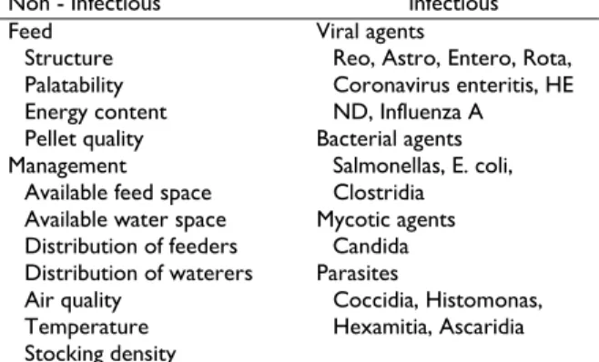
Table 1: Some possible causes of enteric disorders in poultry
Non - Infectious Infectious
Feed Viral agents
Structure Reo, Astro, Entero, Rota,
Palatability Coronavirus enteritis, HE
Energy content ND, Influenza A
Pellet quality Bacterial agents
Management Salmonellas, E. coli,
Available feed space Clostridia
Available water space Mycotic agents
Distribution of feeders Candida
Distribution of waterers Parasites
Air quality Coccidia, Histomonas,
Temperature Hexamitia, Ascaridia
Stocking density
Non-infectious factors involved in intestinal disorders Nutrition
Enteric health and nutrition are closely related. Poor enteric health can adversely affect food digestion, gut motility and nutrient absorption by several means. Likewise, poor nutrition and feed quality can either increase the bird’s susceptibility to enteric disorders or directly cause them (Ferket, 1996; Hafez, 1998). Nutritional factors that influence gut health include feed intake, palatability, feed ingredient quality, feed formulation and pellet quality. In addition, mistakes in feeding technique, the amount of fibre in the feed, the content and quality of the raw materials as well as sudden feed changes or restriction, can result in changes in the intestinal flora and/or in the enzymatic activity which lead to digestive disorders (Nixey, 1989; Kaldhusdal and Skjerve, 1996; D’Mello, 1997; Corless and Sell, 1999; Annett et al., 2002; Batal and Parsons, 2002; Kocher, 2003; McReynolds et al., 2004). Also, excessive levels of mycotoxins and biogenic amines in feed lead to enteric disorders (Smith and Hamilton, 1970; Burditt et al., 1983; Brown et al., 1992; Dwivedi and Burns, 1986; Hoerr, 2008).
Feed contains a little dust; however, excess dust can adversely affect the palatability and lead to reduction of feed intake. Badly stored feed can contain fungal spores, and fat may go rancid which results in reduction of the feed intake and may negatively affect the content of some vitamins in the feed (Dhand et al., 1998; FAO, 2004). Inadequate feeder space and false distribution can result in competition and stronger birds dominating the feeder with a consequent variation in feed intake within the flock.
Feed can contain undesirable substances such as mycotoxins, which adversely affect the immune system, increase the susceptibility of the birds to infectious diseases and cause poor response to vaccine (Uraguchi and Myamazaki, 1978; Campbell et al., 1981; Burns and Dwivedi, 1986; El-Karim et al., 1991; D’Mello et al., 1993, D’Mello and MacDonald, 1998; Devegowda et al., 1998). Proper adjustment of feeder is a factor that should receive constant attention (Hafez, 1998).
Management and environmental factors
Good rearing management is the starting point for healthy, productive and profitable poultry production in agreement with animal welfare. Rearing management mean all factors which influence the birds health include several factors such as house structure, climatic conditions (ventilation, temperature, litter condition), stocking density, feed and water supply, hygienic condition as well as the knowledge’s and qualification of the stockman. These factors affect each other and can promote or inhibit the health condition of the flock. In aim to achieve desired performance results, managers of turkey flocks should integrate good environment, husbandry, nutrition and disease control programmes (Sundrum, 1995; Hafez, 1996; 1998). The rearing management must be directed to satisfy the bird’s requirements, to promote the production and to prevent diseases condition (Morgen and Avens, 1985). Any disturbance will cause stress, which will reduce the resistance of the birds, increase their susceptibility to infections and reduce their immune-response to vaccines (Sainsbury, 1992; Hafez, 1998). Infectious diseases
Several infectious agents such as viruses, bacteria, fungus and parasites are involved in intestinal disorders (Fig. 1). These infectious agents can introduce and spread in poultry farms by different routes. It occurs by vertical and/or horizontal route. At early days of age the main disease problems are related to vertically transmitted infections such as salmonella, E. coli and improper hatchery management (Hafez, 1999; Hafez, 2005; Bermudez and Stewart-Brown, 2008). Those and other infectious agents can also be transmitted horizontally (laterally) by direct contact between infected and non-infected susceptible birds, and through indirect contact with contaminated feed, water, equipment, environment and dust through ingestion or inhalation (Hafez, 1996; Bermudez and Stewart-Brown, 2008).
Infections with Clostridium perfringens
Infections with Clostridium perfringens in poultry can cause several clinical manifestations and lesions include necrotic enteritis, necrotic dermatitis, cholangiohepatitis as well as gizzard erosion. However, subclinical infection can take place too. In addition, C. perfringens type A has been showed to cause food poisoning in humans (Løvland and Kaldhusdal, 2001; McClane et al., 2006; Novoa-Garrido et al., 2006).
Etiology
diseased and healthy birds (Char et al., 1986; Frame and Bickford, 1986; Gazdzinski and Julian, 1992; Branton et al., 1997). The optimum growth occurs within temperature range of 12-50°C and pH between 6.0 and 7.0 (Adams and Moss, 1995). Under optimal conditions, 43−47°C, C. perfringens grows extremely rapidly, with a generation time of 8-10 min, and growth is accompanied by abundant gas production (Bryant and Stevens, 1997). The bacterial spores are very resistant to heat, desiccation, acids and many chemical disinfectants (Willis, 1977).
C. perfringens is divided into 5 biotypes A, B, C, D, and E based on the synthesis of four major lethal toxins: alpha, beta, epsilon, and iota. Along with these four major toxins, enterotoxin (CPE) and beta2 (CPB2) toxins produced by C. perfringens are considered as important toxins for enteric diseases (McDonel, 1986; Songer, 1996; Waters et al., 2003, Smedley et al., 2004, McClane et al., 2006). However, it is not clear whether CPE and CPB2 are involved in C. perfringens-associated avian enteric diseases (Crespo et al., 2007).
The infections in poultry are mostly caused by C. perfringens type A, and to a lesser extent by type C (Songer and Meer, 1996; Engström et al., 2003). Because C. perfringens type A is highly prevalent in the intestines of healthy animals, controversy exists about its real pathogenic role (Smedley et al., 2004; McClane et al., 2006). Additionally, it was shown that strains isolated from necrotic enteritis outbreaks did not produce more alpha toxin compared to isolates from the gut of clinically healthy broilers (Gholamiandehkordi et al., 2006). Timbermont et al. (2009) examined the ability of C. perfringens isolates from both healthy and diseased poultry, and from calf hemorrhagic enteritis cases, producing different concentrations of alpha toxin in vitro, to induce necrotic enteritis in broilers. The obtained results revealed that induction of necrotic lesions in the broiler gut is not associated with the ability to produce alpha toxin in vitro. Moreover, the results also suggest that the virulence of C. perfringens strains is to some extent host specific since two C. perfringens strains isolated from calf hemorrhagic enteritis were not able to produce necrotic lesions in chickens.
Keyburn et al. (2008) were able to identify a novel toxin (netB) in a C. perfringens type A strains isolated from chickens suffering from necrotic enteritis. According to the authors this novel toxin is the first definitive virulence factor to be identified in avian C. perfringens strains capable of causing necrotic enteritis. However, netB strain could be also found in healthy chickens and turkeys (Gad et al., 2011a) as well as in other animal species such bovine (Martin, 2010). On the hand, Martin (2010) reported that the majority (58%) of chickens with NE were caused by C. perfringens isolates that were NetB positive. Under experimental condition they found that only strains that possess NetB were capable of producing NE regardless of the source of the isolate. NetB negative strains including those isolated from cases of NE were unable to produce NE in the disease model. Martin and Smyth (2009) also found a strong correlation between the detection of the cpb2 gene and netb gene. However, when interpreting the results it has to be kept in mind that the presence of the gene of a toxin does not necessarily mean,
that the toxin is produced, as it was shown for netb toxin (Abildgaard et al., 2010) or cpb2 (Crespo et al.,2007).
Necrotic enteritis (NE)
NE is an acute disease caused by Clostridium perfringens when proliferates to high numbers in the small intestine and produces toxins responsible for damaging the intestinal lining (Long and Truscott, 1976; Shane et al., 1985). It was firstly described by Parish (1961). The disease has been observed in several domestic and wild birds world wide. Recently several reviews were published (Van Immerseel et al., 2004; Williams, 2005; Wilson et al., 2005; McDevitt et al., 2006; Opengart, 2008). Beside clinically manifested disease, subclinical infections may take place and are mostly accompanied with reduction of performance.
Mode of infection
The most important source of infection in poultry appears to be contaminated feed, litter, water and the environment (Wijewanta and Seneviratna, 1971; Komnenov et al., 1981; Craven et al., 2001a). In addition, some reports about the possible vertical transmission have been published (Köhler et al., 1974a, b; Shane et al., 1984; Craven et al., 2001b, 2003). Recently, Martin (2010) were able to demonstrate under experimental condition, that factors such as co-infection with Eimeria species, genotype of chicken and the strain of C. perfringens were the most critical factors involved in disease development, while other factors such as age of chickens, contact with litter and protein content of the diet played a lesser role.
Clinical signs and lesions
After experimental infection, the first mild clinical signs are evident approximately 24 to 36 hours after administration of a pure C. perfringens culture to broiler chickens (Bains, 1968; Helmboldt and Bryant, 1971; Balauca et al., 1976; Al-Sheikhly and Truscott, 1977; Balauca, 1978). The clinical signs appear suddenly; apparently healthy birds may become acutely depressed and die within hours (Long, 1973; Tsai and Tung, 1981; Shane et al., 1985). Mortality ranges between 2 and 10%. Affected birds show ruffled feathers, marked depression, in-appetence, tendency to huddle, watery droppings and diarrhoea (Long, 1973; Porter, 1998; Gazdzinski and Julian, 1992).
consumption of high fibre litter and wheat based diet (Branton et al., 1987; 1997; Kaldhusdal and Skjerve, 1996; Ficken and Wages, 1997; Kocher, 2003). Panneman (2000) demonstrated that the proximal intestine of normal birds has very low levels of bacteria, whereas birds affected with dysbacteriosis have substantially higher bacterial counts. Clostridium spp. has been shown to contribute to this overgrowth. Dysbacteriosis is defined as the presence of a qualitatively and/or quantitatively abnormal flora in the small intestine causing a clinical disorder and/or malabsorption. It is seen in broilers after 21 days of age with wet faeces and a reduction in feed intake (Fabri, 2004). Furthermore, Siegel et al. (1993) reported that genetic susceptibility could be an additional factor, which can influence the course of infection, since a significant difference in mortality rate between different major histocompatibility complex genotypes was observed. An outbreak of necrotic enteritis occurred in chickens that were B13B13 or B21B21 at the MHC in sublines of lines selected for high (HA) and low (LA) antibody response to sheep erythrocytes. Percentage mortality and hen-day egg production, although similar for both background genomes, were different for MHC genotypes. Mortality was 6% for B21B21 and 15% for B13B13 types. Although hen-day egg production for both types declined from about 76 to 50%, the decrease occurred earlier but recovery of survivors was faster in B13B13 than in B21B21 pullets (Siegel et al., 1993).
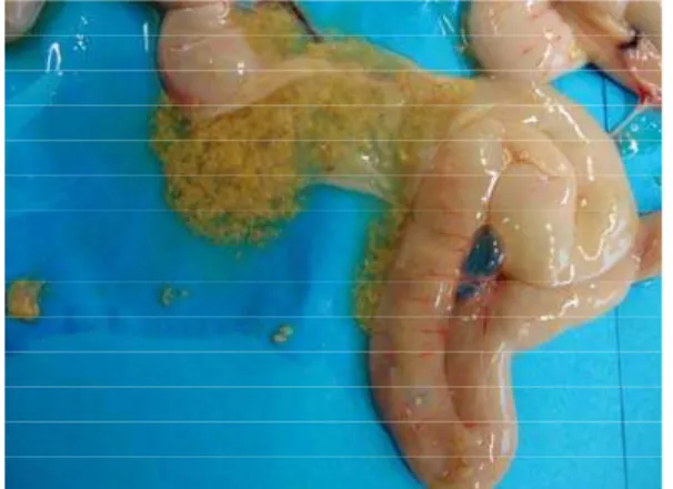
On autopsy dehydration is the most common finding. Breast muscles are dark red and gizzards are full of litter. Severe inflammation in the duodenum and jejunum is the most predominant finding, but in some instances the entire length of the intestinal tract is involved (Bains, 1968; Helmboldt and Bryant, 1971; Long et al., 1974; Tsai and Tung 1981; Ficken and Berkhoff, 1989). The intestine is distended thin walled and filled with gas and contains dark offensive fluid (Broussard et al., 1986). The mucosa is covered with green or brown diphteroid membrane, which can be easily separated from the lining (Fig. 1). Varying degrees of sloughing of the intestinal mucosa could also be observed (Fig. 2). As the condition progresses, areas of necrosis can be recognized from outside of the intestine (Helmboldt and Bryant, 1971; Long et al., 1974; Balauca, 1978; Shane et al., 1985).
Initial microscopic lesions develop at the apices of villi and are characterized by sloughing of epithelium and colonization of the exposed lamina propria with bacilli, accompanied by coagulation necrosis. Progression of lesions usually occurs from villi apices to crypts. Necrosis may extend into the submucosa and muscular layers of the intestine (Fig. 3) (Nairn and Bamford, 1967; Helmboldt and Bryant, 1971; Long et al., 1974; Tsai and Tung, 1981; Opengart, 2008). Large numbers of gram-positive bacilli can be seen (Fig. 4 and 5) within the necrotic debris (Randall, 1991). In per acute cases there is little inflammatory cell infiltrate although, if the animal survives, there is a progression to heterophil and mononuclear cell infiltration followed by fibrosis (Shane et al., 1985; Ficken and Wages, 1997).
Cholangiohepatitis
Cholangiohepatitis causes severe economic losses due to high liver condemnation rate on the processing and
downgraded of the slaughtered carcasses. Clostridium perfringens is usually isolated in association with the disease. The hepatitis characterized by an enlarged firm liver sometimes with a slightly knobby surface and a medium tan colour. Histopathological lesions consist of hyperplasia of the bile duct, fibrinoid necrosis, cholangitis and occasionally focal granulomatous inflammation (Onderka et al., 1990; Løvland and Kaldhusdal, 1999; Sasaki et al., 2000). Onderka et al. (1990) experimentally reproduced the condition by either tying off the bile ducts or injecting C. perfringens into the bile duct. It seems that the presence of C. perfringens in many of the gall bladders of affected livers suggested some involvement of either the bacterium or its toxin which interfere with the liver function.
Gizzard erosions
Gizzard erosions has been observed in commercial broiler chickens and several non-infectious factors such as mycotoxin-contaminated feed, vitamin B6 and E deficiency, inadequate levels of sulphur-containing dietary amino acids, high levels of dietary copper, pelleted feed as well as inclusion of certain fish meals in the diets and were discriminated as possible cause. Ono et al. (2003) reported on Outbreaks of adenoviral gizzard erosion in slaughtered broiler chickens in Japan and Novoa-Garrido et al. (2006) found a significant association between gizzard lesions and increased caecal C. perfringens counts in broiler chickens.
Diagnosis
A presumptive diagnosis may be made from the case history, clinical signs, lesions and staining fresh smears of upper part of the intestinal tract with Gram stain showing an abundant number of clostridia organisms (Ficken and Wages, 1997; Hafez and Jodas, 1997). This should be confirmed by the isolation of the causative agent. For isolation several media are available such as sheep blood agar supplemented with neomycin or tryptose-sulfite-cycloserine agar (TSC). The identification can be carried out using biochemical tests. Most of isolates ferment lactose, glucose, maltose, hydrolyze gelatin, and reduce nitrate. This bacterium is non-motile, indole and catalase negative (Ficken and Berkhoff, 1989). In addition, PCR was developed to detect of alpha toxin (Heikinheimo and Korkeala, 2005) as well as a real-time PCR for quantitative detection of C. perfringens in gastrointestinal tract of poultry (Wise and Siragusa, 2005). Also ELISAs for direct detection of C. perfringens major toxins and enterotoxin are commercially available.
Treatment
flocks using a commercially available broth micro-dilution test kit. No isolates were resistant against β -lactam antibiotics (amoxicillin, oxacillin, and penicillin), lincospectin, tylosin, doxycyclin, tetracycline, enrofloxacin, trimethoprim/sulfamethoxazole, lincomycin, and tilmicosin. A low frequency of resistance was detected against erythromycin and tiamulin with 5 and 20%, respectively. Spectinomycin, neomycin and colistin showed the highest incidence of resistance with 74, 94 and 100%, respectively.
According to Brennan et al. (2000) administration of dietary Tylan® for seven consecutive days following
Fig. 1: Necrotic enteritis: The mucosa is covered with green or brown diphtheroid membrane, which can be easily separated from the lining.
Fig. 2: Severe necrotic enteritis with necrotic pseudomem- brane covering the intestinal mucosa.
Fig. 3: Severe, acute, necrotizing enteritis with extensive transmural spreading as well as associated, necrotizing and granulomatous steatites/serositis; large number of Gram-Positive bacilli, located multifocal within lesions (H & E 20X) (Courtesy: Dr. Olivia Kershaw, Berlin).
Fig. 4: Necrotic enteritis: note large number of Gram-Positive bacilli, located multifocal on the surface as well as within lesions (Gram X200) (Courtesy: Dr. Olivia Kershaw, Berlin).
Fig. 5: Necrotic enteritis: note large number of Gram-Positive bacilli, located multifocal on the surface as well as within lesions (Gram X600) (Courtesy: Dr. Olivia Kershaw, Berlin).
confirmation of an NE field outbreak reduced the NE mortality and lesion score and improved overall growth as well as feed conversion in broilers. The optimum dose of Tylan to control NE was 100 ppm.
No resistance to the ionophorous anticoccidial drugs such as Narasin has been found (Watkins et al., 1997; Martel et al., 2004). Brennan et al. (2001) reported that Narasin, when administrated at 70 ppm in feed from Day 0 to 41 prevents morbidity, mortality and suppression of growth and feed conversion associated with NE in broilers.
20,000 birds raised to final body weights ranging from 4.63 to 7.94 lb. The incidence of SNE was assumed to occur at 20% based on the literature. SNE resulted in a loss to producers ranging from US$878.19 to US$1480.52 per flock. When feed costs required to obtain SNE flocks having a total live body weight equal to equivalent healthy flocks at market age were calculated, the increased cost to producers ranged from US$370.49 to US$739.38 per flock (Skinner et al., 2010).
Strategies to reduce the incidence of clostridial infections are necessary help to increase the profitability of the poultry production and several further approaches are generally used to combat the infection and as alternatives to AGPs. Investigations indicate that competitive exclusion, prebiotics, probiotics, enzymes and acids can impact the incidence and severity of NE in poultry (Fukata et al., 1991; Elwinger et al., 1992; Hofacre et al., 1998; Kaldhusdal et al., 2001). The data suggest that these products may provide the poultry industry with an alternative management tool that has the potential to promote better intestinal health and decrease monetary losses due to C. perfringens (McReynolds et al., 2009).
According to Langhout (2007),these approaches will need adaptations in the feeding program and/or feed production. The practical relevance of these approaches may vary between the different areas in the world. At this moment it is difficult to evaluate novel strategies developed to antibiotic-free feeding concepts. Combination of different approaches is necessary to enhance the performance and reduced health status of the birds such as:
i) Selection of highly digestible feed ingredients to reduce nutrients for microbial degradation.
ii) Improvement in the balance of the essential amino acids resulting in lower total dietary protein levels. This will reduce the risk for clostridium problems, since this bacterium in particular increases during proteolytic fermentation.
iii) Improvement in the physical form of the diet, for example via the inclusion of coarse particles in the diet. Coarse particles will improve the passage rate of the feed through the intestinal tract and as a consequence increase digestion and reduce bacterial fermentation in the intestinal tract.
iv) Introduction of a special prestarter diet in the feeding program. The main objective of this prestarter diet should be to stimulate the development of the immune system and the development of an optimal micro flora.
v) Improvement of climate control in the broiler house to avoid stress in the animal and keeping litter quality in optimal condition.
vi) Improvement of disease control in broilers. This disease control focuses much more on the prevention of health problems than on treatment of diseases.
Vaccination
Active and passive immunity using vaccination against C. perfringens and its toxins appears to offer protection. Heier et al. (2001) found out that broiler flocks with high titres of maternal antibodies against C. perfringens alpha-toxin had lower mortality during the
production period than flocks with low tiers. Also Løvland et al. (2004) use toxoids vaccines based on C. perfringens type A and C toxoids to vaccinate breeder flocks. The IgG responses in vaccinated parent hens were distinct and the levels of antibodies to C. perfringens alpha - toxin in progeny of the vaccinated hens was high enough to protect the progeny against subclinical C. perfringens associated necrotic enteritis. On the other hand several recent investigations showed that immunity to NE after oral infection using virulent strain and subsequent treatment is much better than using avirulent C. perfringens strains and they identified immunogenic secreted proteins apparently uniquely produced by virulent C. perfringens isolates and concluded that there are certain secreted proteins beside to alpha-toxin, that are involved in immunity to NE in broiler chickens (Thompson et al., 2006; Kulkarni et al., 2007). Further additional study showed the ability of oral immunisation against C. perfringens in broiler chickens using an attenuated Salmonella vaccine vector (Kulkarni et al., 2008).
Conclusions
Implementation of several approaches such as improvement of management, feed formulation and use of alternative products to modulate the intestinal flora led to an improvement of the situation. Limiting exposure to infectious agents through biosecurity, vaccination, supportive therapy, cleaning and disinfection are essential. In addition, early recognition in managing the enteric disorders is very important.
Finally, use of an effective anticoccidial drug in the ration is helpful to minimise the effect of enteritis. Since, recent investigations showed that the use of some alternative products might be able to reduce the intestinal colonization with pathogenic bacterial agents. This could be an additional tool to reduce enteric disorders in future.
Acknowledgements
I would like to thank Dr. Olivia Kershaw, Institute of Animal Pathology, Faculty of Veterinary Medicine, Free University Berlin for providing the histopathological figures.
REFERENCES
Abildgaard L, TE Sondergaard, RM Engberg, A Schramm, O Højberg, 2010. In vitro production of necrotic enteritis toxin B, NetB, by netB-positive and netB-negative Clostridium perfringens originating from healthy and diseased broiler chickens. Vet Microbiol, 144: 231-235.
Adams MR and MO Moss, 1995. Bacterial agents of foodborne illness. The Royal Society of Chemistry. Guildford. pp. 364.
Al-Sheikhly F and A Al-Saieg, 1980. Role of coccidia in the occurrence of necrotic enteritis of chickens. Avian Dis, 24: 324-333.
Annett CB, JR Viste, M Chirino-Trejo, HL Classen, DM Middleton and E Simko, 2002 Necrotic enteritis: effects of barley, wheat and corn diets on proliferation of Clostridium perfringens type A. Avian Path, 31: 599-602.
Baba E, AL Fuller, JM Gilbert, SG Thayer and LR McDougald, 1992. Effects of Eimeria brunetti infection and dietary zinc on experimental induction of necrotic enteritis in broiler chickens. Avian Dis, 36: 59-62.
Bains BS, 1968. Necrotic enteritis of chickens. Aust Vet J, 44:40.
Balauca N, 1978. Experimentelle Untersuchungen über die Clostridien Infektion und Intoxikation bei Geflügel, unter besonderer Berücksichtigung der Kokzidiose. Arch Vet, 13:127-141.
Balauca N, B Köhler, F Horsch, R Jungmann and E. Prusas, 1976. Experimentelle Reproduktion der nekrotischen Enteritis des Huhnes. II. Mitteilung. Weitere Mono- und Polyinfektionen mit Cl. perfringens und Kokzidien unter besonderer Berücksichtigung der Bodenhaltung. Arch Exp Veterinärmed, 30:913-923.
Batal AB and CM Parsons, 2002. Effects of age on nutrient digestibility in chicks fed different diets. Poult Sci, 81:400-407.
Bermudez AJ and B Stewart-Brown, 2008. Disease prevention and diagnosis. In: Diseases of Poultry.12th Ed, Saif YM, Fadly AM, Glisson JR, McDougald LR, Nolan LK and Swayne DE, eds. Iowa State University Press. Iowa, USA, pp: 5-42.
Branton SL, BD Lott, JW Deaton, WR Maslin, FW Austin, LM Pote, RW Keirs, MA Latour and EJ Day, 1997. The effect of added complex carbohydrates or added dietary fibre on necrotic enteritis lesions in broiler chickens. Poult Sci, 76: 24-48.
Branton SL, FN Reece and WM Hagler, 1987. Influence of a wheat diet on mortality of broiler chickens associated with necrotic enteritis. Poult Sci, 66: 1326-1330.
Brennan JJ, G Moore, S. Poe, G. Vessie, J Wilson, DA Barnum, A Zimmerman and P Dick, 2000. Efficacy of dietary tylosin phosphate (Tylan) for control of necrotic enteritis in broiler chickens. 89th Meeting Poultry Science Assoc., Montreal, Canada (Abs.). Brennan JJ, R Bagg, DA Barnum J. Wislon and P Dick,
2001. Efficacy of Narasin in the prevention of necrotic enteritis in broiler chicks. Avian Dis, 45: 210-214.
Broussard, CT, Hofacre, CL, Page, RK & Fletcher, OJ 1986. Necrotic enteritis in cage-reared commercial layer pullets. Avian Dis, 30: 617-619.
Brown TP, GE Rottinghaus and ME Williams, 1992. Fumonisin mycotoxicosis in broilers: Performance and pathology. Avian Dis, 36: 450-454.
Bryant AE and LS Stevens, 1997. The Pathogenesis of Gas Gangrene. Academic Press, San Diego, USA, pp: 186-187.
Burditt SJ, WM Hagler Jr. and PB Hamilton, 1983. Survey of molds and mycotoxins for their ability to cause feed refusal in chickens. Poult Sci, 62: 2187-2191.
Burns RB and P Dwivedi, 1986. The natural occurrence of ochratoxin A and its effects in poultry: A review. II. Pathology and immunology. World Poult Sci J, 42: 48-55.
Campbell MLJr, JA Doerr and RD Wyatt, 1981. Immune status in broiler chickens during citrinin toxicosis [abst]. Poult Sci; 60:1634.
Char NL, DI Khan, MRK Rao, V Gopal, and G Narayana, 1986. A rare occurrence of clostridial infections in poultry. Poult Advis, 19: 59-62.
Corless AB and JL Sell, 1999. The effects of delayed access to feed and water on the physical and functional development of the digestive system of young turkeys. Poult Sci, 78: 1158-1169.
Cowen BS, LD Schwartz, RA Wilson and SI Ambrus, 1987. Experimentally induced necrotic enteritis in chickens. Avian Dis, 31: 904-906.
Craven SE, NA Cox, JS Bailey and DE Cosby, 2003. Incidence and tracking of Clostridium perfringens through an integrated broiler chicken operation. Avian Dis, 47: 707–711.
Craven SE, NJ Stern, JS Bailey and NA Cox, 2001a. Incidence of Clostridium perfringens in broiler chickens and their environment during production and processing. Avian Dis, 45: 887-896.
Craven, SE, NA Cox, NJ Stern and JM Mauldin, 2001b. Prevalence of Clostridium perfringens in commercial broiler hatcheries. Avian Dis, 45: 1050-1053. Crespo R, DJ Fisher, HL Shivaprasad, ME
Fernandez-Miyakawa and FA Uzal, 2007. Toxinotypes of Clostridium perfringens isolated from sick and healthy avian species J Vet Diag Invest, 19: 329-333. D’Mello JPF and AMC MacDonald, 1998. Fungal toxins
as disease elicitors. In: Environmental toxicology: Current developments, Rose J, ed. Gordon and Breach Science Publishers Amsterdam, the Netherlands, pp: 253-289.
D’Mello JPF, 1997. Handbook of Plant and Fungal Toxicants. CRC Press, Boca Raton, New York. D’Mello JPF, AMC MacDonald and MP Cochrane, 1993.
A preliminary study of the potential for mycotoxin production in barley grain. Aspects of Applied Biology, 36: 375-382.
Devegowda G, MVLN Raju, N Afzali and HVLN Swamy, 1998. Mycotoxin picture worldwide: novel solutions for their counteraction. Proc Alltech Ann Symp, 14: 241-255.
Dhand NK, DV Joshi and SK Jand, 1998. Fungal contaminants of dairy feed and their toxigenicity. Indian J Anim Sci, 68: 1095-1096.
Dwivedi P and R B. Burns, 1986. The natural occurrence of ochratoxin A and its effects in poultry. A review. Part I. Epidemiology and toxicity. World Poult Sci J, 42:32-47.
El-Karim SA, MS. Arbid, AH Soufy, M Bastamy and MM Effat, 1991. Influence of metabolite ochratoxin A on chicken immune response. Egypt J Comp Pathol Clin Pathol, 4: 159-172.
Engström BE, C Fermer, A Lindberg, E Saarinen, V Båverud and A Gunnarsson, 2003. Molecular typing of isolates of Clostridium perfringens from healthy and diseased poultry. Vet Microbiol, 94: 225-235. Fabri THF, 2004. Necrotic enteritis, Clostridial enteritis or
dysbacteriosis?. Proceedings of the 5th International Symposium on Turkey Diseases, Berlin, Germany (ed. H.M. Hafez), DVG-Verlag, (ISBN 3-938026-15-4). pp: 225-230.
FAO, 2004. Assessing quality and safety of animal feeds. FAO Animal Production and Health Paper 160. FAO, Rome. http://www.fao.org/docrep/007/y5159e/y515 9e00.HTM
Ferket PR, 1996. Nutritional effects on enteric disorders. Proceedings of Enteric Disease Control. 39th Annual Meeting of the American Association of Avian Pathologists. Louisville, KY, USA, pp: 17-21.
Ficken MD and DP Wages,1997. Necrotic enteritis. In Diseases of Poultry. 10th Ed, Calnek BW, Barnes HJ, Beard CW, McDougald LR and Saif YM, eds. Iowa State University Press, Iowa, USA, pp: 261- 264. Ficken MD and HA Berkhoff, 1989. Clostridial
infections. In: Isolation and Identification of Avian Pathogens, 3rd Ed. Purchase HG, Arp LH, Domermuth CH and JE Pearson, eds., American Association of Avian Pathologists: Kennett Square, PA,USA, pp: 47-51.
Frame DD and AA Bickford, 1986. An outbreak of coccidiosis and necrotic enteritis in 16-week-old cage-reared layer replacement pullets. Avian Dis, 30:601-602.
Fukata T, Y Hadate, E Baba and A Arakawa, 1991. Influence of Bacteria on Clostridium perfringens Infections in Young Chickens. Avian Dis, 35: 224-227.
Gad W, R Hauck, M Krüger and HM Hafez, 2011a. Prevalence of Clostridium perfringens in commercial turkey and layer flocks. Archiv für Geflügelkunde, 75: 74–79.
Gad W, R Hauck, M Krüger and HM Hafez, 2011b. Determination of antibiotic sensitivities of Clostridium perfringens isolates from commercial turkeys in Germany in vitro. Archiv für Geflügelkunde, 75: 80-83
Gazdzinski P and RJ Julian, 1992. Necrotic enteritis in turkeys. Avian Dis, 36: 792-798.
Gholamiandehkordi A, R Ducatelle, M Heyndrickx, F Haesebrouck and F Van Immerseel, 2006. Molecular and phenotypical characterization of Clostridium perfringens isolates from poultry flocks with different disease status. Vet Microbiol, 113:143-152.
Hafez HM and S. Jodas, 1997. Nekrotisierende Enteritis In: Putenkrankheiten, Hafez HM and Jodas S ,eds. Ferdinand Enke Verlag Stuttgart Germany, pp: 80-84. Hafez HM, 1996. Übersicht über Probleme der haltungs-
und zuchtbedingten Erkrankungen bei Mastputen. Archiv für Geflügelkunde, 60: 249-256.
Hafez HM, 1998. Management and disease related problems in turkeys. Proceedings of the 2nd International Congress for Veterinarian and Farmers- Quality assurance and animal health management. DLG- Publisher. ISBN 3-7690-0567-8, pp: 63-77.
Hafez HM, 1999. Poultry meat and food safety: pre- and post-harvest approaches to reduce food borne pathogens. World Poult Sci J, 55: 269-280.
Hafez HM, 2001. Enteric diseases of turkeys. In: Turkey Production in Europe in the New Millennium, Hafez HM, ed. Ulmer Verlag Stuttgart, Germany, pp: 164-181.
Hafez HM, 2005. Governmental regulations and concept behind eradication and control of some important poultry diseases. World Poult Sci J, 61: 569-581. Hafez HM, 2008. Ban of antibiotic growth promoters in
the EU: The consequences on poultry health and alternative approaches. Faculty of Veterinary Medicine, Universidad Austral de Chile. Proceeding of the XI Seminario Internacional de Producción y Patología Aviar, 24.10.2008, Valdivia, Chile, pp: 92-100.
Heier BT, A Løvland, KB Soleim, M Kaldhusdal and J Jarp, 2001. A field study of naturally occurring specific antibodies against Clostridium perfringens alpha toxin in Norwegian broiler flocks. Avian Dis, 45: 724-732.
Heikinheimo A and H Korkeala, 2005. Multiplex PCR assay for toxinotyping Clostridium perfringens isolates obtained from Finnish broiler chickens. Letters Appl Microb, 40: 407-411.
Helmboldt CF and ES. Bryant, 1971. The pathology of necrotic enteritis in domestic fowl. Avian Dis, 15: 775-780.
Hoerr FJ. 2008. Mycotoxicoses. In: Diseases of Poultry.12th Ed, Saif YM, Fadly AM, Glisson JR, McDougald LR, Nolan LK and Swayne DE, eds. Iowa State University Press, Iowa, USA, pp., 1197-1229.
Hofacre CL, R. Froyman, B Gautrias, B George, MA Goodwin and J Brown, 1998. Use of Aviguard and other intestinal bioproducts in experimental Clostridium perfringens-associated necrotizing enteritis in broiler chickens. Avian Dis, 42: 579-584. Kaldhusdal M and A Løvland, 2000. The economical
impact of Clostridium perfringens is greater than anticipated. World Poult, 16: 50–51.
Kaldhusdal M and E Skjerve, 1996. Association between cereal contents in the diet and incidence of necrotic enteritis in broiler chickens in Norway. Prev Vet Med, 28: 1-16.
Kaldhusdal M, C Schneitz, M Hofshagen and E Skjerve, 2001. Reduced incidence of Clostridium perfringens -associated lesions and improved performance in broiler chickens treated with normal intestinal bacteria from adult fowl. Avian Dis, 45: 149 -156. Kaldhusdal M, M Hofshagen, A Løvland, H Langstrand
and K Redhead, 1999. Necrotic enteritis challenge models with broiler chickens raised on litter: evaluation of preconditions, Clostridium perfringens strains and outcome variables. FEMS Microbiol Med Immunol, 24: 337-343.
Kocher A, 2003. Nutritional manipulation of necrotic enteritis outbreak in broilers. Recent Adv Anim Nutr (Australia), 14: 111-116.
Köhler B, G Marx, S Köbach and E Böttcher, 1974a. Untersuchungen zur nekrotischen Enteritis der Hühner. 1. Mitteilung: Diagnostik und Bekämpfung. Monatsh für Veterinämedizin, 29: 380 -384.
`Köhler B, S Kölbach and J Meine, 1974b. Untersuchungen zur nekrotischen Enteritis der Hühner. 2. Mitteilung: Mikrobiologische Aspekte. Monatsh für Veterinärmedizin, 29: 385-391.
Komnenov V, M Velhner and M Katrinka, 1981. Importance of feed in the occurrence of clostridial infections in poultry. Vet Glas, 35:245-249.
Kulkarni RR, VR Parreira, S Sharif and JF Prescott, 2007. Immunization of Broiler Chickens against Clostridium perfringens -Induced Necrotic Enteritis. Can Vet J, 14: 1070–1077.
Kulkarni RR, VR Parreira, S Sharif and JF Prescott, 2008. Oral immunization of broiler chickens against necrotic enteritis with an attenuated Salmonella vaccine vector expressing Clostridium perfringens antigens. Vaccine, 26: 4194-4203.
Langhout P, 2007. Broilers nutrition optimisation. Afma Matrix, 16(2): 33-37.
Long JR and RB Truscott, 1976. Necrotic enteritis in broiler chickens III. Reproduction of the disease. Can J Com Med, 40: 53-59.
Long JR, 1973. Necrotic enteritis in broiler chickens. I. A review of the literature and the prevalence of the disease in Ontario. Can J Comp Med, 37: 302-308. Long JR, JR. Pettit and DA Barnum, 1974. Necrotic
enteritis in broiler chickens. II. Pathology and proposed pathogenesis. Can J Comp Med, 38:467-474.
Løvland A and M Kaldhusdal, 1999. Liver lesions seen at slaughter as an indicator of necrotic enteritis in broiler flocks. FEMS Microbiol Med Microbiol, 24: 345-351.
Løvland A and M Kaldhusdal, 2001. Severely impaired production performance in broiler flocks with high incidence of Clostridium perfringens-associated hepatitis. Avian Path, 30: 73-81.
Løvland A, M Kaldhusdal, K Redhead, E Skjerve and A Lillehaug, 2004. Maternal vaccination against subclinical necrotic enteritis in broilers. Avian Path, 33: 81-90.
Martel A, LA Devriese, K Cauwerts, K De Gussem, A Decostere and F Haesebrouck, 2004. Susceptibility of Clostridium perfringens strains from broiler chickens to antibiotics and anticoccidials. Avian Path, 33: 3-7. Martin TG and JA Smyth, 2009: Prevalence of netB
among some clinical isolates of Clostridium perfringens from animals in the United States. Vet Microbiol, 136: 202-205.
Martin TG, 2010. The importance of the strain of Clostridium perfringens in the development of necrotic enteritis of poultry. Dissertations Collection for University of Connecticut. Paper AAI3402008. http://digitalcommons.uconn.edu/dissertations/AAI34 02008. Accessed on January 1, 2010.
McClane BA, FA Uzal, MEF Miyakawa, D Lyerly and T Wilkins, 2006. The Enterotoxic Clostridia. In: The
Prokaryotes: A handbook on the biology of bacteria. Dworkin M and Falkow S. eds. Springer, pp: 763-778.
McDevitt RM, JD Brooker, T Acamovicand and NHC Sparks, 2006. Necrotic enteritis: A continuing challenge for the poultry industry. World Poult Sci J, 62: 221-247.
McDonel JL, 1986. Toxins of Clostridium perfringens types A, B, C, D, and E. In: Pharmacology of Bacterial toxins. Dorner F. and Drews H, eds. Oxford Pergamon Press, pp: 477-517.
McReynolds J, C Waneck, J Byrd, K Genovese, S Duke and D Nisbet, 2009. Efficacy of multistrain direct-fed microbial and phytogenetic products in reducing necrotic enteritis in commercial broilers. Poult Sci, 88: 2075-2080.
McReynolds JL, JA Byrd, RC Anderson, RW Moore, TS Edrington, KJ Genovese, TL Poole, LF Kubena and DJ Nisbet, 2004. Evaluation of immunosuppressants and dietary mechanisms in an experimental disease model for necrotic enteritis. Poult Sci, 83: 1948-1952. Moore PR, A Evensson, TD Luckey, E Mccoy, CA
Elvehjam and EB Hurt, 1946. Use of sulfasuxidine, stretothrian and streptomycin in nutritional studies with chick. J Biol Chemist, 165: 437-441.
Morgen RE and JS Avens,1985. Poultry Sceince and production. Reston Publishing Company, Inc. Reston, Verginia.
Nairn MEB and VW Bamford, 1967. Necrotic enteritis of broiler chickens in Western Australia. Aust Vet J, 43:49-54
Nixey C,1989. Nutritional responses of growing turkeys. In: Recent advance in turkey science, Nixey C and Grey TC, eds. Butterworth, London, pp: 183-199. Novoa-Garrido M., S Larsen and M Kaldhusdal, 2006.
Association between gizzard lesions and increased caecal Clostridium perfringens counts in broiler chickens. Avian Path, 35: 367-372.
Onderka DK, CC Langevin and JA Hanson, 1990. Fibrosing cholehepatitis in broiler chickens induced by bile duct ligations or inoculation of Clostridium perfringens. Can J Vet Res, 54: 285-290.
Ono M., Y Okuda, S Yazawa, I Shibata, S Sato. and K Okada, 2003. Outbreaks of adenoviral gizzard erosion in slaughtered broiler chickens in Japan. Vet Rec, 153: 775-779.
Opengart K, 2008. Necrotic enteritis. In: Diseases of Poultry.12th Ed, Saif YM, Fadly AM, Glisson JR, McDougald LR, Nolan LK and Swayne DE, eds. Iowa State University Press. Iowa, USA, pp: 872-879.
Panneman H, 2000. Clostridial enteritis/dysbacteriosis, fast diagnosis by T-RFLP, a novel diagnostic tool. Proc Elanco Symposium, Montreal, Canada, pp: 19 -29.
Parish WE, 1961. Necrotic enteritis in the fowl (Gallus gallus domesticus). I. Histopathology of the disease and isolation of a strain of Clostridium welchii. J Comp Path, 71: 377-393.
Porter RE,1998. Bacterial enterides of poultry. Poult Sci, 77: 1159-1165.
Sainsbury D, 1992. Poultry health and management. Blackwell Scientific Publications, London.
Sasaki J, M Goryo and K Okada, 2000. Cholangiohepatitis in chickens induced by bile duct ligations and inoculation of Clostridium perfringens. Avian Path, 29: 405 -410.
Shane SM, DG Koetting and KS Harrington, 1984. The occurrence of Clostridium perfringens in the intestine of chicks. Avian Dis, 28: 1120-1124.
Shane SM, JE Gyimah, KS Harrington and TG Snider, 1985. Etiology and pathogenesis of necrotic enteritis. Vet Res Commun, 9: 269-287.
Siegel PB, AS Larsen, CT Larsen and EA Dunnington, 1993. Resistance of chickens to an outbreak of necrotic enteritis as influenced by major histocompatibility genotype and background genome. Poult Sci, 72: 1189-1191.
Skinner JT, S Bauer, V Young, G Pauling and J. Wilson, 2010. An Economic Analysis of the Impact of Subclinical (Mild) Necrotic Enteritis in Broiler Chickens. Avian Dis, 54:1237-1240.
Smedley JG3rd, DJ Fisher, S Sayeed, G Chakrabarti. and BA McClane, 2004. The enteric toxins of Clostridium perfringens. Rev Phys Biochem Pharm, 152: 183-204.
Smith JW and PB Hamilton, 1970. Aflatoxicosis in the broiler chicken. Poult Sci 49:207-215.
Songer JG and RR Meer, 1996. Genotyping of Clostridium perfringens by polymerase chain reaction is a useful adjunct to diagnosis of clostridial enteric disease in animals. Anaerobe, 2: 197-203.
Songer JG, 1996. Clostridial enteric diseases of domestic animals. C Microb Rev, 9: 216-234.
Sundrum A, 1995. Zur Beurteilung der Tiergerechtheit von Haltungsbedingungen. Schriftreihe der DVG, Tagung der Fachgruppe ”Tierschutzrecht und Gerichtliche Veterinärmedizin”- 9/10 März , Stuttgart Hohenheim, Germany, pp: 23-34.
Thompson DR, VR Parreira, RR Kulkarni and JF Prescott, 2006. Live attenuated vaccine-based control of necrotic enteritis of broiler chickens. Vet Microbiol, 113: 25–34.
Timbermont l, A Lanckriet, AR. Gholamiandehkordi, F Pasmans, A Martel, F Haesebrouck, R Ducatelle and
F Van Immerseel, 2009. Origin of Clostridium perfringens isolates determines the ability to induce necrotic enteritis in broilers. Comp Immunol Microbiol Infect Dis, 32: 503–512.
Tsai SS and MC Tung, 1981. An outbreak of necrotic enteritis in broiler chickens. Journal of the Chinese Society Vet Sci, 7: 13-17.
Uraguchi K and Myamazaki, 1978. Toxicology, Biochemistry and Pathology of Mycotoxins. Halsted Press, John Wiley and Sons, New York, pp: 1-106. Van Immerseel F, J De Buck, F Pasmans, G Huyghebaert,
F Haesebrouck and R Ducatelle, 2004. Clostridium perfringens in poultry: an emerging threat for animal and public health. Avian Path, 33: 537-549.
Waters M, A Savoie, HS Garmory, D Bueschel, MR Popoff, JG Songer, RW Titball, BA McClane and MR Sarker, 2003. Genotyping and phenotyping of beta-2 toxigenic Clostridium perfringens faecal isolates associated with gastrointestinal diseases in piglets. J Clin Microb, 41: 3584-3591.
Watkins KL, TR Shryock, RN Dearth and YM Saif, 1997. In vitro antimicrobial susceptibility of Clostridium perfringens from commercial turkey and broiler chicken origin. Vet Microbiol, 54: 195-200.
Wijewanta EA and P Seneviratna, 1971. Bacteriological studies of fatal Clostridium perfringens type-A infection in chickens. Avian Dis, 15: 654-661.
Williams RB, 2005. Intercurrent coccidiosis and necrotic enteritis of chickens: rational, integrated disease management by maintenance of gut integrity. Avian Path, 34: 159-180.
Willis AT, 1977. Anaerobic-bacteriology: clinical and laboratory practice. (3rd ED) Butterworths, London, pp: 360.
Wilson J, G Tice, ML Brash and S St Hilaire, 2005. Manifestations of Clostridium perfringens and related bacterial enteritides in broiler chickens. World Poult Sci J, 61: 435-449.