Sialyl Tn-expressing bladder cancer cells induce a tolerogenic
phenotype in innate and adaptive immune cells
Myl
ene A. Carrascal
a, Paulo F. Severino
a,b, M. Guadalupe Cabral
a,c,
Mariana Silva
a, Jos
e Alexandre Ferreira
d,e, Fernando Calais
f,
Herm
ınia Quinto
f, Cl
audia Pen
f, D
ario Ligeiro
g, L
ucio Lara Santos
e,h,
Fabio Dall’Olio
b, Paula A. Videira
a,*
aCEDOC, Faculdade de Ci^encias Medicas, Universidade NOVA de Lisboa, Lisbon, Portugal
bDepartment of Experimental, Clinical and Specialty Medicine (DIMES), University of Bologna, Bologna, Italy c
Faculdade de Engenharia, Universidade Lusofona de Humanidades e Tecnologias, Lisbon, Portugal
dQOPNA, Mass Spectrometry Center, Department of Chemistry, University of Aveiro, Aveiro, Portugal eExperimental Pathology and Therapeutics Group, Portuguese Institute of Oncology, Porto, Portugal fCentro Hospitalar de Lisboa Central, EPEeServic¸o de Anatomia Patologica, Lisbon, Portugal gCentro de Histocompatibilidade do Sul, Lisboa, Portugal
hDepartment of Surgical Oncology, Portuguese Institute of Oncology, Porto, Portugal
A R T I C L E I N F O
Article history:
Received 2 January 2014 Received in revised form 20 February 2014
Accepted 21 February 2014 Available online 6 March 2014
Keywords: Dendritic cells Sialyl-Tn
Immunological potency T cells
CD44 Mucins
A B S T R A C T
Despite the wide acceptance that glycans are centrally implicated in immunity, exactly how they contribute to the tilt immune response remains poorly defined. In this study, we sought to evaluate the impact of the malignant phenotype-associated glycan, sialyl-Tn (Ssialyl-Tn) in the function of the key orchestrators of the immune response, the dendritic cells (DCs). In high grade bladder cancer tissue, the STn antigen is significantly overex-pressed and correlated with the increased expression of ST6GALNAC1 sialyltransferase. Bladder cancer tissue presenting elevated expression of ST6GALNAC1 showed a correlation with increased expression of CD1a, a marker for bladder immature DCs and showed concomitant low levels of Th1-inducing cytokines IL-12 and TNF-a.In vitro, human DCs co-incubated with STnþ
bladder cancer cells, had an immature phenotype (MHC-IIlow,
CD80lowand CD86low) and were unresponsive to further maturation stimuli. When
contact-ing with STnþ
cancer cells, DCs expressed significantly less IL-12 and TNF-a. Consistent with a tolerogenic DC profile, T cells that were primed by DCs pulsed with antigens derived from STnþ
cancer cells were not activated and showed a FoxP3highIFN-glowphenotype.
Blockade of STn antigens and of STnþ
glycoprotein, CD44 and MUC1, in STnþ
cancer cells was able to lower the induction of tolerance and DCs become more mature.
Overall, our data suggest that STn-expressing cancer cells impair DC maturation and endow DCs with a tolerogenic function, limiting their capacity to trigger protective anti-tumour T cell responses. STn antigens and, in particular, STnþ
glycoproteins are potential targets for circumventing tumour-induced tolerogenic mechanisms.
ª2014 Federation of European Biochemical Societies.
Published by Elsevier B.V. All rights reserved.
* Corresponding author. Immunology Department, CEDOC, Faculdade de Ci^encias Medicas, Universidade Nova de Lisboa, Campo Martires da P atria 130, 1169-056 Lisboa, Portugal. Tel.: þ351 218 803 045; fax:þ351 218853480.
E-mail addresses:[email protected],[email protected](P.A. Videira).
a v a i l a b l e a t
w w w . s c i e n c e d i r e c t . c o m
ScienceDirect
w w w . e l s e v i e r . c o m / l o c a t e / m o l o n c
http://dx.doi.org/10.1016/j.molonc.2014.02.008
1.
Introduction
The sialyl-Tn (STn) antigen is one of the most common tumour-associated carbohydrate antigen, expressed by more than 80% of human carcinomas but rarely observed in normal tissues (Cao et al., 1996). STn is a posttranslational modifica-tion of cell surface glycoproteins commonly resulting from the overexpression of the ST6GALNAC1 sialyltransferase.
This enzyme transfers a sialic acid to theO-6 position of
N-acetylgalactosamine residue linked to a serine or a threonine (GalNAca-O-Ser/Thr) on a given protein, blocking the typical
elongation of O-glycosidic chains in glycoproteins. STn
expression in cancer is associated with adverse outcome
and decreased overall survival of the patients (Itzkowitz
et al., 1990; Werther et al., 1996). It has been reported that STn expression by cancer cells is associated with many malig-nant features such as invasiveness (Julien et al., 2012; Pinho et al., 2007), and with epithelial to mesenchymal transition (Lin et al., 2009), a loss of cell differentiation that is an impor-tant milestone towards cancer metastasis.
STn is highly expressed in high-grade bladder tumours, which present elevated proliferation rates and high risk of recurrence/progression. In bladder cancer, STn enhances motility and invasive capacity of the cancer cells, thus being associated with malignancy (Ferreira et al., 2013).
The expression of STn is clinically relevant, not only as a marker for diagnosis and prognosis in cancer (the CA72-4 serological test), but also as target for therapeutic strategies. One example is a vaccine consisting of STn-clustered epi-topes, Theratope, that has been meaningfully used in clinical trials to immunize breast cancer patients (Adis International, 2003; Holmberg and Sandmaier, 2004; Julien et al., 2012). Other STn-based vaccines have also been developed for application in clinical trial, which includes multi-epitope vaccines con-taining STn and other tumour-associated carbohydrates and
STn-expressing glycopeptides (Heimburg-Molinaro et al.,
2011; Madsen et al., 2013; Niederhafner et al., 2008; Slovin et al., 2007). Nevertheless, STn-based vaccines have had limited success (Miles et al., 2011) probably because the path-ophysiological role of STn-epitopes in cancer cells remains unclear. One of the factors that has been highlighted by many authors is the low immunogenicity of STn-based
vac-cines (Julien et al., 2012; Lakshminarayanan et al., 2012;
Monti et al., 2004). Thus, the elucidation of the immune mech-anisms affected by the expression of STn by cancer cells will contribute to improve anti-STn immunotherapy approaches.
Glycans are involved in multiple biological functions, con-trolling many features of the immune response. In fact the di-versity of glycans and of glycan-recognizing receptors, known as lectins, is highly regulated by the immune cells. One of the examples is the modulation of dendritic cell (DC) functions by changes of their glycan phenotype during differentiation and maturation (Crespo et al., 2009; Videira et al., 2008).
DCs play a unique and decisive role in tumour immunity, being capable of activating antigen-specific T cells against cancer cells (Banchereau and Steinman, 1998). To efficiently prime T cells, DCs undergo maturation, which includes the downregulation of the antigen-uptake machinery, the upregu-lation of antigen presenting molecules, class I and class II
MHC; costimulatory molecules, such as CD80 and CD86 and
the synthesis of immune-enhancing cytokines (Langenkamp
et al., 2000). However, the degree of DC maturation is depen-dent on the type of stimulus and tumour cells usually prevent maturation, through many immunosuppressive strategies
employed in situ. Tumours render DC tolerogenic and bias
the immune response in favour of their own progression (Almand et al., 2001; Vicari et al., 2002). The presence of imma-ture DCs at tumour sites is consistent with DC involvement in the tumour progression, most likely by inducing immune un-responsiveness, i.e. tolerance against cancer (Almand et al., 2001). We have previously reported, in bladder cancer tissue, that the lower expression of markers of DC maturation, such
as MHC-II, was correlated with risk for recurrence (Videira
et al., 2009a). Tumour-residing DCs showing limited matura-tion or anergy, hold back strategies to induce immune re-sponses and create one important obstacle to the efficacy of immune-based-therapies. Concordantly, in bladder cancer tissue, lower levels of mature DCs are associated with low
re-sponses to theBacillus Calmette-Guerin(BCG) immunotherapy,
the gold standard treatment for the prophylaxis and
manage-ment of non-muscle invasive bladder cancer (Beatty et al.,
2004; Videira et al., 2009a).
It has been described that STn epitope may confer, to different cancer cell, protection from immune defence thus contributing to malignant phenotype and cancer progression (Monti et al., 2004; Ozaki et al., 2012). Mucins, and in particular STnþ
MUC1 mucins released by cancer cells inhibited DC maturation and modulate DCs towards IL-10highIL-12low
regu-latory antigen presenting cells with a limited capacity to
trigger protective T helper type 1 (Th1) responses (Monti
et al., 2004). Interestingly, soluble aberrantly glycosylated MUC1 has also been described to elicit maturation, yet unable to promote Th1 responses (Carlos et al., 2005). While the effect of glycoproteins secreted by tumours is becoming more eluci-dated, the role of the overall STn expression at tumour cell surface in immunomodulation, remains unknown. Therefore, in this study, we further investigated the influence of STn expression by bladder cancer cells on the immune potency and functionality of human DCs.
2.
Material and methods
2.1. Reagents
Netherlands). All other reagents were from Sigma (St. Louis, MO, USA) unless otherwise stated.
2.2. Patient and tissue specimens
This study involved 49 patients, from Hospital S~ao Jose in Lis-bon, who underwent transurethral resection of the bladder tu-mours. Matched pairs of histologically verified bladder tumours and normal appearing mucosa remote from the tumour were collected and analysed individually. Based on urothelial carcinoma grading and staging criteria of the World Health Organization 132 (WHO), three different groups were
considered (Table 1), low-grade (LG, n¼ 22) and high-grade
HG non muscle-invasive (NMIBC, n ¼ 21) and
muscle-invasive (MIBC,n¼6) bladder cancers. None of these patients had received prior adjuvant therapy. Patients withcarcinoma in situ(CIS) were not included, as well as patients with presence of upper tract malignancy, other malignancies, and chronic infections, women expectant or lactating and patients with congenital or acquired immunodeficiency. Prior patient con-sent and approval from the institute research ethics commit-tee were obtained.
2.3. Histological analysis
The immunohistochemical analysis was performed in
auto-mated equipment (Ventana BenchMark
ULTRA). All reagents were from Ventana, USA, unless otherwise stated. Briefly, the
slides were heated at 72
C, deparaffinized with EZ Prep and
antigenic recovery at 97
C. Endogenous peroxidase was blocked with 3% of hydrogen peroxide and the slides were incubated with TKH2 mAb (1:10). This was followed by ampli-fication with HRP UltraView Universal Multimer, revelation with UltraView Universal DAB Chromogen and DAB H202 and the intensification was achieved with UltraView Univer-sal DAB Copper. The nuclear contrast was performed with he-matoxylin and bluing. After the immunohistochemical technique, the slides were washed, dehydrated, treated with increasing concentrations of alcohol (75%, 90% and 99%) for 1 min each, cleared in xylene and mounted with synthetic
mounting medium (Quick-D-M-Klinipath). A
semi-quantitative approach was established to score STn expres-sion based on the percentage of tumour that stained positively in comparison to the tumour bulk. The STn expression was assessed double-blindly by two independent observers and validated by an experienced pathologist. Whenever there was a disagreement, the slides were reviewed, until a consensus was reached.
2.4. Cell isolation and culture
Monocytes were isolated by positive selection using anti-CD14 coated magnetic beads (Miltenyi Biotech, Germany) from pe-ripheral blood mononuclear cells (PBMCs) of healthy volun-teers, provided and ethically approved by the Portuguese Blood Institute. Monocytes were differentiated into immature mo-DCs (mo-DCs) as described (Videira et al., 2008). Whenever needed, maturation of mo-DCs (mmo-DCs) was induced, at day 5, with 1mg/ml of lipopolysaccharide (LPS), for 24 h.
2.5. Flow cytometry
Cell purity, differentiation and maturation of mo-DCs was assessed by staining with fluorescein isothiocyanate (FITC)-labelled mAbs against CD14, BDCA-1, CD80 and CD86 or Allo-phycocyanin (APC)-labelled anti-HLA-DR. Unlabelled mouse anti-STn, -CD44 or -MUC1, followed by anti-mouse Ig-FITC were used to characterize MCR cells. Flow cytometry acquisi-tion was performed, using a FacsCalibur Flow Cytometer. File data was analysed using the CELL QUEST (BD Biosciences) and FLOWING (Turku, Finland) software to discriminate specific populations, and to determine the mean fluorescence inten-sity (MFI) of the cells.
2.6. Cell lines
The human bladder cancer cell line variants, MCRcont and
MCRSTn, were generated as described (Ferreira et al., 2013)
and were grown in Dulbecco’s modified Eagle medium (DMEM) (Sigma), supplemented with foetal bovine serum, glutamine, penicillin and streptomycin.
2.7. Confocal laser scanning microscopy
Cells were cultured in cover glasses, fixed with 3.7% parafor-maldehyde and permeabilized with 0.1% TritonX-100. After blocking with 1% bovine serum albumin (BSA), cells were stained using anti-STn, clone TKH2, or anti-ST6GALNAC1, clone 2C3 (Marcos et al., 2011), followed by fluorescent poly-clonal anti-Ig antibody. The cell nuclei were stained with 1mM TO-PRO-3 dye (Molecular Probes, Leiden, Netherlands). Images were acquired with a Leica TCS SP2 AOBS confocal microscope
(Leica Microsystem, Mannheim, GmbH). Representative
confocal cross-section images were selected after Z-stacking.
2.8. Establishment of DC: bladder cancer cell lines cocultures
Cancer cell lines were incubated at 0.2106cells/ml in a 6-well
plate, at 37C. After 24 h, mo-DCs were added in the proportion
of 1:5 (cancer cell: mo-DC) in adhesion buffer (20 mmol/l of trizma hydrochloride, 150 mmol/l of sodium chloride, 1 mmol/l calcium chloride, 1 mmol/l magnesium chloride and 0.5% BSA, pH 8.0) at 37C. After 2 h incubation, the
non-adherent mo-DCs were washed and the coculture of MCR with adherent mo-DCs was stained with mAbs against MHC-II, which is expressed by DCs, but not by MCR cell lines, thus allowing to assess the percentage of adhering mo-DCs in the coculture. The cell viability was measured by annexin-V
Table 1eInformation of patients included in this study.
Number of cancer patients
Age, years median
(range)
Total number of cases 49
Muscle invasive (MIBC) 6 65.1 (56e79) Non muscle invasive (NMIBC)
High grade 21 68.7 (47e84)
staining. Some experiments were performed in adhesion buffer without calcium or magnesium, to assess the impact of divalent ions. In other experiments, mo-DCs were incubated with 24 h supernatants obtained from MCR cell cultures to assess the requirement for cell: cell interactions. When testing function blocking mAb, to avoid unspecific Fc receptor-mediated mAb binding to DCs, the Fc receptors from mo-DCs were blocked with 10% human serum. In addition, the blocking antibodies were added to the MCR cells and not to
mo-DCs. We used 20 mg/ml of anti-CD44 mAb (clone IM7),
1:100 of anti-STn mAb (clone HB-STn1), 1:20 of anti-MUC1 (clone HMFG-2) or isotype control for 30 min, washed and then cocultured the mAb coated MCR cells with mo-DCs, as described above.
2.9. Gene expression analysis by real-time PCR
RNA extraction from formalin-fixed, paraffin embedded sec-tions was performed after deparaffinization of tissues, using Absolutely RNA FFPE kit (Agilent technologies) while RNA from the cocultures was isolated using the GenElute Mamma-lian Total RNA Purification kit (Sigma), according to the man-ufacturer’s instructions. After DNAase treatment, 1mg of total RNA was reverse transcribed with random primers and the real-time PCR was performed with Master Mix, TaqMan probes and primers from Applied Biosystems. The assay ID provided by the manufacturer were: Hs00300842_m1 (ST6GAL-NAC1); Hs00168405_m1 (IL-12a); Hs00174128_m1 (TNF-a);
Hs00174086_m1 (IL-10); Hs00372324_m1 (IL-23),
Hs00203958_m1 (FoxP3) and Hs00174143_m1 (IFN-g). The rela-tive mRNA levels were normalized against the arithmetic
mean of theb-actinand GAPDH expression and calculated by
adapted formula 2 DCt 1000 which infers the number of
mRNA molecules of the gene of interest per 1000 molecules of the endogenous controls (Videira et al., 2009b).DCt stands for the difference between the cycle threshold of the target gene and that of the endogenous control genes. The efficiency for each primer/probe was above 95% (as determined by the manufacturer). When analysing the gene expression of mo-DCs in the coculture, the contamination with MCR cells was disregarded since the analysis of cytokine gene expression in both MCR cell line variants showed a completely absence ofIL-12a,TNF-a, IL-10,IL-23,FoxP3andIFN-ggene expression.
2.10. Phagocytosis assay
Cell lines were labelled with CFSE according to the manufac-turer’s instructions and then induced to apoptosis with
10mM of camptothecin. After 48 h, cells were incubated with
mo-DCs in the proportion of 1:2, in a 48-well plate, for 6 h at 37C or 4C. After incubation, cells were stained with
anti-MHC-II mAb and the percentage of anti-MHC-IIþ
/CFSEþ
cells (mo-DCs that phagocytosed MCR cells) was calculated by flow cytometry and confirmed by confocal microscopy. The values
obtained at 4C were subtracted from the 37C values.
2.11. T cell activation
Human T cells were obtained during monocyte isolation pro-cedure (CD14 PBMC fraction) and maintained in complete
RPMI medium until complete mo-DC differentiation. Autolo-gous T cells were then incubated with mo-DCs following phagocytosis of MCR cells, as described above, in the propor-tion of 8:1, in a 96-well round bottom plate, throughout 11 days. As controls, similar experiments were performed with unstimulated mo-DC or with the MCR cell lines (negative con-trols) or with phytohaemagglutinin (positive control). T cell
activation was assessed by the percentage of CD69þ
cells
within the CD3þ
population (T cells) and their MFI values were evaluated by flow cytometry.
2.12. Protein extraction, immunoprecipitation and western blotting
Proteins were isolated from cell lines using RIPA buffer
(Sig-maeAldrich) and bladder tumour proteins were extracted
from formalin-fixed paraffin embedded tissues using the Qproteome FFPE tissue kit (Qiagen). The amount of protein was estimated with RC protein assay kit (BioRad). CD44 pro-tein was immunoprecipitated from total propro-tein extracts (IP) with anti-CD44 monoclonal antibody (2C5 clone; R&D Sys-tems) using Pierce Direct IP Kit (Thermo Scientific). Protein samples were separated in reducing SDS-PAGE gels, using 20mg per lane, transferred to 0.45mm nitrocellulose membrane (GE Healthcare Life Sciences) and blocked with 1% Carbo-Free Blocking Solution (Vector Laboratories). TKH2 and goat anti-mouse IgG1 heavy chain horseradish peroxidase conjugate (Abcam) were used as primary and secondary antibodies, respectively. The Amersham ECL Prime Western Blotting Detection Reagent (GE Healthcare Life Sciences) was used as developing reagent. Protein extracts treated with sialidase were used as controls.
2.13. Statistical analysis
Statistical analyses were conducted using GraphPad Prism software, version 5.0 (GraphPad Software, La Jolla, CA). Paired
student’st-test was used when data was normally distributed,
and the results expressed as the mean valuesSDs.
Alterna-tively, Wilcoxon signed-rank test was used in the case of non-normal distribution, and the results were plotted individ-ually or as box and whiskers plot. The correlations were ana-lysed using Spearman and Pearson methods. Tests were considered statistically significant whenp<0.05 (*),p<0.01 (**) andp<0.005 (***) and marginal significance was considered
forp<0.1.
3.
Results
3.1. The STn antigen expression and DCs are increased in bladder cancer
(Figure 1B) either in low and high grade tumours. To assess whether STn expression was correlated with altered immune function, we quantified the expression of ST6GALNAC1 and CD1a, a marker for immature DC subtype predominant in bladder carcinoma (Figure 1S) (Troy et al., 1999). We also investigated the expression of interleukin (IL)-12, and tumour necrosis factor (TNF)-a, cytokines naturally expressed by DCs and involved in inducing Th1-immune responses. We have analysed tumour tissue and adjacent normal urothelium, and our data showed that both CD1a and ST6GALNAC1 are upregulated in tumour tissue, as compared with correspond-ing normal urothelium. Interestcorrespond-ingly, the upregulation was more pronounced and significantly correlated in muscle inva-sive and high grade non muscle invainva-sive tumours rather than
low grade tumours (Figure 1C). By contrast IL-12 and TNF-a
expression, which is associated with DC maturation and Th1-inducing, showed an inverse correspondence with
significantly lower expression in high grade tumour tissue than low grade tumour tissue. A correlation was observable
between CD1a and ST6GALNAC1 (r ¼ 0.41 and p ¼ 0.003)
expression, pointing out for an association between STn expression by cancer cells and the presence of immature DCs in tumour tissue.
3.2. Mo-DCs tend to adhere to STnþ
bladder cancer cell lines and show significant less mature phenotype
To further investigate the functional implications of STn-expressing cancer interaction on DC functionality, we
estab-lished in vitro coculture models with STnþ
bladder cancer
cell lines and human mo-DCs. The transduction of
ST6GAL-NAC1sialyltransferase cDNA in MCR bladder cancer cell lines,
that natively do not express STn, induced a dramatic expres-sion of STn (Figure 2A), corroborating the role of ST6GALNAC1
Figure 1eBladder cancer tissues show differential expression of STn antigen, CD1a, IL-12 and TNF-acytokines. A: Analysis of STn expression
in high grade tumour bladder cancer tissue. Representative image of a paraffin embedded section, processed for immunohistochemical staining with anti-STn mAb. STn expression was detected in tumour tissue. B: Association between ST6GALNAC1 and STn expression in bladder tumours. STn expression was determined in low and high grade bladder tumours, by immunohistochemistry, using TKH2 mAbs. Specimens were then selected and distributed into three groups, according to their expression of STn related with the tumour bulk: 0%, 0e15% and more than 15% of tissue expressing STn. The relative mRNA levels ofST6GALNAC1gene, in the paraffin-embedded sections, was analysed individually, by real time PCR and paired compared with data regarding STn expression. Values infer the number of mRNA molecules of aST6GALNAC1gene, per 1000 molecules of the average of the endogenous controls. C: Gene expression analysis in bladder tissue. The relative mRNA levels ofCD1a,IL-12
andTNF-acytokines andST6GALNAC1genes were analysed by real time RT-PCR in specimens from low and high grade non-muscle invasive bladder cancer (NMIBC) (n[22 andn[21, respectively) and muscle invasive bladder cancer (MIBC) (n[6) tissues and also from matched
normal urothelium (n[49). Values infer the number of mRNA molecules of each gene per 1000 molecules of the average of the endogenous
controls.CD1aandST6GALNAC1expression was significantly increased in all tumour tissue, as compared with matched urothelium; whileIL-12
andTNF-acytokines were decreased (p<0.05 (*) and
in STn biosynthesis by bladder cancer cells (Ferreira et al.,
2013). The obtained STnþ
(MCRSTn) and mock transduced STn (MCRcont) cell lines were used throughout this study. Co-incubation of human mo-DCs together with MCR cells showed that a significant number of mo-DCs adhered to the cancer cells. The mean percentage of adhering mo-DCs was significantly higher in the coculture with MCRSTn (1.27-fold
more) than with MCRcont (Figure 2B). We also investigate
whether the observed cellular interactions were dependent on divalent ions, a characteristic of many intercellular inter-actions. In the absence of divalent ions, mo-DC adhesion to bladder cancer cells was reduced to an average of 10% adherent cells and it was not statistically different between the cocultures with each of two cell line variants (data not shown), suggesting that the differences in adhesion of mo-DCs to MCR cells were dependent on divalent cations.
To characterize the maturation phenotype of the mo-DCs adhering to the bladder cancer cells we assessed the expres-sion of the MHC-II antigen presenting molecule, and co-stimulatory molecules, CD80 and CD86. While the mo-DCs co-incubated with MCRcont cells showed increased expres-sion of MHC-II, CD80 and CD86, mo-DCs adhering to MCRSTn
presented an expression level similar to the non-stimulated mo-DCs, suggesting that no maturation was induced (Figure 2C, D and E). To verify whether the immature pheno-type of mo-DCs incubated with MCRSTn was dependent on cell contact, parallel experiences were performed where mo-DCs were incubated in the presence of conditioned media, ob-tained from MCRcont or MCRSTn cell cultures. However, in these conditions the phenotype of mo-DCs incubated with the conditioned medium from either MCR variants was similar (data not shown). The viability of the mo-DCs in culture was approximately 97%, indicating that the observed differences in adhesion and maturation were not due to loss of mo-DC viability. Effects on the MCR cells was also observed, but only when the cocultures were prolonged until 24 h. Specifically, we observed that the proliferative capacity of MCRSTn cells increased 26% (p¼0.007), as compared with the proliferative capacity of this cell line in the absence of mo-DCs (Figure 2S).
In order to confirm that the effects on maturation and adhesion to mo-DCs were specifically caused by the enhanced expression of STn, the adhesion assays were performed with a different cell line, namely the breast cancer cell line,
MDA-MB-231STn, overexpressing the STn antigen (Julien et al.,
Figure 2eMo-DCs adhere preferentially to STnD
MCR cell line and show a less mature phenotype. A: Overexpression of ST6GALNAC1 in MCR bladder cancer cells. MCR cells were transduced or not with a retroviral vector expressing the whole coding region of humanST6GALNAC1. Both negative control (MCRcont) and ST6GALNAC1-transduced (MCRSTn) cell lines were stained with anti-STn or anti-ST6GALNAC1 mAbs and then analysed by confocal microscopy. The MCRSTn cell line, but not MCRcont cells expressed the STn antigen and ST6GALNAC1. B: Characteristics of DCs adherent to MCR cell lines. Mo-DCs were cocultured with MCR cell lines and after 2 h incubation, non-adherent mo-DCs were washed and the percentage of mo-mo-DCs adherent to MCR cell lines was estimated by flow cytometry, as the total of MHC-IID
cells in the coculture, following staining with APC-labelled anti-MHC-II mAb. The mean number of mo-DCs adhering to MCR cells was significantly higher in the co-incubation with MCRSTn than with MCRcont (p[0.045 (*),n[14). C-E: Analysis of mo-DC maturation and co-stimulatory profile.
Adherent mo-DCs were stained with anti-MHC-II (C), anti-CD80 (D) or CD86 (E) mAbs and then analysed by flow cytometry. The expression level of the antigens was inferred from the mean fluorescence intensity (MFI) of the cells. Values are displayed as box-and-whisker plot with mean and quartiles ± maximum/minimum of 5 independent assays. The expression of MHC-II and CD80 was significantly different in mo-DCs co-incubated with MCRSTn, as compared with MCRcont (p[0.0043 (**) andp[0.0438 (*), respectively). MFI values in DCs alone were comparable to
2001). Similarly to MCRSTn, mo-DCs adhered significantly more to MDA-MB-231STn cells when compared to control cells, and these mo-DCs expressed significantly less MHC-II on their surface, showing a less mature phenotype (Figure 3S). Therefore, the same negative effects is obtained
after co-incubation of mo-DCs with STnþ
cancer cells, irre-spective of the cancer type, corroborating the immunomodu-latory role specifically for STn.
We then investigated the effect of STnþ
cancer cells in semi-matured mo-DCs. We stimulated mo-DCs with LPS, a canoni-cal inducer of mo-DC maturation, which resulted in a remark-able increase of MHC-II expression by DCs (Figure 4SA). In our conditions, LPS-matured DCs could still respond to secondary maturation stimuli and were therefore referred semi-mature. We observed that semi-matured mo-DCs adhere significantly less to both MCRcont and MCRSTn, when compared with immature mo-DCs (55% and 61% less, respectively, data not shown). After co-incubation of semi-matured mo-DCs with MCRcont, a significant increased expression of MHC-II was observed (p¼0.043), while no significant effect was obtained by a parallel incubation with MCRSTn cells (Figure 4SA).
To assess whether the contact with MCR cancer cells influ-enced further mo-DC responses to maturation stimulus, we stimulated mo-DCs with LPS, after coculture with MCR cells. In the presence of the cancer cells, mo-DCs showed defective LPS-induced maturation, while in their absence mo-DCs
un-derwent maturation (Figure 4SB). The resistance to
matura-tion was more evident when mo-DCs were previously incubated with MCRSTn, as compared with MCRcont cancer
cells. Thus, the data suggest that the contact with STnþ
bladder cancer cells not only hinders maturation, but also pre-vents further mo-DC maturation.
3.3. Contact with STnþ
cancer cells compromises mo-DC cytokine expression and phagocytosis
Next, we investigated the function of mo-DCs following con-tact with STnþ
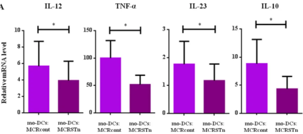
cancer cells. We analysed the expression of several cytokine genes and found that the expression of IL-12, TNF-a, IL-23 and IL-10 was significantly decreased in
mo-DCs co-incubated with MCRSTn, as compared with controls (Figure 3A). Analysis of the level of phosphorylated extracel-lular signal-regulated kinase (ERK), which is involved in cyto-kine expression, was reduced by 21% in mo-DCs following contact with MCRSTn cells (data not shown).
Since mo-DCs physiologically phagocyte apoptotic cancer cells, we then compared the capacity of mo-DCs to phagocyte both MCR cancer cell line variants. As shown inFigure 4A and B, the percentage of mo-DCs which phagocytosed MCRSTn is significantly higher (1.25-fold more) than those that phagocy-tosed MCRcont. Confocal microscopy analysis confirmed the internalization of MCRSTn cells by mo-DCs (Figure 4B).
3.4. T cells activated with mo-DC loaded with STnþ
cancer cells show defective activation profiles
We next analysed the capacity of mo-DCs to activate autolo-gous T cells by measuring the expression of the T cell activa-tion marker CD69. We observed that the mo-DCs that phagocytosed MCRSTn tended to activate significantly less number of T cells, and more weakly, than mo-DCs phagocy-tosing MCRcont (Figure 5A and B). T cell activation was also
lower in T cells primed by mo-DCs that adhered to STnþ
can-cer cells (Figure 5S), as compared to control MCR cells. T cells alone and T cells incubated with each apoptotic MCR cell lines (with no mo-DCs) were not significantly activated and lost viability (less than 5% viable cells after 11 days). By contrast, after 11 days, more than 35% of the T cells cocul-tured with DCs remained viable, suggesting that mo-DCs stimulus is necessary to maintain T cell viability. Inter-estingly, in the T cell: mo-DC (MCRSTn) coculture, the
expres-sion of interferon (IFN)-g and the transcription factor
forkhead box P3 (FoxP3) was significantly affected. Namely,
IFN-g was decreased by 54%, while FoxP3 was increased
39% compared with control (Figure 5C and D). While the
dif-ferences were marginally significant (p<0.1), they indicated a clear tendency for less Th1-skewed T cell response. These data are in agreement with the above mentioned defective
maturation profile of mo-DCs, following contact with STnþ
cancer cells.
Figure 3eCo-incubation with STnDMCR cell line downregulates cytokine expression levels. A: The expression ofTNF-a,IL-23, IL-12and IL-10cytokine genes was evaluated by quantitative real-time PCR. The relative mRNA levels for each cytokine are expressed as the permillage (&) of the expression of the endogenous positive controls. Values represent the means of at least 5 independent assays. The expression ofTNF-a, IL-12, IL-23andIL-10were significantly decreased (p[0.015 (*),p[0.031 (*),p[0.034 (*) andp[0.015 (*) respectively) in mo-DCs co-incubated
3.5. STnþ
glycoproteins blockade restore DC maturation
MUC1 and CD44 are glycoproteins equally expressed by both MCR cells (Figure 6A and B) and described as the most likely candidates for being modified by STn by human cancer cells (Julien et al., 2006). The Western blot analysis of MCRSTn cell lysates, using anti-STn mAb, identified one prominent band of approximately 75 KDa, and two weak bands of 150 KDa and 260 KDa each. In all cases, the blot staining was completely abolished after sialidase treatment, proving the STn staining specificity. Immunoprecipitation assays led us to identify the most prominent band (75 KDa) as CD44. Bladder tumours also showed STn in CD44 proteins, as
shown by the detection of a STnþ
w75 KDa band in the West-ern blot analysis (Figure 6C). In order to confirm the role of the STnþ
protein scaffold, we blocked STnþ
glycoproteins in MCRSTn cells and conducted coculture assays with mo-DCs similar to those described above. In the cocultures, whenever the CD44 was blocked in the MCRSTn cells, they gained the capacity to significantly increase, in mo-DCs, the expression
of MHC-II and cytokines, in particular of IL-12 and TNF-a
(Figure 6D). Blocking of MUC1, as well as STn antigen in MCRSTn cells also upregulated MHC-II and cytokine expres-sion, indicating a tendency, although not statistically signifi-cant (Figure 6D).
4.
Discussion
We have recently reported that the majority of high-grade bladder tumours, presenting elevated proliferation indexes and high risk of recurrence/progression and invasion,
expressed STn (Ferreira et al., 2013). This glycan is not
expressed by normal epithelium and it has been associated with a poor prognosis, and the invasive capacity of the
tumours (Ferreira et al., 2013). Similar observations were
made for breast and gastric cancers and other solid tumours, supporting the ubiquitous association of STn with malignancy. The expression of the STn antigen is also accepted to be implicated in the immunogenicity of cancer cells and this has been explored in STn-based vaccines to induce protective
immune responses against STnþ
cancers. Humoral immune responses against STn have been reported in vaccinated
pa-tients and mouse models (Holmberg and Sandmaier, 2004;
Julien et al., 2009), demonstrating that the immune system re-acts against STn antigens. However, the potency of the im-mune response is not clinical relevant to provide patients with robust anti-tumour protection.
While, glycans are known to be implicated in several im-mune responses, the involved biological processes are diverse and complex and thus still poorly understood. Lectin-glycan ligand interactions are implicated not only in mechanisms that establish immune protection, but also in those that estab-lish immunological tolerance. Given the clinical relevance of STn antigens, a deeper investigation on the interaction of STn expressing cancer cells with the immune system is war-ranted to improve and develop novel STn-based therapeutics. Here we have studied the influence of STn antigen in the modulation of DCs, which are recognized by their pivotal role in the definition of immune responses. In the tumour tis-sue, we observed a significant correlation between STn expression and an immature profile of DCs. Indeed, the expression of ST6GALNAC1, which is correlated with the expression of the STn epitopes, in bladder cancer, is correlated with CD1a, a marker of immature DCs. Interestingly, high grade bladder tumours presented not only higher levels of ST6GALNAC1 and CD1a, but also lower levels of the
pro-inflammatory, Th1-inducing cytokines, IL-12 and TNF-a,
thus supporting the association between STn and DC imma-ture profile.
Figure 4eSTnD
MCR cell lines are better phagocytosed by mo-DCs. Both MCRcont and MCRSTn cell lines were labelled with CFSE and then induced to apoptosis. Cells were then incubated with mo-DCs to allow phagocytosis, in the proportion of 1:2 for 6 h at 37C or 4C and then
stained with anti-MHC-II mAb. A: Flow cytometric analysis of percentage of DCs that phagocytosed MCR cells lines. The percentage of mo-DCs that phagocytosed MCR cells was calculated based on the positivity for both MHC-II and CFSE staining. The values obtained at 4
C were subtracted from the 37
C values. MCRSTn cells were significantly more phagocytosed than MCRcont (p[0.001 (**),n[4). B: Microscopic
analysis of mo-DCs that phagocytosed MCR cells lines. Representative confocal microscopy image showing mo-DCs that phagocytosed MCRSTn cells [MHC-IID
(red)/CFSED
Maturation is a crucial process that enables DCs to effec-tively prime T cells to mount responses against malignant cells (Steinman et al., 2003). To confirm a possible inverse
as-sociation between STnþ
cancer cells and DC maturation and to investigate if STn antigen played a role in DC maturation, we usedin vitromodels of STnþ
cancer cells cocultured with human DCs. We have found that DCs tend to adhere more to STnþ
cancer cells and that this contact inhibits DC matura-tion and co-stimulamatura-tion, when compared with DCs cocultured with STn-control cells. The higher cell adhesion to STnþ
can-cer cells, which could be due to either stronger or longer cell contacts, was responsible for the defective maturation of
mo-DCs. After coculture with STnþ
cancer cells, the lower expression of MHC-II and of co-stimulatory molecules could not be rescued by LPS stimulation, suggesting that mo-DC become resistant to further maturation stimuli.
An hallmark of immature DCs is a high phagocytic capac-ity, which is lowered upon maturation. In agreement with the fact that STn-expressing cancer cells prevents the matura-tion of mo-DCs, mo-DCs showed better phagocytic capacity for STnþ
cancer cells than for control cells. Nevertheless, the effects seen might not only be caused by STn but also some general feature of the MCR cells, since MCRcont cells also induced small maturation arrest.
Mo-DCs incubated with STnþ
cancer cells showed
defec-tive expression of TNF-aand IL-12, which is consistent not
only with DC immature phenotype but also with data observed in bladder tumour tissues. These observations were also in agreement with other reports showing that tumour environment lowers the expression of inflammatory cytokines by DCs (Ishida et al., 2008). We have not detected
significantly altered expression of anti-inflammatory
Figure 5eT cell activation is reduced when primed with mo-DCs that phagocytosed STnDcancer cells. Mo-DCs were allowed to phagocytose MCR cells and then incubated with autologous T cells (1:8 proportion). AeB: The coculture was analysed by flow cytometry for the expression of the T cell early activation marker CD69. A: Graphical representation of the percentage of CD69D
T cells. Data was determined by the percentage of CD69D
, within the CD3 population (n[3). Mo-DCs that phagocytosed MCRSTn cells induce significantly less activation in T cells than
mo-DCs that phagocytosed MCRcont cells (p[0.026 (*)). B: A representative CD69 histogram for T cells following priming with mo-DCs that
phagocytosed MCRcont (solid line) or MCRSTn (dashed line) cell line. Expression of CD69 by resting T cells is shown as staining control (grey filled peak). CeD: The coculture was analysed by quantitative real-time PCR regarding the expression ofIFN-gandFoxP3genes. The relative mRNA levels for each gene are expressed as the permillage (&) of the expression of the endogenous positive controls. C: The expression of IFN-g
was reduced by 54% (p[0.074) in T cells co-incubated with mo-DCs that phagocytosed MCRSTn as compared to controls (n[4). D: T cells
Figure 6eSTnD
glycoproteins blockade restore mo-DCs maturation. AeB: MCR cell lines were analysed by flow cytometry regarding the expression of possible scaffolds of STn, CD44 and MUC1 glycoproteins. MCRcont (dashed line) and MCRSTn (solid line) cell lines were stained with anti-CD44 (A) or anti-MUC1 (B) mAb (not filled peaks) or only with secondary mAb (grey filled peak) as staining control. Both MCR cell lines express similarly CD44 and MUC1 glycoproteins. C: Analysis of STnD
proteins in cancer cells. Total protein lysates from MCRSTn cell line (left image) and primary bladder tumour samples (right image) were treated (T) or not (NT) with sialidase. CD44 immunoprecipitation (IP) from MCRSTn total proteins was performed using Pierce Direct IP Kit (middle image). Cell lysates and CD44 IP were separated and the proteins were transferred to nitrocellulose membrane and stained with anti-STn mAb (clone TKH2). MCRSTn cells showed three proteins (z75 KDa, 150 KDa and 260 KDa) decorated with STn and the most
prominent STnDprotein showed a molecular weight ofz75 KDa. As control, when the lysate was treated with sialidase, staining with anti-STn was
completely abrogated. The CD44 IP analysis showed that CD44 protein is decorated with STn in MCRSTn cells. D: The expression ofMHC-II,IL-12
andTNF-ais restored when blocking STn and STnDglycoproteins. Gene expression was evaluated by quantitative real-time PCR in mo-DCs incubated with MCRSTn cell line in presence of anti-CD44, -MUC1 or -STn blocking mAbs. Mo-DC Fc receptors were previously blocked to avoid non-specific Fc receptor-mediated antibody binding. The expression values were calculated as described in the Material and Method section and correspond to the ratio between the expression of mo-DCs incubated with MCRSTn cell line in presence of blocking mAbs and the expression of mo-DCs incubated with MCRSTn in presence of isotypic control. CD44 blockade was able to increase the expression of MHC-II, IL-12 and TNF-a(p[0.0294 (*),p[0.0357
cytokines, with the exception of IL-10. IL-10 downregulation may be a balanced consequence of the downregulation of pro-inflammatory cytokines, playing a dual proliferative and inhibitory effect (Ogden et al., 2005).
The negative effect of STnþ
cancer cells on the maturation and Th1-inducing profile of mo-DCs were observable in less
than 2 h of coculture. On the other hand, STnþ
cancer cells could themselves be influenced by mo-DCs. This was evi-denced by the significant increase proliferation of MCRSTn cells in the presence of mo-DCs that was not observable in MCRcont cells. Yet these differences were only observable af-ter 24 h coculture, and the mechanism behind the alaf-tered pro-liferation may be dissociated from the ones implied on the altered mo-DC phenotype. However, in the future, the effect of mo-DCs in tumour cell proliferation should be further clar-ified and it might be associated with the fact that STnþ
bladder tumours present elevated proliferation indexesin situ.
The fact that using other STnþ
cell line, i.e., the MDA-MB-231STn breast cancer cell line, also rendered mo-DCs with an immature phenotype, with an extent similarly to the phenotype induced by MCRSTn bladder cancer line, strongly suggested that the specific STn overexpression in cancer cell was responsible for the induction of tolerogenic DCs reported here. Nevertheless, we cannot disregard the fact that ST6GAL-NAC1 overexpression in cell lines also alters other glycan structures beyond the STn (Table IS).
To further confirm the role of STn expression by cancer cells in DC maturation, we used different strategies to abro-gate the MCRSTn cancer cell interaction with DCs. Blocking of STn antigen in MCR cells, by means of the HB-STn antibody,
described to block STn interactions (Pinho et al., 2007),
resulted in tendency of MCRSTn cells to induce DC matura-tion. This suggested that blocking STn antigen may reverse
the propensity of STnþ
cancer cells to induce tolerogenic DCs. The fact that we were not able to obtain statistical signif-icant differences may have to do with the role of the protein scaffolds that also participate in the recognition or interaction of glycans (Padler-Karavani et al., 2008). Therefore, anti-glycan mAbs may not have efficient function-blocking properties, as anti-scaffolds have.
We have observed that the major protein scaffold in the MCRSTn cells and bladder tumours is CD44. Moreover CD44 is also decorated with STn in MDA-MB-231STn breast cancer
cells (Julien et al., 2006), which in this work induced the
same DC immunomodulation as MCRSTn cells. These findings
suggested us a possible involvement of STnþ
CD44 in the acquisition of the immature profile by DCs. Concordantly, blockade of MCR CD44 by means of functional blocking anti-bodies was able to restore the capacity of the mo-DCs to become matured.
CD44 is a rather ubiquitously expressed adhesion molecule, involved in several processes including migration and cell sig-nalling and recognition. The role of CD44 in governing the
pro-gression of tumours, including bladder carcinoma (Golshani
et al., 2008), is well documented and its expression by leuko-cytes also plays a role in modulating immune responses (Hegde et al., 2008; Jacobs and Sackstein, 2011; Weiss et al.,
1997). However the role of its recognition by immune cells,
such as DCs, is unclear. In our model, it is possible that DC re-ceptor for CD44 may recognize differently the protein due to
substitution of normal glycosylation by STn in cancer cells. CD44 is a receptor for hyaluronic acid and other extracellular proteins, such as osteopontin and collagens. Very recently it has been reported that the mannose receptor interacts with CD44 (Salmi et al., 2013). Although, mannose receptor primar-ily binds mannose and fucose, it is possible that in our model, the STn expression affected the CD44 recognition by mannose receptor, leading to the described immunological implications. Nevertheless, other receptors expressed by DCs may also be involved, such as the Sialic acid-binding immunoglobulin-type lectins (Siglec)-9 that have also been reported to recognize STn antigen (Ohta et al., 2010). In addition, the participation of other STn scaffolds such as MUC1 (Monti et al., 2004) should not be excluded and as matter of fact, we also observed that blockade of MUC1 results in a slight induction of DC matura-tion. Further studies are therefore necessary to better charac-terize the mechanisms of STn antigen recognition by DCs.
As mentioned above, previous studies were controversial in demonstrating that STn mucins were implicated in
inducing the immature DC phenotype (Carlos et al., 2005;
Monti et al., 2004). While this controversy may be due to the complexity of the involved mechanisms, these studies were in agreement, when showing that STn expression has a nega-tive influence on the capacity of DCs to activate T cells, result-ing in DCs that do not support T cell commitment to Th1
phenotype, which is important for tumour rejection (Carlos
et al., 2005; Monti et al., 2004).
DCs are currently used as anti-tumour cell-based vaccines, where one of the most common strategies is the upload of pa-tient’s DCsex vivowith the tumour cell antigens (lysates, cells fusion or apoptotic bodies). Tumour antigens are phagocy-tosed by DCs and these cells are then used to induce anti-tumour T cells responses and further anti-tumour rejection (Palucka and Banchereau, 2013). It has been reported that DCs pulsed with apoptotic cancer cells are able to activate cytotoxic T cells against poorly immunogenic cancer cells (Goldszmid et al., 2003) and in bladder cancer patients in-creases survival and cure rate (Nair et al., 1997). In the present
study, DCs pulsed with immunosuppressive STnþ
cancer cells showed a tolerogenic/regulatory profile, resistant to further maturation stimuli and limited capacity to activate T cells. In these conditions, mo-DCs express lower levels of Th1-inducing cytokines and preferentially polarize T cells towards a FOXP3high
IFN-glowphenotype, typical of regulatory T cells. On the other hand, it has been recently reported by us that STn expression in bladder cancer is associated with better re-sponses to complementary immunotherapy with Bacillus
Calmette-Guerin, a potent inducer of Th1 responses (Lima
et al., 2013). Therefore, the combination of Th1-inducing ther-apies with DC-based therapy against STn-bearing cancer cells could be a promising strategy to induce anti-tumour protec-tive responses. Nevertheless, a better understanding of the mechanisms behind the tolerogenic/regulatory profile of DCs
induced by STnþ
cancer cells is still necessary.
5.
Conclusions
ability to down-regulate the anti-cancer immune-response through different mechanisms. First, it hinders the expression of MHC-II and co-stimulatory molecules by DCs, resulting in impaired ability to present cancer-associated antigens to T cells and making DCs unresponsive to successive activation stimuli. Second, it hinders the expression of inflammatory, Th1-inducing cytokines in DCs, which may result in an atten-uation of the Th1 microenvironment. Third, DCs pulsed with STn-expressing cancer cells show a reduced ability to activate and polarize T cells towards the Th1 phenotype, resulting in impaired ability to mount an effective anti-tumour response. Finally, blockade of STn antigens and STn protein may reverse tolerance induced by cancer cells. Altogether, these results highlight the expression of STn by cancer cells as a crucial event in the establishment of the tolerogenic microenviron-ment which allows cancers to escape from the attack of innate and adaptive immunity.
Acknowledgements
This work was supported by the Portuguese Foundation for
Science and Technology (FCT) e PTDC/SAU-MII/67561/2006
and Premio Santander Totta - UNL (Paula A. Videira), LPCC/
Pfizer2011 (Mylene A. Carrascal), SFRH/BPD/21619/2005 (M.
Guadalupe Cabral), SFRH/BD/81860/2011 (Mariana Silva), SFRH/BD/45120/2008 (Paulo F. Severino) and SFRH/BPD/ 66288/2009 (Jose Alexandre Ferreira). FCT is co-financed by Eu-ropean Social Fund (ESF) under Human Potential Operation Programme (POPH) from National Strategic Reference Frame-work (NSRF).) and European Union, QREN, FEDER, COMPETE, for funding the Organic Chemistry Research Unit (QOPNA)
(project PEst-C/QUI/UI0062/2013;
FCOMP-01-0124-FEDER-037296).
The MDA-MB-231STn and MDA-MB-231cont cell lines were kindly gifted by Professor Philippe Delannoy (Lille University, France) and the anti-STn (clone TKH2) and the anti-MUC1 (clone HMFG-2) antibodies kindly gifted by Professor Celso Reis (Porto University, Portugal). We thank Manuela Correia
for her technical assistance and Helio Crespo for drawing
the graphical abstract.
Appendix A.
Supplementary data
Supplementary data related to this article can be found at http://dx.doi.org/10.1016/j.molonc.2014.02.008.
R E F E R E N C E S
Adis International, L., 2003. Cancer vaccine THERATOPE-Biomira. Drugs R&D 4, 236e240.
Almand, B., Clark, J.I., Nikitina, E., van Beynen, J., English, N.R., Knight, S.C., Carbone, D.P., Gabrilovich, D.I., 2001. Increased production of immature myeloid cells in cancer patients: a mechanism of immunosuppression in cancer. J. Immunol. 166, 678e689.
Banchereau, J., Steinman, R.M., 1998. Dendritic cells and the control of immunity. Nature 392, 245e252.
Beatty, J.D., Islam, S., North, M.E., Knight, S.C., Ogden, C.W., 2004. Urine dendritic cells: a noninvasive probe for immune activity in bladder cancer? BJU Int. 94, 1377e1383.
Cao, Y., Stosiek, P., Springer, G.F., Karsten, U., 1996. Thomsen-Friedenreich-related carbohydrate antigens in normal adult human tissues: a systematic and comparative study. Histochem. Cell Biol. 106, 197e207.
Carlos, C.A., Dong, H.F., Howard, O.M., Oppenheim, J.J.,
Hanisch, F.G., Finn, O.J., 2005. Human tumor antigen MUC1 is chemotactic for immature dendritic cells and elicits
maturation but does not promote Th1 type immunity. J. Immunol. 175, 1628e1635.
Crespo, H.J., Cabral, M.G., Teixeira, A.V., Lau, J.T., Trindade, H., Videira, P.A., 2009. Effect of sialic acid loss on dendritic cell maturation. Immunology 128, e621ee631.
Ferreira, J.A., Videira, P.A., Lima, L., Pereira, S., Silva, M., Carrascal, M., Severino, P.F., Fernandes, E., Almeida, A., Costa, C., Vitorino, R., Amaro, T., Oliveira, M.J., Reis, C.A., Dall’Olio, F., Amado, F., Santos, L.L., 2013. Overexpression of tumour-associated carbohydrate antigen sialyl-Tn in advanced bladder tumours. Mol. Oncol. 7, 719e731.
Goldszmid, R.S., Idoyaga, J., Bravo, A.I., Steinman, R., Mordoh, J., Wainstok, R., 2003. Dendritic cells charged with apoptotic tumor cells induce long-lived protective CD4þ
and CD8þ
T cell immunity against B16 melanoma. J. Immunol. 171, 5940e5947.
Golshani, R., Lopez, L., Estrella, V., Kramer, M., Iida, N.,
Lokeshwar, V.B., 2008. Hyaluronic acid synthase-1 expression regulates bladder cancer growth, invasion, and angiogenesis through CD44. Cancer Res. 68, 483e491.
Griffiths, A.B., Padmanabhan, N., Shepherd, P., Lazarus, C., Maisey, M., Fentiman, I., Rubens, R., Taylor-Papadimitriou, J., 1986. Monoclonal antibody HMFG-2 retains activity in vivo and binds to high molecular weight components expressed by metastatic breast cancers. Mol. Biol. Med. 3, 425e435.
Hegde, V.L., Singh, N.P., Nagarkatti, P.S., Nagarkatti, M., 2008. CD44 mobilization in allogeneic dendritic cell-T cell immunological synapse plays a key role in T cell activation. J. Leukoc. Biol. 84, 134e142.
Heimburg-Molinaro, J., Lum, M., Vijay, G., Jain, M., Almogren, A., Rittenhouse-Olson, K., 2011. Cancer vaccines and
carbohydrate epitopes. Vaccine 29, 8802e8826.
Holmberg, L.A., Sandmaier, B.M., 2004. Vaccination with theratope (STn-KLH) as treatment for breast cancer. Expert Rev. Vaccines 3, 655e663.
Ishida, A., Ohta, M., Toda, M., Murata, T., Usui, T., Akita, K., Inoue, M., Nakada, H., 2008. Mucin-induced apoptosis of monocyte-derived dendritic cells during maturation. Proteomics 8, 3342e3349.
Itzkowitz, S.H., Bloom, E.J., Kokal, W.A., Modin, G., Hakomori, S., Kim, Y.S., 1990. Sialosyl-Tn. A novel mucin antigen associated with prognosis in colorectal cancer patients. Cancer 66, 1960e1966.
Jacobs, P.P., Sackstein, R., 2011. CD44 and HCELL: preventing hematogenous metastasis at step 1. FEBS Lett. 585, 3148e3158.
Julien, S., Adriaenssens, E., Ottenberg, K., Furlan, A., Courtand, G., Vercoutter-Edouart, A.S., Hanisch, F.G., Delannoy, P., Le Bourhis, X., 2006. ST6GalNAc I expression in MDA-MB-231 breast cancer cells greatly modifies their O-glycosylation pattern and enhances their tumourigenicity. Glycobiology 16, 54e64.
Julien, S., Picco, G., Sewell, R., Vercoutter-Edouart, A.S., Tarp, M., Miles, D., Clausen, H., Taylor-Papadimitriou, J., Burchell, J.M., 2009. Sialyl-Tn vaccine induces antibody-mediated tumour protection in a relevant murine model. Br. J. Cancer 100, 1746e1754.
Julien, S., Videira, P.A., Delannoy, P., 2012. Sialyl-Tn in cancer: (how) did we miss the target? Biomolecules 2, 435e466.
Kjeldsen, T., Clausen, H., Hirohashi, S., Ogawa, T., Iijima, H., Hakomori, S., 1988. Preparation and characterization of monoclonal antibodies directed to the tumor-associated O-linked sialosyl-2e–6 alpha-N-acetylgalactosaminyl
(sialosyl-Tn) epitope. Cancer Res. 48, 2214e2220.
Lakshminarayanan, V., Thompson, P., Wolfert, M.A., Buskas, T., Bradley, J.M., Pathangey, L.B., Madsen, C.S., Cohen, P.A., Gendler, S.J., Boons, G.J., 2012. Immune recognition of tumor-associated mucin MUC1 is achieved by a fully synthetic aberrantly glycosylated MUC1 tripartite vaccine. Proc. Natl. Acad. Sci. U S A 109, 261e266.
Langenkamp, A., Messi, M., Lanzavecchia, A., Sallusto, F., 2000. Kinetics of dendritic cell activation: impact on priming of TH1, TH2 and nonpolarized T cells. Nat. Immunol. 1, 311e316.
Lima, L., Severino, P.F., Silva, M., Miranda, A., Tavares, A., Pereira, S., Fernandes, E., Cruz, R., Amaro, T., Reis, C.A., Dall’olio, F., Amado, F., Videira, P.A., Santos, L., Ferreira, J.A., 2013. Response of high-risk of recurrence/progression bladder tumours expressing sialyl-Tn and sialyl-6-T to BCG
immunotherapy. Br. J. Cancer 109, 2106e2114.
Lin, J.C., Liao, S.K., Lee, E.H., Hung, M.S., Sayion, Y., Chen, H.C., Kang, C.C., Huang, L.S., Cherng, J.M., 2009. Molecular events associated with epithelial to mesenchymal transition of nasopharyngeal carcinoma cells in the absence of Epstein-Barr virus genome. J. Biomed. Sci. 16, 105.
Madsen, C.B., Pedersen, A.E., Wandall, H.H., 2013. Glycan-mediated modification of the immune response. Oncoimmunology 2, e23659.
Marcos, N.T., Bennett, E.P., Gomes, J., Magalhaes, A., Gomes, C., David, L., Dar, I., Jeanneau, C., DeFrees, S., Krustrup, D., Vogel, L.K., Kure, E.H., Burchell, J., Taylor-Papadimitriou, J., Clausen, H., Mandel, U., Reis, C.A., 2011. ST6GalNAc-I controls expression of sialyl-Tn antigen in gastrointestinal tissues. Front. Biosci. 3, 1443e1455.
Miles, D., Roche, H., Martin, M., Perren, T.J., Cameron, D.A., Glaspy, J., Dodwell, D., Parker, J., Mayordomo, J., Tres, A., Murray, J.L., Ibrahim, N.K., Theratope Study, G., 2011. Phase III multicenter clinical trial of the sialyl-TN (STn)-keyhole limpet hemocyanin (KLH) vaccine for metastatic breast cancer. Oncologist 16, 1092e1100.
Monti, P., Leone, B.E., Zerbi, A., Balzano, G., Cainarca, S., Sordi, V., Pontillo, M., Mercalli, A., Di Carlo, V., Allavena, P., Piemonti, L., 2004. Tumor-derived MUC1 mucins interact with differentiating monocytes and induce IL-10highIL-12low regulatory dendritic cell. J. Immunol. 172, 7341e7349.
Nair, S.K., Snyder, D., Rouse, B.T., Gilboa, E., 1997. Regression of tumors in mice vaccinated with professional antigen-presenting cells pulsed with tumor extracts. International journal of cancer. J. Int. Cancer 70, 706e715.
Niederhafner, P., Reinis, M., Sebestik, J., Jezek, J., 2008. Glycopeptide dendrimers, part III: a review. Use of
glycopeptide dendrimers in immunotherapy and diagnosis of cancer and viral diseases. J. Peptide Science Off. Pub. Eur. Peptide Soc. 14, 556e587.
Ogden, C.A., Pound, J.D., Batth, B.K., Owens, S., Johannessen, I., Wood, K., Gregory, C.D., 2005. Enhanced apoptotic cell
clearance capacity and B cell survival factor production by IL-10-activated macrophages: implications for Burkitt’s
lymphoma. J. Immunol. 174, 3015e3023.
Ohta, M., Ishida, A., Toda, M., Akita, K., Inoue, M., Yamashita, K., Watanabe, M., Murata, T., Usui, T., Nakada, H., 2010. Immunomodulation of monocyte-derived dendritic cells through ligation of tumor-produced mucins to Siglec-9. Biochem. Biophy. Res. Commun. 402, 663e669.
Ozaki, H., Matsuzaki, H., Ando, H., Kaji, H., Nakanishi, H., Ikehara, Y., Narimatsu, H., 2012. Enhancement of metastatic ability by ectopic expression of ST6GalNAcI on a gastric cancer cell line in a mouse model. Clin. Exp. Metastasis 29, 229e238.
Padler-Karavani, V., Yu, H., Cao, H., Chokhawala, H., Karp, F., Varki, N., Chen, X., Varki, A., 2008. Diversity in specificity, abundance, and composition of anti-Neu5Gc antibodies in normal humans: potential implications for disease. Glycobiology 18, 818e830.
Palucka, K., Banchereau, J., 2013. Dendritic-cell-based therapeutic cancer vaccines. Immunity 39, 38e48.
Pinho, S., Marcos, N.T., Ferreira, B., Carvalho, A.S., Oliveira, M.J., Santos-Silva, F., Harduin-Lepers, A., Reis, C.A., 2007. Biological significance of cancer-associated sialyl-Tn antigen:
modulation of malignant phenotype in gastric carcinoma cells. Cancer Lett. 249, 157e170.
Salmi, M., Karikoski, M., Elima, K., Rantakari, P., Jalkanen, S., 2013. CD44 binds to macrophage mannose receptor on lymphatic endothelium and supports lymphocyte migration via afferent lymphatics. Circ. Res. 112, 1577e1582.
Slovin, S.F., Ragupathi, G., Fernandez, C., Diani, M.,
Jefferson, M.P., Wilton, A., Kelly, W.K., Morris, M., Solit, D., Clausen, H., Livingston, P., Scher, H.I., 2007. A polyvalent vaccine for high-risk prostate patients: “are more antigens better?”. Cancer Immunol. Immunother. CII 56, 1921e1930.
Steinman, R.M., Hawiger, D., Nussenzweig, M.C., 2003.
Tolerogenic dendritic cells. Annu. Rev. Immunol. 21, 685e711.
Troy, A.J., Davidson, P.J., Atkinson, C.H., Hart, D.N., 1999. CD1a dendritic cells predominate in transitional cell carcinoma of bladder and kidney but are minimally activated. J. Urol. 161, 1962e1967.
Vicari, A.P., Caux, C., Trinchieri, G., 2002. Tumour escape from immune surveillance through dendritic cell inactivation. Semin. Cancer Biol. 12, 33e42.
Videira, P.A., Amado, I.F., Crespo, H.J., Alguero, M.C., Dall’Olio, F., Cabral, M.G., Trindade, H., 2008. Surface alpha 3- and alpha 2-6-sialylation of human monocytes and derived dendritic cells and its influence on endocytosis. Glycoconj. J. 25, 259e268.
Videira, P.A., Calais, F.M., Correia, M., Ligeiro, D., Crespo, H.J., Calais, F., Trindade, H., 2009a. Efficacy of bacille Calmette-Guerin immunotherapy predicted by expression of antigen-presenting molecules and chemokines. Urology 74, 944e950.
Videira, P.A., Correia, M., Malagolini, N., Crespo, H.J., Ligeiro, D., Calais, F.M., Trindade, H., Dall’Olio, F., 2009b. ST3Gal.I sialyltransferase relevance in bladder cancer tissues and cell lines. BMC Cancer 9, 357.
Weiss, J.M., Sleeman, J., Renkl, A.C., Dittmar, H., Termeer, C.C., Taxis, S., Howells, N., Hofmann, M., Kohler, G., Schopf, E., Ponta, H., Herrlich, P., Simon, J.C., 1997. An essential role for CD44 variant isoforms in epidermal Langerhans cell and blood dendritic cell function. J. Cell Biol. 137, 1137e1147.