Ana Rita Silva Martins Nunes
Faculdade de Ciências Médicas | Universidade Nova de Lisboa
Lisboa, 2013
Carotidbody
Carotid artery
cAMP
PKA tmAC
ATP
sAC
ATP
PDE4 AMP
O
2
/CO
2
-sensitive cyclic AMP-signalling
Ana Rita Silva Martins Nunes
Lisboa, 2013
O
2
/CO
2
-sensitive cAMP-signalling
Dissertation presented to obtain the PhD degree in “Ciências da Vida
- Especialidade de Farmacologia” at the Faculdade de Ciências
Médicas, Universidade Nova de Lisboa.
This work was supported by:
Doctoral degree scholarship SFRH/BD/39474/2007
155/07 one year scholarship (2007)
!
The author of this thesis performed all the methodology and
analyzed all the experimental data included in this thesis, except the
carotid nerve recordings (results included in the
Results Chapter 3-
“Bicarbonate-sensitive soluble adenylyl cyclase (sAC) and
transmembrance adenylyl cyclase in peripheral chemoreceptors”).
Andrew Holmes performed carotid nerve recordings under the
supervision of Professor Prem Kumar at College of Medical and Dental
Sciences from University of Birmingham, UK.
The scientific content of the present thesis has been included in the
publication of the following book chapters and international scientific
periodicals with referees:
Book chapters:
Nunes A.R.
, Batuca J.R. and Monteiro E.C., Functional characterization
of phosphodiesterases 4 in the rat carotid body: effect of oxygen
concentration. In The arterial chemoreceptors, 129-136, Gonzalez et al
(eds.), Springer, Netherlands, 2009.
Nunes A.R.
, Monteiro E.C., Johnson S.M., Gauda E.B.,
Bicarbonate-regulated soluble adenylyl cyclase (sAC) expression and activity in
peripheral chemoreceptors. In The arterial chemoreceptors, 129-136,
Gonzalez et al (eds.), Springer, Netherlands, 2009.
Nunes A.R.
, Sample V., Xiang Y.K., Monteiro E.C., Gauda E.B. and
Zhang J. Effect of oxygen on phosphodiesterases (PDE) 3 and 4
isoforms and PKA activity in the Superior Cervical Ganglia. In Arterial
chemoreceptors: From Molecules to systems, 758; 287-294. C.A. Nurse
et al (eds.), Springer, Netherlands, 2012.
Papers in International Scientific Periodicals with Referees
Nunes A.R.
, Batuca J.R. and Monteiro E.C., Different effects of hypoxia
Gauda E. B.
*, Bicarbonate-sensitive soluble adenylyl cyclase (sAC) and
transmembrane adenylyl cyclase in peripheral chemoreceptors (in
submission).
Nunes A.R.
, Monteiro E.C., Zhang J., Gauda E. B.
*,
Bicarbonate-sensitive soluble adenylyl cyclase (sAC) and transmembrane adenylyl
cyclase in central chemoreceptors (in preparation).
Other manuscripts published during the author’s doctoral studies
not related with the content of the present thesis:
Papers in International Scientific Periodicals with Referees
Nunes AR
, Chavez-Valdez R, Ezell T, Donnelly DF, Glover JC, Gauda
EB. Effect of development on [Ca2+]i response kinetics to ATP in petrosal
ganglion neurons: a pharmacological approach using optical recording.
Journal Applied Physiology 112:1393-1402, 2012.
Chavez-Valdez R*, Mason A*,
Nunes AR
, Northington FJ, Tankersley C,
Ahlawat R, Johnson S, Gauda EB. Effect of hyperoxic exposure during
early development on neurotrophin expression in the carotid body and
nucleus tractus solitarii. Journal Applied Physiology 112:1762-1772, 2012.
The work presented in this thesis was awarded with the 1
stDe
Castro-Heymans-Neil Award, by
The XVIIIth International Society for
Arterial Chemoreception
(ISAC) meeting held in Hamilton, Ontario,
Canada, July 10
th-15
th, 2011.
Table of Contents
i
Table of Contents
List of tables
iii
List of Figures
iv
Acknowledgments
ix
Abbreviations
xi
Abstract
xv
Resumo
xix
Introduction
1
Overview
3
Control of breathing: A role of peripheral and central chemoreceptors
4
Peripheral chemoreceptors- Carotid body
5
CO
2central chemoreceptors
8
Integration of peripheral, central chemoreceptors and respiratory
neuron
12
Chemotransduction mechanisms for O
2and CO
2in carotid body
15
O
2sensing mechanism
16
CO
2sensing mechanism
17
Role of cAMP in O
2and CO
2detection in the carotid body
20
cAMP/signaling pathway
22
cAMP Pathway- Adenylyl Cyclases
22
cAMP Pathway- Phosphodiesterases
26
cAMP Pathway- Effectors
29
Extracellular and intercellular cAMP signaling
31
Hypothesis and Aims
35
General Methods
39
Surgical Procedures
41
Animals used
41
Dissection of the biological preparations
45
Preparations
45
Whole organ
45
Dissociated primary cell cultures
45
Primary cultures of carotid body cells
46
Primary cultures of superior cervical ganglia neurons
48
Organotypic slice cultures
50
Methodologies
52
Quantitative Real-time Polymerase Chain Reaction (RT-PCR)
57
Fluorescence resonance energy transfer (FRET)
61
Intracellular pH measurements
65
Iridium oxide pH sensor microelectrodes
65
Fluorescent intracellular pH measurement
68
Statistical analysis
69
Methods- Diagram
69
Results
71
Chapter 1
73
Functional characterization of phosphodiesterases 4 in the rat carotid
body: effect of oxygen concentrations.
68
75
Acute hypoxia modifies cAMP level induced by inhibitors of
phosphodiesterase-4 in rat carotid bodies, carotid arteries and
superior cervical ganglia
83
Chapter 2
93
Effect of oxygen on phosphodiesterases (PDE)3 and 4 isoforms and
PKA activity in the Superior Cervical Ganglia
94
Chapter 3
103
Bicarbonate-regulated soluble adenylyl cyclase (sAC) mRNA
expression and activity in peripheral chemoreceptors
105
Bicarbonate-sensitive soluble adenylyl cyclase (sAC) and
transmembrane adenylyl cyclase in Peripheral chemoreceptors
113
Bicarbonate-sensitive soluble adenylyl cyclase (sAC) and
transmembrane adenylyl cyclase in central CO
2chemoreceptors- a
comparative study with peripheral chemoreceptors
141
Discussion
157
Attachments
171
Resumo alargado
173
Tabelas
179
List of Tables
iii
List of Tables
Introduction
Table 1
Neurotransmitter/ neuromodulator present in the carotid body
7
Table 2
Receptors for neurotransmitter/ neuromodulator/ neuropeptide in
the carotid body (CB)
9
Table 3
Characteristics of the CO
2central chemoreceptors
12
Table 4
Effects of different conditions on cAMP levels
21
General Methods
Table 1
In vitro whole tissue experimental conditions
54
Table 2
Primer sequences
58
Results
Chapter 1
Functional characterization of phosphodiesterases 4 in the rat carotid
body:effect of oxygen concentrations
Table 1
Comparison between the efficacies and potencies of PDE inhbitors
in normoxia and hypoxia
78
Acute
hypoxia
modifies
cAMP
levels
induced
by
inhibitors
of
phosphodiesterase-4 in rat carotid bodies, carotid arteries and superior cervical
ganglia
Table 1
Comparison
between
the
efficacies
and
potencies
of
phosphodiesterase inhibitors (IBMX, rolipram, Ro 20-1724) in
normoxia (20%O
2) and hypoxia (5%O
2)
88
Chapter 3
Bicarbonate-regulated soluble adenylyl cyclase (sAC) mRNA expression and
activity in peripheral chemoreceptors
Table 1
sAC mRNA expression in peripheral chemoreceptors and
non-chemoreceptors relative to CB
108
Table 2
Effect of HCO
3-/CO
2on sAC mRNA relative gene expression
(relative expression to 0mM HCO
3for each tissue)
108
Table 3
Effects of changes in HCO
3-/CO
2on cAMP levels
109
Discussion
Table 1
Effects of cAMP in the carotid body 166
Attachments
Table 1
cAMP
168
Table 2
Adenylate cyclase isoforms 171
Table 3
PDE families 172
Figure 1
Carotid body (CB) peripheral chemoreceptors 6
Figure 2
CO
2Central chemoreceptor localization 11
Figure 3
Chemoreceptor control of breathing
14
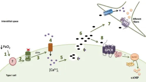
Figure 4
Steps in O
2chemotransduction mechanism in CB type I cells
17
Figure 5
CO
2chemotransduction mechanism in CB type I cells 19
Figure 6
Transmembrane adenylate cyclase (tmAC) structure
24
Figure 7
Soluble adenylate cyclase (sAC) structure
25
Figure 8
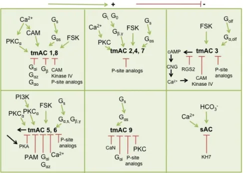
Regulation of adenylate cyclase isoforms
26
Figure 9
Cyclic nucleotide phosphodiesterase (PDE) structure
27
Figure 10 cAMP effectores
29
Figure 11 Extracellular cAMP pathways
32
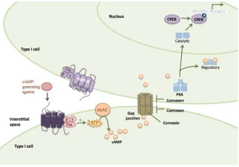
Figure 12 Intercellular cAMP pathways
33
Methodology
Figure 1
Dissection of peripheral chemo- and non-chemoreceptor
tissues from a rat
42
Figure 2
Representation of the CO
2central chemoreceptor tissues
from a rat
44
Figure 3
Primary culture of cells dissociated from CB from SD rats at
P10.
48
Figure 4
Primary cultures from superior cervical ganglia from SD rats
at P10.
49
Figure 5
Culture of organotypic SCG/CB slices 51
Figure 6
Evaluation of cell death in slice culture
51
Figure 7
Identification of SCG neurons in organotypic slices from rat
at P6
52
Figure 8
Set up for in vitro experiments
53
Figure 9
Specificity of the primers 60
Figure 10
PNA-labelled type I cells expressing A-kinase activity
reporter (AKAR3)
62
Figure 11
Primary superior cervical ganglia (SCG) cells expressing
A-kinase Activity reporter (AKAR3) can be identified by
expression of tyrosine hydroxylase (TH, a specific marker of
these cells).
62
Figure 12
Expression of A-Kinase Activity Reporter (AKAR3) visualized
by FITC channel in organotypic slices
62
Figure 13
Perfusion system for FRET imaging. 63
Figure 14
SCG organotypic slices expressing AKAR3 contain high
basal PKA activity.
65
Figure 15
Potentiostat system
67
Figure 16
Calibration curves of the iridium oxide microelectrode and
commercial glass electrode.
67
List of Figures
v
Results
Chapter 1
Functional characterization of phosphodiesterases 4 in the rat carotid
body: effect of oxygen concentrations
Figure 1
Effects of rolipram, Ro 20-1724 and IBMX on cAMP levels in
rat carotid bodies in (a) normoxia (20%O2) (0% effect-
0.75±0.08 pmol cAMP/mg) and (b) hypoxia (5%O2) (0%
effect- 0.63±0.05 pmol cAMP/mg).
78
Figure 2
Effects of hypoxia in the presence of selective (rolipram an
Ro 20-1724) and non-selective (IBMX) PDE inhibitors on
cAMP levels in rat carotid bodies.
79
Acute hypoxia modifies cAMP levels induced by inhibitors of
phosphodiesterase-4 in rat carotid bodies, carotid arteris and superior
cervical ganglia
Figure 1
Effect of different pre-incubation conditions on cAMP levels
of carotid body (CB, n=6-9), superior cervical ganglia (SCG,
n=2-5) and carotid artery (CA, n=4-5) in response to hypoxia
(5%O2/5%CO2)
and
in
the
presence
of
isobutylmethylxanthine (IBMX).
86
Figure 2
Effect of hypoxia on cAMP levels (expressed as pmol/mg
tissue) in rat carotid bodies (CBs, n=15), superior cervical
ganglia (SCG, n=11) and carotid arteries (CAs, n=11-12) in
the absence of PDE inhibitors.
86
Figure 3
Effects of PDE4 inhibitors on cAMP levels in rat carotid
bodies (CBs). Concentration-response curves of (A)
rolipram, (B) Ro 20-1724 and (C) isobutylmethylxanthine
(IBMX) for effects on cAMP levels induced during normoxia
(20%O2) [0%effect- 0.75 ±0.08 pmol cAMP/mg (n=15)] and
hypoxia (5%O2).
87
Figure 4
Effects of PDE4 inhibitors on cAMP levels in rat carotid
arteries (CAs) incubated in normoxia [20%O2, 0% of effect
corresponds to 0.19 ± 0.02 pmol.mg-1 (n=12)] and hypoxia.
88
Figure 5
Concentration-response curves for the effects of (A)
rolipram, (B) Ro 20-1724, (C) isobutylmethylxanthine (IBMX)
and (D) erythro-9-(2-hydroxy-3-nonyl)adenine (EHNA) on
cAMP levels in rat superior cervical ganglia in normoxia
(20%O2) [0%effect- 0.36 ±0.03 pmol cAMP/mg (n=11)] and
in hypoxia (5%O2).
89
Chapter 2
Effect of oxygen on phosphodiesterases (PDE)3 and 4 isoforms and PKA
activity in the Superior Cervical Ganglia
Figure
39.1
a)Pseudocolor images (400X) and (b) representative response
curves of AKAR3 to rolipram (Rol, 1
µ
M), milrinone (Mil, 10
Figure
39.2
Heterogeneous responses in PKA activity induced by PDE
inhibitors in SCG neurons
99
Figure
39.3
Effect of different oxygen concentrations on PDE3 and PDE4
isoform gene expression in the whole superior cervical
ganglia (SCG).
100
Chapter 3
Bicarbonate-regulated soluble adenylyl cyclase (sAC) mRNA
expression and activity in peripheral chemoreceptors
Figure 1
Immunoblot showing sAC protein expression in CB, SCG
and testis.
109
Bicarbonate-sensitive soluble and transmembrane adenylyl cyclases in
peripheral chemoreceptors
Figure 1
Effect of forskolin and MDL-12,330A on cAMP levels
123
Figure 2
Concentration-response curves of HCO
3-/%CO
2for effects
on cAMP levels in the CB and non-chemosensitive tissues
124
Figure 3
BCECF dye fluorescent intensity representing changes in
intracellular pH (pHi) in the superfused CB
125
Figure 4 Effects of activators of sAC and tmAC on PKA activity in
type I cells using FRET-based reporters.
128
Figure 5
sAC and tmAC mRNA expression in the carotid body (CB)
and petrosal ganglia using qRT-PCR
129
Figure 6
Effect of tmAC inhibitors.
131
Figure 7 Effect of tmAC 2’5’-ddADO (100uM), KH7 (10-50 uM) or both
on cAMP production in response to different concentrations
of HCO
3/CO
2.
131
Figure 8 Effect of different concentrations of HCO
3/CO
2on the levels
of cAMP in the carotid body during 5-39 minutes of
incubation, in the absence of PDE inhibitors.
133
Figure 9
Acidic hypercapnia but not isohydric hypercapnia induces
chemoafferent stimulation.
133
Figure 10 tmAC but not sAC modulates the basal normocapnic carotid
body chemoafferent discharge frequency and the response
to acidic hypercapnia.
134
Bicarbonate-sensitive soluble and transmembrane adenylyl cyclases in
central CO
2chemoreceptors- a comparative study with the carotid body
Figure 1
sAC mRNA expression in carotid body and central
chemoreceptors and non-chemoreceptor tissues
148
Figure 2
Effect of A)MDL-12,330A and B)2’5’-ddADO on cAMP
production induced by forskolin the locus coeruleus
148
Figure 3 Concentration-response curves of HCO
3-/CO
2for effects on
cAMP levels in central chemo and non chemosensitive
tissues
150
List of Figures
vii
using qRT-PCR
Figure 5 Effect of MDL-12330A, a tmAC inhibitor and KH7, a sAC
inhibitor, on cAMP levels in normocapnia and hypercapnia
151
Figure 6
Effect of forskolin (FSK) on sAC gene expression in locus
coeruleus (LC) and cortex
Acknowledgments
ix
Acknowledgments
During the 5 years of my PhD, I met wonderful people from all over the
world, since supervisors, collaborators, co-workers and friends, who I have to
thank for the encouragement and inspiration, and for all that I’ve learnt from them.
The first person I would like to thank is Professor Dr. Emilia Monteiro.
About 6 years ago she received me in her laboratory and about one year after she
gave me the possibility to start my PhD and go abroad. As a strong, successful
woman, she has inspired me all of these years. I thank her for her
guidance/insight, encouragement and very important, optimism.
I am also very grateful to Professor Dr. Estelle Gauda. She gave me the
opportunity to work in a great institution: The Johns Hopkins University. She
received me in her lab and in her family! I thank her for her contagious passion for
research and travelling! She gave me the opportunity to attend and participate into
several scientific meeting, and connect and collaborate with fantastic people.
Throughout these years I had the fantastic opportunity to meet and
collaborate with Dr. Cristine Kranz, Dr. Heinz Steiner, Dr. Shree Johnson, Dr.
Glover, Dr. Donnelly, Dr. Kim Insook and be mentored by Dr. Jin Zhang. Thanks
for the insight, good suggestions and advices. I thank Prof. Constancio Gonzalez
for the opportunity to spend 3 months in his laboratory, prior to the start of this
PhD. I would like to thank Dr. Prem Kumar and Andrew for all the insight and help
in this project.
All the hours in the lab would not be as fun as they were without my lab
mates. Ariel, besides her excellent lab skills and her promptly help, she is also a
great friend and a great roomy. Tarrah, thank you for your help and your hugs!
Thank you Dr. Raul for all your help since I came to Baltimore and thank you for
my first American beer! Thank you, Shree and Ruth. Special thanks to Dr.
Mclemore for being always available and helping me reviewing the english of this
Joana, Lucilia, Inês, Dr. Teresa, Teresa, Daniela. A special thanks to Joana for
teaching me how to dissect carotid bodies when I first came to the laboratory,
Sílvia for some CB discussions, for teaching me some techniques over the three
months I spent in Valladolid with her and the great time we had there, and also
Daniela and Lucília for the help, support and friendship over the last months.
A big thanks to all my friends that have helped me pass through all these
good and bad, easy and difficult roller coaster times that were part of this PhD. To
all my friends in Portugal that encouraged me to go to USA and they received me
back and made me feel at home again. Special thanks to mana Catarina, Miguel,
Ines, Ivo (o quarteto Guerreiro!), Joao e Sonia Quaresma, Catia, Sergio. My
friends that I met in the Charm city (Baltimore), in special, Veronica, Susy and
Colin, Eva, Nisha, Mariana, Recep, Mateus, Lia, Maarje, Vinny, and so many
more…. I would like to thank Eva and Jenny for letting me stay in their house the
last 3 months I was in Baltimore and thank you everybody for the great times we
had together!
I would like also to thank the Centre for Metabolism and obesity research
(CMOR), Dr. Wolfgang and Susana Rodrigues for letting me use the speed
vacuum and the fluorescent microplate reader.
And at last, but so important: my FAMILY: Mãe e Pai, Pedro e Lina e
Miguel… não há palavras que cheguem para vos agradecer! Vocês sempre me
apoiaram e estão sempre presentes comigo seja onde for! Mãe e pai, obrigada
pela vossa paciência, pelas horas intermináveis ao telefone, pelo apoio, por toda
a força que me deram e acima de tudo, motivação!
I also would like to thank Fundacao para a Ciência e tecnologia (FCT),
xi
Abbreviations
ANOVA
Analysis of variance
AM
Adrenomedullin peptide
∆
ER
Change in emission ratio
5-HT
Serotonin (5-Hydroxytryptamine).
AC
Adenylyl cyclases
Ach
Acethylcholine
ADO
Adenosine
AKAR
A-kinase Activity Reporter
ANP
Atrial Natriuretic Peptide
ATP
Adenosine triphosphate
BCECF
2’7’-Bis(2-carboxyethyl)-5(6)-carboxyfluoresceinacetoxy methyl
ester
CA
Catecholamines
Ca
2+Calcium ion
CAH
Carbonic Anhydrase
CaM
Calmodulin
cAMP
cyclic adenosine monophosphate
CB
Carotid Body
CCK
Cholescystokinin
CFP
Cyan fluorescent protein
CGRP
Calcitonin gene-related peptide
CNG
Cyclic nucleotide gated channel
CNS
Central nervous system
CO
Carbon Monoxide
CO
2Carbon dioxide
COPD
Chronic Obstructive Pulmonary Disease
CSF
Cerebrospinal fluid
CSN
Carotid sinus nerve
DA
Dopamine
DRG
Dorsal respiratory group
EIA
Enzyme immunoassay
EPAC
Exchange proteins activated by cAMP
FRET
Flourescence resonance energy transfer
FSK
Forskolin
GPCR
G-protein coupled receptors
H
2O
Water
HCO
3-Bicarbonate ion
K
+Potassium ion
KMM
Krebs modified medium
LC
Locus coeruleus
LHA
Lateral Hypothalamus
Mg
2+Magnesium ion
MIL
Milrinone
mmHg
millimetre of mercury
NA
+Sodium ion
NADH
Nicotinamide adenine dinucleotide reduced form
NE
Norepinephrine
NO
Nitric Oxide
NG
Nodoso ganglia
NHE
Na
+/H
+exchanger
NM
Neuromodulator
NP
Neuro peptide
NT
Neurotransmitter
nTS
Nucleus Tractus Solitarius
O
2Oxygen
OSA
Obstructive Sleep Apneia
P
Posnatal day of life
PaCO
2Partial pressure of carbon dioxide in arterial blood
PaO
2Partial pressure of oxygen in arterial blood
PBS
Phosphate buffered saline
PDE
Phosphodiesterases
PG
Petrosal ganglia
pH
iintracellular pH
PI
Propidium iodide
PKA
Protein Kinase A
PNA
Fluoresceinated peanut agglutinin
preBotC
pre-Botzinger complex
PRG
Pontine Respiratory Group
RT-qPCR
Real-time quantitative polymerase chain reaction
RN
Raphe Nucleus
ROL
Rolipram
RTN
Retrotrapezoid nuclei
Abbreviations
xiii
SCG
Superior cervical ganglia
SD
Sprague-Dawley rats
SEM
Standard error of the mean
TASK
TWIK-related acid-sensitive potassium
TH
Tyrosine Hydroxylase
tmAC
transmembrane adenylyl cyclase
VIP
Vasointestinal peptide
VLM
Ventrolateral medulla
VRG
Ventral respiratory group
Abstract
xv
Abstract
The carotid body (CB) is a small-paired organ sensitive to changes in
blood PaO
2, PaCO
2and pH. Type I (glomus) cells of the CB, the sensor units,
detect the stimulus and release neurotransmitters. These neurotransmitters bind
either to pos-synaptic receptors in the terminals of the carotid sinus nerve (CSN)
to trigger the hyperventilatory reflex, or to pre-synaptic receptors located in cells to
modulate their activity. These receptors can be ionotropic or metabotropic; the
later are coupled to transmembrane adenylyl cyclases (tmAC). The exact
mechanism by which changes in O
2/CO
2are detected in the CB is not fully
understood, but changes in cAMP levels have been associated with the O
2/CO
2transduction mechanism of this organ. cAMP levels are regulated by their
synthesis via two types of adenylyl cyclase: neurotransmitter-sensitive
transmembrane (tmAC) and bicarbonate sensitive-soluble adenylate cyclase
(sAC), and by their hydrolysis mediated by phosphodiesterases (PDE). The work
present in this dissertation was aimed to investigate the role of cAMP in the rat CB
chemotransduction mechanisms, how specific is cAMP signaling pathways in the
CB mainly in different CO
2conditions, and to determine whether the enzymes that
participate in cAMP signal transduction in the CB are regulated by O
2/CO
2.To
achieve this aim we characterized pharmacologically PDE4 in the CB and
non-chemoreceptor tissues and studied the effects of acute hypoxia on the cAMP
accumulation induced by PDE inhibitors. Concentration-response curves for the
effects of a non-specific (IBMX) and specific PDE2 and PDE4 inhibitors (EHNA,
Rolipram and Ro 20-1724, respectively) on cAMP levels were performed in
normoxic (20%O
2/5%CO
2) and hypoxic conditions (5%O
2/5%CO
2). We further
explored the characterization of PDEs in the superior cervical ganglia (SCG) using
FRET-based sensors in primary dissociated neurons in the presence of
non-specific (IBMX) and non-specific PDE3 and PDE4 inhibitors (milrinone and rolipram,
concentrations of HCO
3/CO
2. Our work was focused on the characterization of
sAC, an enzyme activated by changes in HCO
3/CO
2, in the CB and peripheral non
chemoreceptor tissues (SCG and petrosal and nodose ganglia). We also
compared the relative contribution of tmAC and sAC to the CO
2-sensing
mechanism in the CB. The experiments assayed for changes in sAC and tmAC
mRNA expression (qRT-PCR), cAMP levels (ELISA), activation of protein kinase
A (PKA, FRET-based sensors in type I cells) and carotid sinus nerve activity
(recording) in the presence and absence of activators or inhibitors of sAC, tmAC
and PKA. Lastly, we investigated sAC and tmAC expression and activity in central
chemoreceptors (locus coeruleus, raphe nuclei and the ventrolateral medulla) and
compared the role of these enzymes in these tissues and CB.
The main findings of this work were: 1) PDE4 isoforms are functional in
rat CBs, SCG and carotid arteries (CA), while PDE2 is only functional in the SCG;
2) The effects of PDE inhibitors on cAMP accumulation were increased by acute
hypoxia in both CB and CA, but reduced in the SCG; 3) a differential pattern of
PDE regulation in the SCG was observed that potentially represents
subpopulations of ganglion cells with different physiological functions; 4) sAC is
ubiquitously expressed and functional in CB and central chemoreceptors, but
apparently does not mediate a role in hypercapnia sensing; instead sAC activity
seems to be maximal under normocapnic conditions; 5) tmAC and sAC apparently
contribute more to cAMP accumulation when situations of low HCO
3/CO
2occur; 6)
Inhibition of tmAC activity decrease the CSN discharge in the same extent
between normocapnic and hypercapnic conditions; 7) The sensibility of the CB to
CO
2seems to be mediated by pH.
Taken together, our results suggest that cAMP has a role in the
homeostasis of the CB, and it is not a specific mediator of hypoxia/hypercapnia
transduction of this organ. The O
2/CO
2regulation of the enzymes studied in this
Abstract
xvii
increases in cAMP described by others in hypercapnic conditions are not
Resumo
xix
Resumo
O corpo carotídeo (CB) é um pequeno órgão sensível a variações na
PaO
2, PaCO
2e pH. As células tipo I (células glómicas) do corpo carotídeo, as
unidades sensoriais deste órgão, libertam neurotransmissores em resposta às
variações dos gases arteriais. Estes neurotransmissores atuam em recetores
pós-sinápticos localizados nas terminações do nervo do seio carotídeo (CSN)
desencadeando hiperventilação, ou em recetores pré-sinápticos localizados nas
células do CB, modulando assim a atividade deste órgão. Estes recetores podem
ser classificados em ionotrópicos ou metabotrópicos, estando estes últimos
acoplados a adenilatos ciclases transmembranares (tmAC). O mecanismo exato
pelo qual as variações dos gases arteriais são detetadas pelo CB ainda não está
completamente elucidado, mas tem sido sugerido que as alterações nos níveis de
cAMP estejam associadas ao mecanismo de deteção de variações de O
2e CO
2.
Os níveis de cAMP podem ser regulados pela sua via de síntese, mediada por
dois tipos de adenilatos ciclases: tmAC sensível aos neurotransmissores e
adenilato ciclase solúvel (sAC) sensível a variações de HCO
3/CO
2; e pela sua via
de degradação mediada por fosfodiesterases.
Os principais objetivos do presente trabalho foram, em primeiro lugar,
esclarecer o papel da via de sinalização do cAMP no mecanismo de
quimiotransdução do CB de rato; foi igualmente nosso propósito investigar qual a
especificidade da via de sinalização do cAMP no CB em resposta a diferentes
concentrações de CO
2e, por fim, determinar se os enzimas que participam na via
de transdução de sinal do cAMP são reguladas por O
2/CO
2.Com este intuito, caracterizamos farmacologicamente a PDE4 no CB e em
tecidos não quimiorecetores e, observámos o efeito de hipóxia aguda na
acumulação dos níveis de cAMP, induzidos pelos inibidores de PDEs. Foram
elaboradas curvas de dose-resposta para os efeitos de inibidores, não
específicos (IBMX) e específicos para a PDE2 e PDE4 (EHNA, Rolipram e Ro
culturas primárias de neurónios na presença de inibidores não específicos (IBMX)
e específicos para a PDE3 e
PDE4 (milrinone e rolipram, respetivamente). Foram ainda estudadas as
alterações na expressão de PDE3A-B e PDE4A-D em resposta a diferentes
percentagens de oxigénio no SCG através de RT-qPCR. Caracterizámos a via de
síntese do cAMP no CB em resposta a variações na concentração de HCO
3/CO
2.
Esta componente do trabalho baseou-se na caracterização da sAC, um enzima
regulado por HCO
3/CO
2, no CB e em tecidos não quimioreceptores periféricos
(SCG, gânglio petroso e gânglio nodoso). Foi ainda comparada a contribuição
relativa da tmAC e sAC no mecanismo de sensibilidade ao CO
2no CB. Para o
efeito, foram estudadas as alterações nos níveis de expressão de sAC e tmAC
(RT-qPCR), nos níveis de cAMP (ELISA), na ativação da proteína cinase A (PKA,
FRET baseado em sensores) e na atividade do CSN (registos) na presença e
ausência de ativadores e inibidores das AC, tmAC e PKA. Por último, a
expressão e actividade da sAC e da tmAC foram estudadas em tecidos
quimioreceptores centrais (locus
ceruleus, rafe e medula ventro-lateral) e não
quimioreceptores (córtex), comparando a sua actividade com a observada no CB.
O nosso trabalho teve os seguintes resultados principais: 1) PDE4 está funcional
no corpo carotídeo, SCG e artérias carótidas (CA) de rato, embora a PDE2 só se
encontre funcional no SCG; 2) Os efeitos dos inibidores de PDE nos níveis de
acumulação de cAMP foram exacerbados em situações de hipóxia aguda no CB
e CA, mas foram atenuados no SCG; 3) No SCG, diferentes tipos de células
apresentaram uma caracterização específica de PDEs, sugerindo uma
subpopulação de células no gânglio com funções fisiológicas distintas; 4) sAC
encontra-se expressa e funcional no CB e nos quimiorecetores centrais (locus
coeruleus, rafe e medula ventrolateral), mas não desempenha qualquer papel na
deteção de situações de hipercapnia; no entanto a sua actividade parece ser
Resumo
xxi
contribuição para o aumento dos níveis de cAMP em condições de baixos níveis
de bicarbonato e CO
2; 6) A inibição da atividade da tmAC diminui a frequência de
descarga do CSN de forma identica quer em condições de nomocapnia quer de
hipercapnia; 7) A sensibilidade do CB às variações de CO
2é provavelmente
mediada por variações de pH.
Em conclusão, os resultados obtidos neste trabalho sugerem que o
cAMP tem um papel importante na homeostase do CB, mas que não é um
mediador específico da transdução à hipóxia e hipercapnia neste orgão. A
regulação por alterações nos níveis de O
2/CO
2dos enzimas estudados neste
trabalho, e que envolvem a via de sinalização do cAMP, não é específica do
corpo carotídeo. O presente trabalho demonstrou ainda que os aumentos de
cAMP, induzidos por condições de hipercápnia, descritos na literatura, não se
I
Introduction
3
I
N
T
R
OD
U
C
T
ION
Overview
Organisms closely maintain the partial pressures of oxygen (PaO
2)
and
carbon dioxide (PaCO
2) in arterial blood within a narrow range in order to maintain
homeostasis. Small changes in PaO
2and PaCO
2are rapidly detected by
specialized chemoreceptors that mediate reflex responses to adjust the levels of
these gases in the blood within the physiological range. Thus, understanding the
O
2and CO
2transduction and regulation mechanisms of these chemoreceptors is
of clinical relevance. Although many advances have been made in the recent
decades, the specific molecules and processes modulating chemotransduction
are not yet fully understood.
Oxygen (O
2) is fundamental to the survival of mammalian cells. O
2enters
in the lungs and passes into the alveoli, and diffuses into the bloodstream in
exchange for CO
2. O
2is transported in the blood by hemoglobin or dissolved in
plasma at a PaO
2of 100 mmHg in most mammalian species, and diffuses into
systemic tissues in exchange for the CO
2produced in the cell. Once in the cell, O
2is required directly as the final electron acceptor in the mitochondrial electron
transport chain, a series of electron carriers embedded in the eukaryotic
mitochondrial membrane. Through a series of redox reactions, high-energy
electrons are passed from cofactors nicotinamide adenine dinucleotide (NADH;
reduced form) and flavin adenine dinucleotide (FADH
2; reduced form) to O
2. In this
process, an electrochemical proton (H
+) gradient is generated and used to
synthesize adenosine triphosphate (ATP), the energy currency of the cell, and
energy-rich organic substrates are broken down into CO
2and water (H
2O). CO
2diffuses readily from cells into the bloodstream, where it can combine with H
2O to
form carbonic acid (H
2CO
3), which dissociates to liberate a H
+and a bicarbonate
ion (HCO
3-) through the following reaction catalyzed by carbonic anhydrases
(CAH, EC 4.2.1.1):
Cells actively transport H
+and HCO
3-
across their cell membranes to
maintain constant intracellular pH (pH
i), and to buffer intracellular and extracellular
fluids. The physiological level of HCO
3-in the blood in healthy individuals is 24-27
mmol/L; higher levels
reflect metabolic alkalosis, while lower concentrations are
indicators of metabolic acidosis. Under normal conditions, CO
2is dissolved in the
blood at a PaCO
2of approximately 35-45mmHg (4.6-5.9% CO
2), the range at
which CO
2diffuses from the blood into the alveoli in exchange for O
2.
Hence, the exchange of the blood gases between the alveoli and the
blood continuously supplies O
2to the cells and reduces CO
2to maintain
homeostasis. This mechanism is regulated by peripheral and central
chemoreceptors that send input to the respiratory center in the brainstem to adjust
ventilation and to maintain the arterial blood gases within a physiological range.
Control of breathing: A role of peripheral and central chemoreceptors
In mammals, classic O
2-sensitive chemoreceptors are found in the
peripheral nervous system: the carotid (CB) and aortic bodies. Both organs trigger
hyperventilation in response to hypoxia (decrease in PaO
2), although the major
contributor to hypoxia-induced hyperventilation is the CB (for a review (Gonzalez
et al.
, 1994). More recently, evidence for O
2sensitivity has been reported in the
central nervous system (CNS) in the C1 sympathoexcitatory region of the rostral
ventrolateral medulla, hypothalamus, pre-Botzinger complex (preBotC) and
nucleus tractus solitarius (nTS) (for a review (Neubauer and Sunderram, 2004).
CO
2-sensitive chemoreceptors are present primarily in the CNS
(brainstem). However, specialized cells within the CB also depolarize in response
to changes in CO
2/H
+, a mechanism that appears to be faster and important in
initiating the central CO
2sensitivity mechanism (Smith
et al.
, 2006).
Introduction
5
I
N
T
R
OD
U
C
T
ION
Peripheral chemoreceptors- Carotid body
The CB is a small (≈ 40
μ
g wet weigh and 600
μ
m length in the rat,
(McDonald, 1980) and heterogeneous organ composed of type I (glomus or
chemosensitive units) and type II (sustentacular) cells, afferent and efferent nerve
endings, blood vessels and connective tissue (Figure 1A). Type I cells are the
primary sensory element within the CB. Type II cells have a glial-like function and
it has been shown that these cells may be dormant stem cells, which proliferate in
response to hypoxia and differentiate into new type I cells (Pardal
et al.
, 2007).
Recently it has been proposed that type II cells may function as ATP amplifiers
during chemotransduction via activation of pannexin-1 (a gap junction like protein)
and P2Y2 receptors (Zhang
et al.
, 2012).
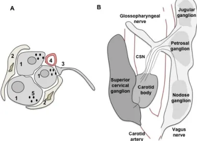
The CB is strategically located bilaterally in the bifurcation of the common
carotid artery (Figure 1B), the artery that provides the main oxygenated blood
supply to the brain. The arterial blood flows into the CB through a short artery, a
branch of the internal or external carotid artery, which ends in a dense capillary
network that supplies a large flow of blood into the organ. Thus, the CB quickly
detects changes in PaO
2and responds to hypoxic (low PaO
2) conditions.
When type I cells detect decreases in PaO
2they activate a transduction
mechanism (in more detail in
Chemotransduction mechanisms for O
2and CO
2in
the carotid body
) that involves calcium (Ca
2+) entry and release of multiple
ganglion (PG) neurons and resultant depolarization of a group of specific neurons
in the NTS in the brainstem.
Figure 1 - Carotid body (CB) peripheral chemoreceptors. A) Representation of the histological structure of the CB representing the four principal components of this organ: type I cells (1) grouped in clusters and surrounded by type II cells (2), sensory nerve endings (3) originated from the carotid sinus nerve and capillaries (4). Synaptic vesicles (5) are a characteristic of type I cells. The CB also contains a large fraction of connective tissue. B) Representation of the carotid artery bifurcation. The CB is at the bifurcation, innervated by sensory fibers originated in the petrosal ganglion via the carotid sinus nerve. The superior cervical ganglion also innervates the CB via ganglioglomerular nerves. (Figure adapted and modified by Gonzalez et al., 1992, Ichikawa, 2002).
The activation of excitatory pre-synaptic receptors (also called autoreceptors)
induces an increase in intracellular Ca
2+, [Ca
2+]
i,and subsequent release of other
neurotransmitters/ neuromodulators from the CB that can thus bind to excitatory
or inhibitory receptors (e.g. the effect of Ach reviewed in (Shirahata
et al.
, 2007)).
Although some of the neurotransmitters are shown to be important mediators of
hypoxic sensitivity, they may also be responsible for the transduction of other
stimuli in the CB (for a review, (Gonzalez
et al.
, 1994)). The CB not only senses
changes in PaO
2, but in PaCO
2, pH and glucose, cytokines, circulating hormones,
extracellular ions (K
+), osmolarity and temperature, thereby acting as a polymodal
Introduction
7
I
N T R OD U C T IONTable 1 - Neuropeptide /Neurotransmitters/ Neuromodulators present in the carotid body.
NP/NT /NM
Localization
Species Ref
TIC CA PG CB G
N eu ro p ep ti d es
SP + + + + Cat, guinea pig, rat, monkey, feline,
rabbit, canine 1
NA + + Cat 2
ENK + + Cat, rat, rabbit, piglet, dog, human 3
NY + + Rat, 4
ANP + Cat, human 5
GA + + + Guinea pig, monkey, rat, chicken 6
CCK + Dog, human 7
VIP + + , Cat, rat, dog, pig, monkey, human 8
CGRP + + Mammals, birds and amphibious 9
AM + Rat, human 10
NTSin + Human and experimental animals 11
ST + Chicken, guinea pig, amphibious 12
BO + Human 13
AT II + + Rat 14
KISS + Rat, human 15
N T / n eu ro m o d u la to rs
DA + + + All mammalian species 16
NE + + + Mouse, rat 17
ADO + Rat 18
5-HT Human, rat, mouse, chicken 19
GABA + Mouse, cat, rat 20
Ach + + Cat, rat, rabbit, cat 21
ATP + Cat 22
NO + Rat, cat 23
CO + Rat, cat 24
NP, neuropeptide; NT, neurotransmitter; NM, neuromodulator; TIC, type I cells; CA, carotid artery; PG, petrosal ganglia; CB, whole carotid body; G, glossopharingeal nerve; SP, Substance P; NA, Neurokinin A; ENK, Leu- and Met- enkephalins; NY,
Neuropeptide Y; ANP, Atrial natriuretic peptide; GA, Galanin; CCK, Cholescystokinin; VIP, Vasointerstinal peptide; CGRP,
Calcitonin Gene-related peptide; AM, Adrenomedullin; NTSin, neurotensin; ST, Somatostatin; BO, Bombesin; ATII, Angiotesin II;
KISS, Kisspeptin1; DA, Dopamine; NE, Norepiniphrine; ADO, Adenosine; 5-HT, Serotonine; GABA, gamma-aminotutyric acid;
Ach, Acetylcholine; ATP, adenosine triphosphate; NO, nitric oxide; CO, carbon monoxide.
References:
1-for a review see Heym and Kummer, 1989; 2-Prabhakar et al, 1989; 3- for a review see Porzionato et al, 2008; 4- Oomori et al, 1991, 2002; 5-Wang et al, 1991,Benvenuti et al, 1996; 6- for a review see Porzionato et al, 2008, Heym and Kummer, 1989; 7- for a review see Porzionato et al, 2008, Heym and Kummer, 1989; 8- for a review see Heym and Kummer, 1989, Smith et al, 1990; 9-for a review see Porzionato et al, 2008; 10- Martinez et al, 2003; Porzionato et al, 2006; 11- for a review see Porzionato et al, 2008; 12- for a review see Porzionato et al, 2008; 13- Smith et al, 1990; 14- Lam and Leung, 2002; 15-Porzionato et al, 2011; 16- For a review see Gonzalez et al, 1994; 17- Oomori et al. 1994; Christie and Hansen, 1983; 18- Conde et al, 2004; 19- Perrin et al, 1986; Oomori et al, 1994; Kameda et al, 1990; 20-Oomori et al. 1994; Igarashi et al, 2009; Fearon et al, 2003; 21
The expression and activity of specific neurotransmitters and their receptors
changes with postnatal development (Bairam and Carroll, 2005), and can
influence CB function during development.
CO
2central chemoreceptors
The central chemoreceptors mainly sense changes in CO
2/pH in the
brain. The levels of CO
2/pH in the brain are determined by changes in PaCO
2that
occur when alveolar ventilation decreases with a constant rate of CO
2production
in the tissues, or vice-versa. The arterial blood reaches the brain through the
carotid and vertebral arteries. Since the CB is located at the bifurcation of the
carotid artery, this organ detects changes in PaCO
2quicker than the brain and
modulates the central chemoreceptors sensitivity. The carotid and vertebral
arteries supply blood to the surface of the ventral medulla and send penetrating
dorsal branches deep into the tissue, where some central CO
2chemoreceptors
have been identified: surface of the ventrolateral medulla (VLM), the raphe nuclei
(RN), the retrotrapezoid nuclei (RTN), the preBötC, the locus coeruleus (LC), and
the NTS (reviewed in(Nattie, 1999))
(Figure 2). Since CO
2is highly diffusible,
when PaCO
2rises, it easily crosses the blood brain barrier (which is relatively
impermeable to H
+and HCO
3-
ions) and diffuses (from the cerebral blood vessels)
into the cerebrospinal fluid (CSF), changing the pH. In this sense, the central
chemoreceptor neurons predominantly sense alterations in pH in response to
CO
2, (i.e., CO
2/H
+sensing, (Lahiri and Forster, 2003, Putnam
et al.
, 2004)).
However, evidence supports the existence of direct CO
2sensors, which have
been demonstrated in the LC and the VLM (Filosa and Putnam, 2003). The levels
of CO
2in the brain are also influenced by the rate of CO
2production in the
Introduction
9
I
N T R OD U C T IONTable 2 - Neuropeptide /Neurotransmitters/ Neuromodulators Receptors.
N T / NM Su b ty p e Me ta b o tr o p ic Io n o tr o p ic D1 Gs ---A C s tim u la tio n , c A M P ---R at , c at , rabbit T ype I c ells /P G/ SC G 1 W ho le C B , P G and SC G, t ype I c ells o f C B , nerv e endings
α2A
Gi /G o ---A C in h ib iti o n , c A M P ; P L A s 2 s tim u la tio n (-) R at , rabbit , c at SC G (rat ), t ype I c ells (rabbit and c at ) 3 M2 /M 4 G i/G o ---c A M P , P K A a n d Ca 2+ fr o m in tr a s to re s ---c at C B , SC G, P G 5 M1 /M 3 Gq /G 11 ---P L C , P iP 2 , IP 3 a n d D A G (+ ) C at C B , SC G, P G 6 α4 , α 7 a n d ß 2 , α4 ß2 h et e r a t ty pe I c ells , P G, C SN af ferent s 7 (+ ) ✓ α3 , α 4, α5 , α 7? ?, ß 2 , ß 4 M ic e (C 57B L/ 6) C B t is s ue s ec tio ns 9 α3 , α 7, ß 2 H u m a n W ho le C B 10 P 2 X2 /3 , P 2 X 3 , P 2 X 2 , ---✓ C a 2+ (+ ) R a t, c a t, h u m a n s , m o u s e P e tr o s a l g a n g lio n a ff e re n ts , w h o le C B , S C G 11 P 2 Y1 Gq /G 11 ,G i /G o ---P L C s tim u la tio n w ith IP 3 (-) ? ? ra t ty p e I c e lls 12 ra t P G 14 ra b b it T y p e I c e lls 15 A2A Gs ---A C s tim u la tio n (+ ) R a t, h u m a n , W h o le C B , p o s t s y n a p tic a lly o n C S N 16 A2B Gs ---A C s tim u la tio n (+ ) ra t W h o le C B 17 5 -H T2A Gq /G 1 1 , Gi /G o ---P L C + , A C -(-) ra t ty p e I c e lls , P G (jus t a few in P G) 18 5 -H T3 ----✓ (+ ) N a + , K + , C a 2 + ra t PG 19 5 -h t5A Gi /G o ----(-) A C in h ib iti o n ra t T y p e I c e lls a n d P G 20 R e c e p to r E ffe c t o n C SN a c ti v ity Sp e c ie s L o c a li za ti o n R e f N a + , K + , C a 2 + f ro m e x tr a c e llu la r α3 , α 4, α7 ?? , ß 2, ß 4 C a t W ho le C B s , SC G, P G 8 R at , rabbit , c at , m o us e C 57B L/ 6, hum an 2 NE ß2 Gs ---A C s tim u la tio n , c A M P (+ ) rat W ho le C B DA D2 Gi /G o ---A C in h ib iti o n , c A M P (-) a 5-H T E ffe c to r/ R e s p o n s e ra t ty p e II c e lls 13 ADO A1 Gi /G o ---A C in h ib iti o n (-) ? ? ATP P 2 Y2 Gq /G 11 ,G i /G o ---P L C s tim u la tio n (-) ? ? 4 Ac h b )
---a) Mainly inhibitory, occasionally excitatory (rabbit; Iturriaga et al, 2004); b) Although in rat there is less characterization of these receptors, in other species, cat and rabbit, the nAchR α3,4,5,7, and ß2,4 are present in type I cell, α7 in the CNS afferents and α3,4,7, and ß2,4 in PG; the mAchR M1 and M2 are in type I cells, M1 in CSN afferents and M1 and M2 in PG neurons (for a revision, Shirahata e tal, 2007);
?, suggested, but no direct evidences; Gs, G protein stimulatory; Gi/Go, G protein inhibitory; AC, adenylyl cyclase; PLA2, phosholipase A2, PLC, phospholipase C, PLD, phospholipase D, PIP2, Phosphatidylinositol4-5-biphosphate; IP3, Inositol triphosphate; DAG, Diacylglycerol, D, (+) excitatory, (-) inhibitory; DA, dopamine; NE, norepiniphrine, Ach, acetylcholine, ATP, adenosine triphosphate, ADO, adenosine,5-HT, serotonine,PG, Petrosal ganglia; SCG, Superior cervical ganglia; CB, carotd body.
Ref: 1 Bairam et al, 1998; Bairam et al, 1997; Czyzyk-KrZeska et al, 1992, Kahlin et al, 2010, Fagerlunde et al, 2010; 2- Bairam et al, 1997; Czyzyk-KrZeska et al, 1992, Kahlin et al, 2010, Fagerlunde et al, 2010; 3- Gauda 2002; Almaraz et al.1997; Kou et al., 1991.; 4- Mir e tal, 1983; 5- Bairam et al, 2006; 6- Bairam et al, 2006; 7-Meza RC, 2010, 2012; Niane et al, 2009; Zhong and Nurse, 1997; He et al, 2005; Gauda, 2002; Conde and Monteiro, 2006; 8- Bairam et al, 2007; 9- Kahin et al, 2010; 10- Ferlunger et al, 2010; 11- Prasal et al 2000, Fagerlun et al, 2010; Bairam et al, 2007; Rong et al, 2003; 12- Xu et al, 2005; 13- Xu et al, 2003; 14- Gauda et al, 2002; 15- Rocher et al, 1999; 16- Kobayashi et al, 2000, Fagerlung et al, 2010; 17- Kobayashi et al, 2000;
Table 2 –cont. N T / NM Su b ty p e Me ta b o tr o p ic Io n o tr o p ic H2 Gq /G11 ---P L C s tim u la tio n ? c a t W h o le C B 22 H3 Gi /G o ---A C in h ib iti o n (-) C a t, h u m a n ty p e II c e lls , P G 23 NK 1 G s ---P L C s tim u la tio n ra t S C G , P G 24 NK 2 , N K3 G s , G q /G11 ---A C s tim u la tio n ? ET ET A Gq /G11 ---P L C , P L A 2 , P L D s tim u la tio n , ? ra t ty p e I c e lls 25 EPO E P O R c ) ---c) ? ra t C B c lu s te rs 26 T ro p h in s T R K B d ) ---d) ? ? C e ll ty p e I 27 Ki s s K is s R Gq /G11 ---P L C s tim u la tio n ? R a t a n d h u m a n T y p e I c e lls , S C G 28 IL -1 B , IL -6 R x , IL -1 R I ---e) (+ ) ra t C B , ty p e I c e lls 29 T N F -R 1 , T N F -R 2 f ) ---f) (-) C a t, r a t G lo m u s c e lls 30 31 O b -R a , O b R b , O B -R C , O b -R f ---? R a t a n d h u m a n T y p e I c e lls 32 ra t 34 G A B A A ( α 2 , α 3, b 3, g 2 ) 36 G A B A A ( α 2 , b 3, g 2) G A B A B G i/G o ---A C in h ib iti o n (-) R a t, m o u s e T y p e I c e lls 38 GA BA G A B A A (+ ) Hist a-min e H1 Gq /G11 ---A C s tim u la tio n R e c e p to r E ffe c t o n C SN a c ti v ity Sp e c ie s R e f 21 S
P ne oki Cyt
T L R 4 g ) ---g) ? ra t A n g io te n s i o n I I A T 1 G q /G 1 1 ---P L C s tim u la tio n (+ ) T y p e I c e lls T y p e I c e lls , P G 33 EN K d ? ? G i/G o ? ? ---A C in h ib iti o n (-) R a t w h o le C B ? ? 37 E ffe c to r/ R e s p o n s e L o c a li za ti o n C a t, h u m a n (+ ) ---✓ In c re a s e s C l-(-) h u m a n W h o le C B 35 ---✓ in c re a s e s C l-(-) c a t T y p e I c e lls , a n d c e ll b o d ie s a n d n e rv e s o f P G ---✓ in c re a s e s C l -(-) R a t S e n s o ry n e rv e ( C S N ) e n d in g s
There is some controversial with the role of histamine: while some considered that it is an excitatory NT in type cells, others (Burlon et al, 2009) do not, considering that the excitatory effect may be mediated by terminal afferents; c) EPOR contains a number of of phosphotyrosines that are phosphorylated by JaK2 and serve as docking sites for a variety of intracellular pathway activators and Stats; d) TrKB has the highest affinity to the binding of brain-derived neurotropjic factor (BDNF) and NT4. TrKB receptor is a tyrosine knase family receptor; ??, suggested, but no direct evidences; GABA, gamma-aminobutyric acid, ENK, enkephalins, SP, substancia P, ET, endothelin
Ref: 21- Lazarov et al, 2009; Del Rio et al, 2008; 22- Del Rio et al, 2008; 23- Lazarov et al, 2009; Del Rio et al, 2008; 24- Gauda et al, 2002; 25- Chen et al, 2002; 26- Lam et al, 2009; 27- Porzionato et al, 2008; 28- Porzionato et al, 2011; 29- Lam et al, 2008; Wang et al, 2002, 2007; 30- Fernandez et al, 2011; 31- Fernandez et al, 2011; 32- Porzionato et al, 2011; 33- Fung et al, 2001;
Introduction
11
I
N
T
R
OD
U
C
T
ION
Some areas of the cerebellum, hypothalamus and midbrain have also
been identified as areas with chemoreceptor properties (Nattie, 1999) (Figure 2).
It has also been demonstrated that glia cells in the brain also detect changes in
CO
2/H
+(Erlichman and Leiter, 2010).
These central chemoreceptor sites have specific characteristics that are
presented in Table 3. The putative contribution of these putative regions to the
overall ventilation may depend on the stimulus intensity, neurotransmitter content,
arousal state, maturation and gender (Table 3; Nattie, 1999).
Figure 2 - CO2 central chemoreceptor localization. LHA, lateral hypothalamus; DR, dorsal raphe; FN, fastigial nucleus; 4v, fourth ventricle; LC, locus coeruleus; 7N, facial nerve; cNTS, caudal nucleus tractus solitarious; AMB, ambiguous; VII, facial nucleus; SO, superior olive; PBC, pre-Botzinge complex; rVRG, rostral ventral respiratory group; cVLM, caudal ventrolateral medulla; RTN/pFRG, retrotrapezoid nucleus/parafacial respiratory group; and Pn, pons. Adapted from Nattie and Li, 2012.
Changes in CO
2are closely linked to changes in O
2. Although the main
O
2sensors are located in the peripheral chemoreceptors in the CB, as previously
mentioned, some central regions also have been identified as O
2-sensitive sites
Table 3- Characteristics of the central CO2 chemoreceptors
CO2 site Location Neuron
type CO2 maturation Stimuli
RTN
Located 130-230 µm from the ventral surface of the medulla and in the marginal
glial layer
Glutaminergic
Fully developed at birth and does not change during the
1st two weeks of life
Extracellular pH (pHe)
Raphe nucleus
Medullary raphe located in close proximity to the VLM surface, while the dorsal
raphe is found at the surface of the midbrain
Serotonergic
Increase in neurons responsive to hypercapnia
during the 1st two weeks. (Wang et al, 1999)
pH
VLM Located in the braistem Purinergic N/A pH, CO2
LC Located at the
ponto-medullary border, underneath the 4th ventricle
in the pons
Noradrenergic/
catecholami-nergic
No activation in newborn (Wtckstrom et al, 2002); mature electrophysiological responses on Postnatal day (P)1 (Struden et al 2001) and reduction of sensitivity
and magnitude of the chemosensory responses around P10 (Nichols et al,
2008).
pH, CO2
NTS
Located superficially in the dorsal medulla and surrounds the 4th ventricle
Glutaminergic Fully developed at birth
(Conrad et al, 2009) pH
Pre botzinger
Site of the inspiratory
rhythm generation Glutaminergic N/A pH
glia Astrocytes of the VLM
Purinergic N/A CO2
Lateral hypo-thalamus
Lateral region of the
hypothalamus Orexinergic N/A pHe
(Adapted from Hempleman and Pilarski, 2011; Huckstepp and Dale, 2011.)