Comparison of the
in vivo
and
in vitro
genotoxicity of glyphosate
isopropylamine salt in three different organisms
Carlos Alvarez-Moya
1, Mónica Reynoso Silva
1, Carlos Valdez Ramírez
1, David Gómez Gallardo
1,
Rafael León Sánchez
2, Alejandro Canales Aguirre
3and Alfredo Feria Velasco
41
Environmental Mutagenesis Laboratory, Departamento de Biología Celular y Molecular,
University of Guadalajara, Zapopan, Mexico.
2
Banco Nacional Genómico de Tilapia, Departamento de Ingeniería de Proyectos,
University of Guadalajara, Guadalajara, Mexico.
3Unit of Medical and Pharmaceutical Biotechnology,
Jalisco Center for Research and Assistance in Technology and Design, Guadalajara, Mexico.
4División de Ciencias Biológicas, University of Guadalajara, Zapopan, Mexico.
Abstract
There is considerable controversy with regard to the genotoxicity of glyphosate, with some reports stating that this compound is non-toxic for fish, birds and mammals. In this work, we used the comet assay to examine the genotoxicity of glyphosate isopropylamine (0.7, 7, 70 and 700 mM) in human lymphocytes, erythrocytes of Oreochromis niloticus and staminal nuclei of Tradescantia (4430) in vitro and in vivo. Cells, nuclei and fish that had and had not been exposed to 5 mM N-nitrosodiethylamine (NDEA) were used as positive and negative controls, re-spectively. Significant (p < 0.01) genetic damage was observedin vivo and in vitro in all cell types and organisms tested. Human lymphocytes andTradescantia hairs showed lower genetic damage in vivo compared to in vitro, pos-sibly because of efficient metabolization of the herbicide. In O. niloticus erythrocytes, significant (p < 0.001) genotoxicity was observed at³7mM, whereasin vitro, glyphosphate was genotoxic in human lymphocytes and Tra-descantia hairs at³0.7mM. These results indicate that glyphosate is genotoxic in the cells and organisms studied at concentrations of 0.7-7mM.
Key words:comet assay, DNA damage, genotoxicity, glyphosate. Received: March 15, 2013; Accepted: September 14, 2013.
Introduction
Glyphosate (N-(phosphonomethyl) glycine) is usu-ally produced as glyphosate isopropylamine salt, one of the most widely used herbicides (Cox, 1998). The US Environ-mental Protection Agency (EPA) classified glyphosate as category E, indicating “evidence of no carcinogenicity for humans” (EPA, 1993) and, according to the US Forest Ser-vice (1997), glyphosate has no adverse effects in humans. Glyphosphate is also reportedly not genotoxic in soil mi-croorganisms (Owczareket al., 1999; Busseet al., 2001;
Conner and Black, 2004; De Rooset al., 2005; Dimitrovet al., 2006). Chronic feeding studies of glyphosate have
pro-vided no evidence of a carcinogenic effect in mice or rats (Williamset al., 2000). The risk of genotoxicity in humans
is low in areas where glyphosate is applied for coca and poppy eradication (Bolognesiet al., 2009), but an
associa-tion with multiple myeloma has been suggested (De Roos
et al., 2005). In contrast, several studies using different
tests have reported high genotoxicity for glyphosate (Sivi-kova and Dianovsky, 2006; Sparlinget al., 2006; Cavas and
Könen, 2007; Alvarezet al., 2011; Guilhermeet al., 2012),
with the genotoxicity observed being related to the test sys-tem used (Zúñiga, 2001),e.g., plants (Dimitrovet al., 2006;
Alvarezet al., 2011; Trutaet al., 2011), fish (Cavas and
Könen, 2007) and human cells (De Roos et al., 2005;
Bolognesi et al., 2009) are reportedly very sensitive to
glyphosate.
Comparisons between the cells of different organisms
in vivoandin vitrousing the comet assay system can be
im-portant in assessing the genotoxicity of glyphosate. The comet assay system, which was first used in human lym-phocytes, is very efficient in detecting genotoxicity (Singh
et al., 1988) because it allows the visualization of damage
directly in the genetic material of individual cells. This test has also been used in plants and fish (Koppen and Vers-chaeve, 1996; Alvarezet al., 2001; Guilhermeet al., 2012).
www.sbg.org.br
Send correspondence to Carlos Alvarez-Moya. Environmental Mu-tagenesis Laboratory km 15, Carr. a Nogales, CP 45020, Zapopan, Jalisco, Mexico. E-mail: [email protected].
In this study, the genotoxicity of glyphosate was as-sessed by applying the comet assay toTradescantia(clone
4430) staminal nuclei, tilapia (Oreochromis niloticus)
ery-throcytes and human lymphocytes. The findings in the latter cells were compared with the genetic damage in lym-phocytes from workers occupationally exposed to glypho-sate based on data from a previous report (Paz-y-Miñoet al., 2007).
Materials and Methods
Chemicals
Glyphosate (N-(phosphonomethyl)-glycine), 96% (CAS No. 1071-83-6, lot 09816 PE) was obtained from Aldrich Chemical Co. (St. Louis, MO, USA) (Cox, 1995). The concentrations tested were 0.7, 7, 70 and 700mM, with
5 mM N-nitrosodiethylamine (NDEA, CAS No. 55-18-5) being used as a positive control. These concentrations were used in a previous investigation (Alvarezet al., 2011).
Organisms studied
For each concentration, eight tilapia (O. niloticus)
and 30Tradescantiaplants were studied, in addition to the
corresponding negative and positive controls,in vivoandin vitro. In the case of human lymphocytesin vitro, cells from
eight individuals were also used. Paz-y-Miñoet al.(2007)
studied the direct effect of glyphosate in occupationally ex-posed humans. Their study was done in similar conditions to ours with respect to the control groups. Blood samples were tested using the alkaline comet assay as described (Singhet al., 1988; Paz-y-Miñoet al., 2007). Comets were
analyzed on a Zeiss fluorescence microscope fitted with a 50 W mercury lamp and an excitation filter of 515-560 nm. While the above cited authors used a calibrated ocular mi-crometer, we used a comet assay program. Nonetheless, the use of either method did not affect the final tail length mea-surements because the comparison was in microns.
Preparation of cells and nuclei
For the preparation of human lymphocytes, periph-eral blood samples were obtained from a finger puncture in young students who initially responded to a questionnaire to rule out exposure to genotoxic agents. Individuals on medical treatment, smokers, drug users and inhabitants of the contaminated area of Guadalajara, Jalisco, Mexico were excluded from the study. Each blood sample was placed in a test tube containing 3 mL of phosphate-buffered saline (PBS; 160 mM NaCl, 8 mM Na2HPO4, 4 mM NaH2PO4and 50 mM EDTA; pH 7) and immediately centrifuged at 3000 rpm for 5 min. The supernatant was removed and the pellet was resuspended in phosphate buffer and immediately stored at 4 °C until used.
Tilapia erythrocytes were from blood collected from specimens 10±3 cm in length obtained from Banco
Nacio-nal Genómico de Tilapia Oreochromis niloticus. The fish
were acclimatized in 5000 L aquaria under a natural photoperiod in aerated, recirculating tap water, with the fol-lowing physicochemical conditions: salinity 0, temperature
20 ± 1 °C, pH 7.3 ± 0.2 and dissolved oxygen 8.1 ±
0.5 mg/L. During this period, the fish were fed with fish roe every other day. The experiment was done in 20 L aquaria, in static mode. The fish were deprived of food for one day before the experiment and during the experimental period. Thirty-two fish were divided into four aquaria (groups of eight fish per treatment) and exposed to different
concen-trations of Roundup® (Monsanto; Roundup is the
trade-mark name for a glyphosphate product) (in vivotreatment). Another two aquaria with clean water served as a negative control and positive control (5 mM NDEA). Each glypho-sate concentration was tested during a 20 h exposure and there was no mortality at any of the concentrations. After each exposure, 0.5 mL of erythrocytes was obtained by branchial puncture and the cells then washed and centri-fuged in ice-cold PBS, as mentioned for human lympho-cytes.
Cell viability was assessed with the Trypan Blue test using 20 mL of peripheral blood lymphocytes and 20 mL of circulating erythrocyte suspension. The mean percentage viability for each group was > 89%.
Tradescantia (clone 4430, a T. subacaulis X T. hirsutiflorahybrid that is highly sensitive to environmental
mutagens) was used to prepare stamen nuclei. The plants were grown under controlled conditions, at a daytime tem-perature of 22 °C and night-time temtem-perature of 16-18 °C. For each concentration, 30 inflorescences (~10 flowers containing 1500-3000 stamen hairs) were immersed for 3 h in 250 mL of the desired concentration of glyphosate iso-propylamine salt (in vivostudy). The same number of
inflo-rescences and time interval were used for the negative and positive controls (Hoaglands solution and 5 mM NDEA, re-spectively). The experiment was done in duplicate. After treatment, the inflorescences were washed with distilled water and placed in Hoagland’s solution. On day 6 after treatment (the point at which the mutation became evident (Underbrinket al., 1973), indicating that it had not been
re-paired), the stamen nuclei from treated plants were used for the comet assay and were separated according to Alvarezet al.(2001).
For thein vitrostudy, stamen nuclei from ten flowers
of untreated plants for each experimental condition were
placed in a cold mortar with 500 mL of Honda buffer
(0.44 M sucrose, 2.5% Ficoll (type 400), 5% Dextran T-40,
25 mM Tris-HCl (pH 8.5), 10 mM MgCl2, 10 mMb
-mer-captoethanol and 2.5% Triton X-100) and homogenized for 2 min, after which the mixture was filtered through a nylon mesh (80mm). The nuclei were separated by centrifugation
solu-tion and stored at -20 °C until electrophoresis (Alvarezet al., 2001).
Slides ofTradescantianuclei, fish erythrocytes and
human lymphocytes were prepared according to Singhet
al.(1988). Slides with nuclei from untreated plants were
exposed to each concentration of glyphosate isopropyla-mine salt for 3 h at 25 °C, washed three times with distilled water and then stored at 4 °C to prevent repair of the in-duced damage. Other slides with nuclei from cells that had or had not been exposed to 5 mM NDEA were used as posi-tive and negaposi-tive controls, respecposi-tively. Slides with stamen nuclei from treated plants (in vivoexperiment) were also
prepared.
Slides of human lymphocytes and fish erythrocytes from thein vitroexperiments were exposed to each
concen-tration of glyphosate isopropylamine salt for 20 h at 25 °C, washed and then stored at 4 °C, as described above. Slides of cells that had or had not been exposed to NDEA were used as positive and negative controls, respectively. Cells from fish that had or had not been exposed to 5 mM NDEA were used as positive and negative controls, respectively, in the in vivo experiments. The entire experiment was
re-peated twice.
Comet assay
The nuclei from Tradescantia stamens and human
lymphocytes were used in the comet assay, as described by Singhet al.(1988) and Alvarezet al.(2001). The slides
were immersed in lysis buffer (2.5 M NaCl, 100 mM
Na2EDTA, 10 mM Tris-HCl, 1% sodium lauryl sarcosine,
1% Triton X-100 and 10% DMSO, pH 10) for 20 h at 4 °C to ensure nuclear lysis and then placed in a horizontal elec-trophoresis system with a high pH buffer (30 mM NaOH,
1 mM Na2EDTA, pH 13) for 45 min to allow DNA
unfold-ing before electrophoresis for 20 min (Tradescantianuclei
and human lymphocytes) or 10 min (fish erythrocytes) at 1.0 V/cm with an amperage of ~300 mA. The same electro-phoretic unit and power supply were used throughout the study (Hartmannet al., 2003). All of the steps described
above were done under yellow light to prevent additional changes to the DNA.
After electrophoresis, the slides were gently washed to remove the alkaline solution and then immersed in neu-tralization buffer (0.4 M Tris base, pH 7.5) for 5 min. The
gels were stained with ethidium bromide (100 mL at
20mg/mL) for 3 min and then rinsed three times with
dis-tilled water. The preparation was subsequently covered with a coverslip and the slides were examined by fluores-cence microscopy using a light microscope equipped with a 515-560 nm excitation filter. Nuclei and cells were ob-served at 10X magnification and tail length during migra-tion was determined by using Comet assay software based on published protocols (Hartmannet al., 2003).
Approxi-mately 50 cells or nuclei per slide and two slides for each experimental point and controls were evaluated.
Data analysis
The results were expressed as the mean±SD and were analyzed by one-way analysis of variance (ANOVA) using
the CoStat program (Ma et al., 1994). All experimental
groups were compared with the corresponding negative con-trol using the Dunnett test. Fifty cells were used for all organ-isms. ForTradescantia, the number of hairs/nuclei tested in the treatments varied from 50 to 250, depending on flower availability. A value of p£0.05 indicated significance.
Results
Comet assay of human lymphocytes
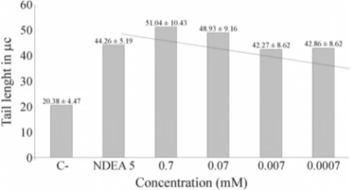
Figure 1 shows that the migration (tail length) of hu-man lymphocyte DNA in the comet assay increased with the concentration of glyphosate and was generally proportional to the latter. The responses to different concentrations of glyphosate differed significantly among themselves (p < 0.0001, ANOVA) and were significantly different from the negative and positive controls (p£0.01, Dunnett test).
Comet assay ofO. niloticuserythrocytes
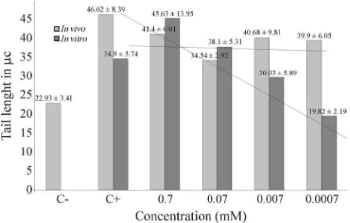
Figure 2 shows the comet assay results for tilapia erythrocytes exposed to different concentrations of glypho-sate isopropylamine saltin vitroandin vivo. When testedin vitro, the increase in DNA migration was proportional to
the glyphosate concentration (p £ 0.001), although no
genotoxicity was observed at 0.0007 mM. The responses to different concentrations of glyphosate differed signifi-cantly among themselves (p < 0.0001, ANOVA) and were significantly different from the negative and positive con-trols (p£0.01, Dunnett test). Glyphosate was also
geno-toxic to fish erythrocytesin vivo(p £0.001), but the
res-ponse was not concentration-dependent.
Comet assay ofTradescantiastamen nuclei
The comet assay results forTradescantiastamen
nu-clei exposed to glyphosatein vitroandin vivoare shown in
Figure 1- Tail length in human lymphocytes exposed to different
Figure 3. In vitro, there was a positive relationship between the glyphosate concentration and the increase in DNA mi-gration (at glyphosate concentrations of 0.0007 to 0.07 mM); there were also significant differences (p£0.0001) among the responses to these three
tions of glyphosate, and between all glyphosate concentra-tions and the negative control (p£0.01). Although
glypho-sate was also significantly (p£0.05) genotoxicin vivo, this response was not proportional to the concentration tested and was significantly lower than that observedin vitro.
Table 1 compares the genotoxicity of glyphosate in human lymphocytes,O. niloticuserythrocytes and Trades-cantia (clone 4430) stamen nuclei in vivo and in vitro.
Glyphosate was clearly genotoxic in all cases.
Discussion
The comet assay is a valuable and sensitive tool for detecting genetic damage in individual cells (Singhet al., 1988). Alvarezet al.(2001) reported a protocol that simpli-fies this assay inTradescantiastamen nuclei. Genetic dam-age induced by glyphosate has been reported (Mañaset al., 2009; Vera-Candiotiet al., 2013) and the comet assay has been used to demonstrate genotoxicity in fish hepatic cells andTradescantianuclei (Alvarezet al., 2011; Guilhermeet al., 2012). Although the evidence of glyphosate genotoxi-city or carcinogenigenotoxi-city fromin vitroand animal studies is
poor (De Rooset al., 2005), a few epidemiological reports
have indicated potential health effects (De Roos et al.,
2003).
In this study, the comet assay clearly showed that glyphosate was genotoxic in the cells examined. Since the in vivo effect of glyphosate in humans was not examined in this work, we compared the data from a study of the direct effect of glyphosate in occupationally exposed humans (Paz-y-Miñoet al., 2007) with the effect of direct exposure of human lymphocytes to glyphosate observed here. As shown in Table 2, glyphosate was genotoxic in both stud-ies. The tail length was very similar in both cases and there was a positive relationship between genotoxicity and gly-phosate concentration/dose.
The high genotoxicity of glyphosate in human lym-phocytes, Tradescantia nuclei and fish erythrocytes in vitro may be indicative of direct DNA damage (Kirkland, 1998; Torstenssonet al., 1989; Alvarezet al., 2011). The geno-toxicity of glyphosate was lowerin vivothanin vitro, per-haps because of efficient herbicide degradation in the former situation. This generalization did not apply to eryth-rocytes ofO. niloticus since these fish were particularly sensitive to glyphosate, perhaps because of poor herbicide metabolism; this finding could also explain the lack of con-centration-dependent genetic damage. Glyphosate has pre-viously been reported to cause DNA damage in liver cells ofAnguila anguila(Guilhermeet al., 2012) but differently, Oreochromis niloticus erythrocytes showed to be more sus-ceptible to lower concentration.
Figure 3- Tail lengths of stamen nuclei fromTradescantiainflorescences exposed to different concentrations of isopropylamine glyphosatein vitro
andin vivo. The lines indicate the relationship between glyphosate con-centration and tail length in thein vivoandin vitrostudies. C and C+
-negative and positive controls, respectively. The values above the col-umns are the mean±SD (n = 30). Tail length (mc).
Figure 2- Tail lengths ofO. niloticuserythrocytes exposed to different concentrations of isopropylamine glyphosate in vitro andin vivo. The di-agonal line and horizontal line indicate the relationship between glypho-sate concentration and tail length in thein vivoandin vitrostudies,
respec-tively. C- and C+ - negative and positive controls, respecrespec-tively. The values above the columns are the mean±SD (n = 8). Tail length (mc).
Table 1- Comparison of the genotoxicity of glyphosate in human lymphocytesin vitroand O. niloticus erythrocytes andTradescantiastamen nuclei (clone 4430)in vivoandin vitro.
Human cells O. niloticusblood cells Tradescantia(clone 4430) stamen nuclei
In vitroexposed cells In vivoexposed fishes In vitroexposed cells In vivoexposedTradescantiaplants In vitroexposed nuclei
PE PE PE PE PE
Ranket al.(1993) investigated the potential
genoto-xicity of glyphosate inA. cepa. The anaphase-telophase Alliumtest showed that glyphosate significantly increased
the genetic damage at concentrations of 1.44 mg/mL and 2.88 mg/mL. Our results suggest that the minimal concen-tration for glyphosate genotoxicity was close to 0.0007 mM (118mg/L), although genotoxicity may start at even lower
concentrations. This finding agrees with other studies that have tested similar concentrations,e.g., 58-116mg/L
(Al-varezet al., 2011; Guilhermeet al., 2012). However, high
concentrations have been reported by Kültiginet al.(2011)
(100-500 mg/L) and Prasadet al.(2009) (25-50 mg/L).
The selection of an adequate bioassay to detect geno-toxicity is a very important factor in obtaining useful results (Zúñiga, 2001). In the present study, the comet assay was clearly sufficiently sensitive to detect the genotoxicity of glyphosate isopropylamine in cells and nuclei of different organisms.
In conclusion, our results indicate that glyphosate is genotoxic, depending on the time and concentration used, as reported by Polettaet al.(2009). Given the extensive use
of this herbicide, it is clear that glyphosate has a potential risk for a variety of organisms, including humans.
Acknowledgments
This study was partially supported by the University of Guadalajara - COECYTJAL de México 401.
References
Alvarez C, Reynoso SM, Villalobos AM, Islas SA, Castañeda VH and González MRM (2011) Evaluation of genetic damage induced by glyphosate isopropylamine salt using Trades-cantiabioassays. Genet Mol Biol 34:127-130.
Alvarez C, Santerre A, Zúñiga G, Torres O, Padilla E and Feria A (2001) Evaluation of the genotoxic activity of maleic hydra-zide, ethyl methane sulfonate, and N-nitrosodiethylamine in Tradescantia. Salud Pública Méx 43:563-569.
Bolognesi C, Carrasquillab G, Volpia S, Solomonc KR and Marshalld EJ (2009) Biomonitoring of genotoxic risk in ag-ricultural workers from five Colombian regions: Associa-tion to occupaAssocia-tional exposure to glyphosate. J Toxicol Envi-ron Health A 72:15-16.
Busse MD, Ratcliff AW, Shestak CJ and Powers RF (2001) Glyphosate toxicity and the effects of a long-term vegetation control on soil microbial communities. Soil Biol Biochem 33:1777-1789.
Cavas T and Könen S (2007) Detection of cytogenetic and DNA damage in peripheral erythrocytes of goldfish (Carassius auratus) exposed to glyphosate formulation using the micro-nucleus test and comet assay. Mutagenesis 22:263-268. Conner DE and Black MC (2004) Evaluation of lethality and
genotoxicity in the freshwater musselUtterbackia imbecillis (Bivalvia, Unionidae) exposed singly and in combination to chemicals used in lawn care. Arch Environ Contam Toxicol 46:362-371.
Cox C (1995) Glyphosate, Part 1-2. Toxicology, human exposure and ecological effects. J Pestic Reform 15:3-4.
Cox C (1998) Glyphosate (roundup). J Pestic Reform 18:3-17. De Roos AJ, Zahm SH, Cantor KP, Weisenburger DD, Holmes
FF, Burmeister LF and Blair A (2003) Integrative assess-ment of multiple pesticides as risk factors for non-Hodg-kin’s lymphoma among men. Occup Environ Med 60:e11. De Roos AJ, Blair A, Rusiecki JA, Hoppin JA, Svec M, Dosemeci
M, Sandler DP and Alavanja MC (2005) Cancer incidence among glyphosate-exposed pesticide applicators in the Ag-ricultural Health Study. Environ Health Perspect 113:49-54. Dimitrov BD, Gadeva PG, Benova DK and Bineva MV (2006)
Comparative genotoxicity of the herbicide roundup, stomp and reglone in plant and mammalian test systems. Mutagen-esis 21:375-382.
EPA (1993) Environmental Protection Agency Registration Eligi-bility Decision (RED) Glyphosate. EPA-738-R-93-014. En-vironmental Protection Agency, Washington DC, 74 pp. Guilherme S, Gaivão I, Santos MA and Pacheco M (2012) DNA
damage in fish (Anguilla anguilla) exposed to a glyphosate-based herbicide – Elucidation of organ-specificity and the role of oxidative stress. Mutat Res 743:1-9.
Hartmann A, Agurell E, Beever C, Brendler-Schwaab S, Bur-linson B, Clay P, Collins A, Smith A, Speit G, Thybaud V,et al.(2003) Recommendations for conducting thein vivo al-kaline comet assay. Mutagenesis 18:45-51.
Kirkland D (1998) Chromosome aberration testing in genetic tox-icology – Past, present and future. Mutat Res 404:173-185. Koppen G and Verschaeve L (1996) The alkaline comet on plant
cells: A new genotoxicity test for DNA breaks inVicia faba root cells. Mutat Res 360:193-200.
Kültigin Ç, Emine Y, Zafer T, Kürsad Y, Kürsat Ç and Figen Ç (2010) Investigation of toxic effects of the glyphosate on Alliumcepa. J Agric Sci 17: 131-142.
Ma TH, Cabrera GL, Cebulska-Wasilewska A, Chen R, Loarca F, Vandenberg AL and Salamone MF (1994)Tradescantia sta-men hair mutation bioassay. Mutat Res 310:211-220. Mañas F, Peralta L, Raviolo J, García OH, Weyers A, Ugnia L,
Gonzalez CM, Larripa I and Gorla N (2009) Genotoxicity of glyphosate assessed by the comet assay and cytogenetic test. Environ Toxicol Pharmacol 28:37-41.
Owczarek M, De Marco A, De Simone C and Ambrosio CD (1999) Evaluation of the toxic and genotoxic activity of
Table 2- Comparison between human lymphocytes from persons occupa-tionally exposed to glyphosate (Paz-y-Miñoet al., 2007) and human lym-phocytes exposed directly to various concentrations of the compound.
Studyin vivo(Paz-y-Miñoet al., 2007) mM Tail length (mc)
Individuals exposed 35.5±6.4 Individuals not exposed 25.9±0.6 Studyin vitro(present study)
Lymphocytes exposed 0.7 51.0±10.4 0.07 48.9±9.2 0.007 42.3±8.6
0.0007 42.9±8.6 Lymphocytes not exposed 20.4±4.1
some pesticides in to soil-plant system. Proceedings of the IX Symposium on Pesticide Chemistry, Cremona, Italy, pp 755-762.
Paz-y-Miño C, Sánchez ME, Arévalo M, Muñoz MJ, Witte T, Oleas G and Leone PE (2007) Evaluation of DNA damage in an Ecuadorian population exposed to glyphosate. Genet Mol Biol 30:456-460.
Poletta GL, Larriera A, Kleinsorge E and Mudry MD (2009) Genotoxicity of the herbicide formulation Roundup®
(gly-phosate) in broad-snouted caiman (Caiman latirostris) evi-denced by the Comet assay and the Micronucleus test. Mutat Res 672:95-102.
Rank J, Jensen A G, Skov B, Pedersen LH and Jensen K (1993) Genotoxicity testing of the herbicide Roundup and its active ingredient glyphosate isopropylamine using the mouse bone marrow micronucleus test, Salmonella mutagenicity test, andAlliumanaphase-telophase test. Mutat Res 300:29-36. Singh N, McCoy M, Tice R and Schneider L (1988) A simple
technique for quantitation of low levels of DNA damage in individual cells. Exp Cell Res 175:184-199.
Sivikova K and Dianovsky J (2006) Cytogenetic effect of techni-cal glyphosate on cultivated bovine peripheral lymphocytes. Int J Hyg Environ Health 209:15-20.
Sparling DW, Matson C, Bickham J and Doelling-Brown P (2006) Toxicity of glyphosate as Glypro (R) and LI700 to red-eared slider (Trachemys scripta elegans) embryos and early hatchlings. Environ Toxicol Chem 25:2768-2774. Prasad S, Srivastava S, Singh M and Shukla Y (2009) Clastogenic
effects of glyphosate in bone marrow cells of Swiss albino mice. J Toxicol 2009:308985.
Torstensson NT, Lundgren LN and Stenström J (1989) Influence of climatic and edaphic factors on persistence of glyphosate and 2,4-D in forest soils. Ecotoxicol Environ Saf 18:230-239.
Truta E, Vochita G, Rosu CM, Zamfirache MM and Olteanu Z (2011) Evaluation of Roundup-induced toxicity on genetic material and on length growth of barley seedlings. Acta Biol Hung 62:290-301.
Underbrink AC, Schairer LA and Sparrow AH (1973) Tradescan-tiastamen hairs: A radiobiological test system applicable to chemical mutagenesis. In: Hollaender A (ed) Chemical Mutagens: Principles and Methods for their Detection. Ple-num Press, New York, pp 71-207.
US Forest Service (1997) Glyphosate: Herbicide Information Pro-file. Pacific Northwest Region. Bulletin United Stated Drug Administration, Washington, 25 pp.
Vera-Candioti J, Soloneski S and Larramendy ML (2013) Evalua-tion of the genotoxic and cytotoxic effects of glyphosate-based herbicides in the ten spotted live-bearer fish Cnes-terodon decemmaculatus (Jenyns, 1842). Ecotoxicol Envi-ron Saf 89:166-173.
Williams GM, Kroes R and Munro IC (2000) Safety evaluation and risk assessment of the herbicide Roundup and its active ingredient, glyphosate, for humans. Regul Toxicol Phar-macol 31:117-165.
Zúñiga GG (2001) Sistemas de detección de daño genético. In: Álvarez C (ed) Genética, Ambiente y Salud. 2nd edition. Universidad de Guadalajara, Guadalajara, pp 127-150.
Associate Editor: Daisy Maria Fávero Salvadori