801 801 801 801 801 Mem Inst Oswaldo Cruz, Rio de Janeiro, Vol. 93(6): 801-805, Nov./Dec. 1998
Identification of Mycobacteria by Thin Layer
Chromatographic Analysis of Mycolic Acids and
Conventional Biochemical Method: Four Years of Experience
Clarice Queico Fujimura Leite/
+, Clovis Wesley Oliveira de Souza*,
Sergio Roberto de Andrade Leite**
Faculdade de Ciências Farmacêuticas, Universidade Estadual Paulista, Caixa Postal 502, 14801-970 Araraquara, SP, Brasil *Departamento de Saúde Pública, Universidade Federal de São Carlos, São Carlos, SP,
Brasil **Instituto de Química, Universidade Estadual Paulista, Araraquara, SP, Brasil
Mycolic acids analysis by thin-layer chromatography (TLC) has been employed by several laboratories worldwide as a method for fast identification of mycobacteria. This method was intro-duced in Brazil by our laboratory in 1992 as a routine identification technique. Up to the present, 861 strains isolated were identified by mycolic acids TLC and by standard biochemical tests; 61% out of these strains came as clinical samples, 4% isolated from frogs and 35% as environmental samples. Mycobacterium tuberculosis strains identified by classical methods were confirmed by their mycolic acids contents (I, III and IV). The method allowed earlier differentiation of M. avium
complex - MAC (mycolic acids I, IV and VI) from M. simiae (acids I, II and IV), both with similar biochemical properties. The method also permitted to distinguish M. fortuitum (acids I and V) from M. chelonae (acids I and II) , and to detect mixed mycobacterial infections cases as M. tuberculosis with MAC and M. fortuitum with MAC. Concluding, four years experience shows that mycolic acids TLC is an easy, reliable, fast and inexpensive method, an important tool to put together conventional mycobacteria identification methods.
Key words: mycobacteria identification - mycolic acids - thin layer chromatography
After years of declining case rates, tuberculo-sis is again a major public health problem in Bra-zil and in other developing countries (Hijjar 1992). The emergency of AIDS augmented the incidence of infections with other mycobacteria that live in the environment and are resistant to antitubercu-losis drugs (Kritske et al. 1995). However the iden-tification of Mycobacterium species through mor-phological, cultural and biochemical tests, requires several weeks or even months of fastidious work (Wayne et al. 1991). DNA probe analysis is a fast and trusty method, but needs a specific probe for each species, resulting expensive. The utilization of different chemical methods has greatly improved the classification and identification of mycobacte-ria (Luquin et al. 1991, Hines & Fraziers 1993, Leite et al. 1995). Chemical analysis of cellular fatty acids, especially of those with high
molecu-lar-weight a-branched b-hydroxylated chain, the
mycolic acids, has been used as a rapid test for mycobacterial systematics (Lévy-Frébault et al.
1986, Butler & Kilburn 1990).
Mycolic acid methyl ester analysis by thin layer chromatography (TLC) has been employed by sev-eral laboratories worldwide as a fast method for identification of clinical (Lévy-Frébault et al. 1987) and environmental (Falcão et al. 1993) isolated mycobacteria. Mycolic acids can be resolved into several classes according to the presence of differ-ent functional groups in their molecules. Partial congruence is found between the distribution of mycolic acids types and the finer taxonomic rela-tionships among the mycobacteria as shown in 16S rRNA studies (Goodfellow & Magee 1998). In spite of its advantages, the method was introduced in Brazil as a routine identification technique only in 1992 by our laboratory (Leite et al. 1995). The purpose of this study is to present our four years experience of using TLC associated to conventional biochemical identification methods in a Brazilian mycobacteriology laboratory.
MATERIALS AND METHODS
Reagents and culture media - Diazomethane was prepared by reaction of nitrosomethylurea
This work was supported by Fapesp (Brazil) grant no. 89/3474-2.
+Corresponding author. Fax: +55-16-232-1576 or
222.0073. E-mail: [email protected] Received 11 March 1998
802 802 802 802
802 Technique Improvement for Mycobacteria Identification Clarice QF Leite et al.
(synthesized in our laboratory) with potassium hydroxide. All the reagents and solvents utilized were of analytical grade. The silica gel plates for TLC chromatography were from Merck and Sigma. Lowenstein-Jensen culture medium was prepared at our laboratory.
Bacterial strains and growing conditions - A total of 861 strains were tested: 532 human lates came from Sanatory Hospital, 56 frog iso-lates (frog raising) and 273 environmental isoiso-lates (swimming pools, rivers, aquarium and drinking waters). The type strains M. avium CIPT 14031001,
M. chelonae CIPT 14042003, M. fortuitum CIPT 14041001 and M. tuberculosis CIPT 14001002 were obtained from Institut Pasteur collection and were used for the preparation of mycolic acid stan-dards. Mycobacteria were maintained on Lowenstein-Jensen medium slants at 4°C. For ex-traction of mycolic acids, the strains were grown on Lowenstein-Jensen medium at 35-37°C. In-cubation temperature for M. marinum, M. gor-donae and M. chelonae was 30°C. The incubation period ranged from 7 to 10 days for rapidly ing species and from 25 to 32 days for slowly grow-ing species.
Identification of mycobacteria by conventional methods - All mycobacteria strains were identified by their rate of growth, colonial morphology, pig-mentation and biochemical properties (David et al.
1989).
Mycolic acid extraction and methylation pro-cedures - The mycolic acid analysis was carried out according to the method proposed by Daffé et al. (1983), with some modifications. About 25-50 mg of mycobacteria, derived from Lowenstein-Jensen medium, were dispersed into a 5% (w/v) potassium hydroxide solution in 2-methoxyethanol. The mixture was maintained at 110°C for 2 hr, cooled and acidified with 1 ml of sulfuric acid so-lution (20% w/w). Mycolic acids were then ex-tracted by shaking the mixture twice with diethyl ether (5 ml). The ether phase was decanted and washed three times with 2 ml of water. Ether was removed on a water bath, and the mycolic acids were methylated by addition of 1 ml of diazo-methane ether solution.
Analytical methods and identification of my-colic acids - For TLC of mycolic acid, their me-thyl esters were spotted onto silica gel G (20 x 20 cm x 0.25 mm plates). Mycolic acids of the refer-ence strains spotted together served as landmarks for the identification of mycolic acids extracted from the studied strains. One dimensional analysis was carried out by using two different elution sys-tems: diethyl ether/petroleum ether (12:88 v/v), with three developments of the chromatogram, and
dichlorometane with a single development (Daffé et al. 1983). The presence of the separated compo-nents was revealed by spraying the chromatograms with 0, 01% (w/v) rhodamine in phosphate buffer (Daffé et al. 1983).
RESULTS
The sources and identification of 861 strains analyzed by conventional biochemical method are presented in Table I. Ninety percent of human’s strains were M. tuberculosis (480 strains). Sev-eral other species of mycobacteria were identified from human infection. Mycobacterium avium com-plex (MAC) was the second in incidence, corre-sponding to 4.5% (24 strains). The major strain isolated from frogs was M. marinum (89.3%) and from the environment was M. fortuitum (38.5%) and other fast growing mycobacteria (FGM) (19.4%).
TABLE I
Sources and species of 861 mycobacteria identified by conventional biochemical method
Humans Frogs Water
MAC a 24 - 5
M. chelonae 5 2 7
M. fortuitum 7 - 105
FGM b - 3 53
M. gordonae 7 1 49
M. kansasii 1 - 2
M. marinum 2 50 2
M. scrofulaceum 2 - 1
M. simiae 1 -
-M. smegmatis - - 22
M. terrae complex - - 27
M. tuberculosis complex 480 -
-Unidentified mycobacteria 3 -
-Total 532 56 273
a: Mycobacterium avium complex; b: fast growing mycobacteria; M: Mycobacterium.
be-803 803803 803803 Mem Inst Oswaldo Cruz, Rio de Janeiro, Vol. 93(6), Nov./Dec. 1998
low the mycolate type I. However, in dichlorometh-ane, mycolate III migrates below the mycolate II and here its Rf value is similar to that of mycolate IV. Typical thin-layer chromatograms of the mycolic acids methyl-esters are shown in Figs 1 and 2.
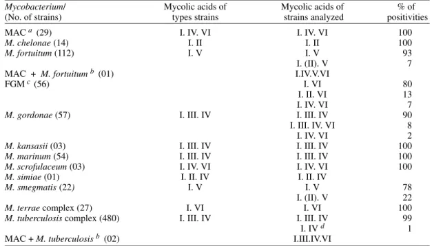
TABLE II
Mycolic acids profiles of 861 strains identified by classical procedures
Mycobacterium/ Mycolic acids of Mycolic acids of % of
(No. of strains) types strains strains analyzed positivities
MAC a (29) I. IV. VI I. IV. VI 100
M. chelonae (14) I. II I. II 100
M. fortuitum (112) I. V I. V 93
I. (II). V 7
MAC + M. fortuitum b (01) I.IV.V.VI
FGMc (56) I. VI 80
I. II. VI 13
I. IV. VI 7
M. gordonae (57) I. III. IV I. III. IV 90
I. III. IV. VI 8
I. IV. VI 2
M. kansasii (03) I. III. IV I. III. IV 100
M. marinum (54) I. III. IV I. III. IV 100
M. scrofulaceum (03) I. IV. VI I. IV. VI 100
M. simiae (01) I. II. IV I. II. IV
M. smegmatis (22) I. V I. V 78
I. (II). V 22
M. terrae complex (27) I. VI I. VI 100
M. tuberculosis complex (480) I. III. IV I. III. IV 99
I. IV d 1
MAC + M. tuberculosis b (02) I.III.IV.VI
a:Mycobacterium avium-intracellulare complex; b: mixed infection; c: fast growing non-pathogenic mycobacteria d:BCG.
1 2 3 4 5 6 7 8 9
Fig. 1: chromatogram of mycolic acid methyl esters. Solvent: dichloromethane. Lines 1 to 4: reference strains. Lines 5 to 9: strains analyzed. 1: Mycobacterium chelonae CIPT 14042003; 2: M. tuberculosis CIPT 14001002; 3: M. avium CIPT 14031001; 4: M. fortuitum CIPT 14041001; 5: M. tuberculo-sis; 6: M. smegmatis; 7: M. scrofulaceum, 8: M. chelonae; 9: M. fortuitum.
1 2 3 4 5 6 7 8 9
Fig. 2: chromatogram of mycolic acid methyl esters. Solvent: diethyl ether-petroleum ether. Lines 1 to 4 references strains. Lines 5 to 9-strains analyzed. 1: Mycobacterium chelonae CIPT 14042003; 2: M. tuberculosis CIPT 14001002; 3: M. avium CIPT 14031001; 4: M. fortuitum CIPT 14041001; 5: M. tuber-culosis; 6: M. smegmatis; 7: M. scrofulaceum; 8: M. chelonae; 9: M. fortuitum.
MAC, M. chelonae, M. kansasii, M. marinum, M. scrofulaceum, M. terrae complex and M. simiae
804 804 804 804
804 Technique Improvement for Mycobacteria Identification Clarice QF Leite et al.
gordonae the mycolic acid profiles were in agree-ment with corresponding species (Minnikin et al.
1984). One percent of M. tuberculosis gave mycolates I and IV that are characteristic of BCG (Calmet-Guérin bacillus)(David et al. 1989). In this group, by additional biochemical tests, we confirmed identification of BCG.
M. gordonae strains have mycolic acids I.III.IV, but in 2% of them we found mycolic acid profile I.IV.VI that is common in M. scrofulaceum and MAC(David et al. 1989). After repeating the bio-chemical tests, we confirmed those strains as M. scrofulaceum. In 8% of M. gordonae we found mycolates I.III.IV.VI and this group was differen-tiated from the type strains (acids I, III and IV) only by their mycolic acids profiles, since both exhibit identical physiological and biochemical properties. Excluding M. chelonae, M. fortuitum
and M. smegmatis, all of other fast growing myco-bacteria (FGM) had mycolic acids I and VI, with or without types II or IV too.
The conventional method was not able to iden-tify 0.6% of the human isolated strains (Table I), which were characterized by mycolate TLC as in-volved with mixed infections, as associations of
M. tuberculosis and MAC or MAC and M. fortuitum.
DISCUSSION
M. tuberculosis is the most common agent of human tuberculosis in Brazil (Kritski et al. 1995) and for this species the identification by the con-ventional method agrees in 98% with the mycolates TLC. Tuberculosis caused by other members of
M. tuberculosis complex is clinically indistinguish-able of the one caused by M. tuberculosis, and their prevalence is unknown in most developing coun-tries because of their limited laboratory facilities (Covisi et al. 1998). In this sense, the TLC analy-sis makes easy the distinction between M. tuber-culosis and M. bovis variety BCG, that was first identified by its mycolate profile (Table II) and then confirmed by classical biochemical tests.
MAC infection is the second most important mycobacteriosis in Brazil, frequently associated to AIDS (Saad et al. 1997). In this study the mem-bers of this complex were easily identified by their mycolic acids I, IV and VI.
Mycolic acids analysis by TLC is becoming a good tool to detect mixed infections, as it may be concluded from this work: two cases of mixed in-fection by M. tuberculosis and MAC and one case by MAC and M. fortuitum. Lévy-Frébault et al. (1987) reported that they could distinguish M. simiae from M. avium by mycolates TLC in a mixed infection. This method also allowed the correct identification of potentially pathogenic M.
scrofulaceum, misidentified as the saprophytic M. gordonae, and to detect M. simiae, a rare species, sometimes misidentified as M. tuberculosis or M. avium by biochemical tests (Wayne et al. 1991). Lévy-Frébault et al. (1986) performing mycolic acids TLC analyses of 133 strains of MAC, found that about 10% of those were in fact M. simiae.
Among the FGM species only M. fortuitum and
M. chelonae are commonly found as human patho-gens (Hijjar 1992). The identification of these mycobacteria is very hard when using the conven-tional method (David et al. 1989). M. fortuitum with mycolic acids I and V and M. chelonae with I and II were easily identified among our samples by TLC. The usefulness of mycolic acid analysis for FGM was described by Butler and Kilburn (1990), which utilized another chromatographic technique for mycolic acids identification, namely the high efficiency liquid chromatography (HPLC).
The conventional biochemical methods were unable to discriminate 8% of our frog and envi-ronmental isolated mycobacteria, which were dif-ferentiated from the type strains of M. gordonae
only by their mycolic acids profiles. This result is interesting because Butler et al. (1996) also de-scribe the occurrence of two different mycolic ac-ids chromatotypes for M. gordonae analyzed by HPLC method. For Leite et al. (1989) the mycolic acids determination is of particular interest for en-vironmental mycobacteria identification, mainly those not frequently isolated from humans.
In our four years experience we concluded that, mycolic acids analysis by TLC joined with con-ventional biochemical method, is a valuable tool for mycobacteria identification. This proceeding has allowed us to detect and identify all strains fre-quently isolated from human, environment and frog. Mycolic acids determination by TLC is a very simple technique and useful for the first step screen-ing in mycobacteria identification. Joined with conventional methods it detects rare or new spe-cies, as well as mixed infections, undetectable by the classical tests alone.
ACKNOWLEDGMENTS
To Prof. Petr Melnikov for language revision.
REFERENCES
Butler WR, Kilburn JO 1990. High-performance liquid chromatography patterns of mycolic acids as crite-ria for identification of Mycobacterium chelonae, Mycobacterium fortuitum and Mycobacterium smegmatis. J Clin Microbiol 28: 2094-2098. Butler WR, Floyd MM, Silcox V, Desmond E, Duffey
Pub-805 805805 805805 Mem Inst Oswaldo Cruz, Rio de Janeiro, Vol. 93(6), Nov./Dec. 1998
lic Health Service-CDC, Atlanta, USA.
Covisi O, Grange JM, Daborn CJ, Raviglione MC, Fujikura T, Cousins D, Robinson RA, Huchzermeyer HFAK, Kantor I, Meslin FX 1998. Zoonotic tuber-culosis due to Mycobacterium bovis in developing countries. Emerg Infect Dis4: 59-70.
Daffé M, Lanéelle MA, Asselineau C, Lévy-Frébault V, David H 1983. Intérêt taxonomique des acides gras des mycobactéries; proposition d’une méthode d’analyse. Ann Microbiol (Paris) 134B: 241-256. David H, Lévy-Frébault V, Thorel MF 1989. Méthodes
de Laboratoire pour Mycobactériologie Clinique, Institute Pasteur, Paris, 85 pp.
Falcão DP, Valentini SR, Leite CQF 1993. Pathogenic and potentially pathogenic microorganisms as con-taminants of fresh water from different sources. Water Res 27:1737-1741.
Goodfellow M, Magee JG 1998. Taxonomy of myco-bacteria, p. 1-71. In PRJ Gangadharam, Mycobac-teria I, Basic Aspects, Chapman & Hall Medical Microbiology Series, New York.
Hijjar MA 1992. Epidemiologia da tuberculose no Brasil. Informe Epidemiológico do SUS, Brasília 1: 53-87. Hines ME, Fraziers KS 1993. Differentiation of myco-bacteria on the basis of chemotype profiles by using matrix solid-phase dispersion acid thin-layer chro-matography. J Clin Microbiol 31: 610-614. Kritski A, Dalcomo MP, Bravo de Souza R, Hollanda,T,
Gontijo Filho PP, Fiuza de Mello FA 1995. Associação tuberculose e infecção pelo HIV no Brasil. Bol Of Sanit Panam, Washington 118: 542-554.
Leite CQF, Barreto AMW, Leite SRA 1995. Thin-layer chromatography of mycobactins and mycolic acids for the identification of clinical mycobacteria. Rev Microbiol 26: 192-199.
Leite CQF, Giannini MJSM, Falcão DP, Lévy-Frébault V, David H 1989. Presence of Mycobacterium marinum and other opportunistic mycobacteria in swimming pool waters in Araraquara, SP. Rev Microbiol 19: 354-359.
Lévy-Frébault V, Goh KS, David H 1986. Mycolic acid analysis for clinical identification of Mycobacterium avium and related mycobacteria. J Clin Microbiol 24: 835-839.
Lévy-Frébault V, Panzon B, Buré A, Katlama C, Marche C 1987. Mycobacterium simiae and Mycobacterium avium-M. intracellulare mixed infection in acquired immune deficiency syndrome. J Clin Microbiol 25: 154-157.
Luquin M, Ausina V, Calahorra FL, Belda F, Barceló MG, Celma C, Prats G 1991. Evaluation of practi-cal chromatographic procedures for identification of clinical mycobacteria. J Clin Microbiol 29: 120-130. Minnikin DE, Minnikin SM, Parlett JH, Goodfellow M, Magnusson M 1984. Mycolic acid patterns of some species of Mycobacterium. Arch Microbiol 139: 225-231.
Saad MHF, Vincent V, Dawson DJ, Palari M, Ferrazoli L, Fonseca LS 1997. Analysis of Mycobacterium avium complex serovars isolated from AIDS patients from southeast Brazil. Mem Inst Oswaldo Cruz 92: 471-475.
806 806 806 806