361 361 361 361 361 Mem Inst Oswaldo Cruz, Rio de Janeiro, Vol. 98(3): 361-365, April 2003
Reproductive System Abnormalities in
Schistosoma mansoni
Adult Worms Isolated from
Nectomys squamipes
(Muridae: Sigmodontinae): Brightfield and Confocal Laser
Scanning Microscopy Analysis
Renata Heisler Neves, Michele Costa-Silva, Elaine Machado Martinez,
Carla de Lamare Biolchini, Henrique Leonel Lenzi*/
++, Delir Corrêa Gomes**/
++,
José Roberto Machado-Silva/
+/
++Departamento de Patologia e Laboratórios, Faculdade de Ciências Médicas, Universidade do Estado do Rio de Janeiro, Av. 28 de Setembro, 87 fundos, 5o andar, 20551-031 Rio de Janeiro, RJ, Brasil *Departamento de Patologia **Laboratório de Helmintos
Parasitos de Vertebrados, Departamento de Helmintologia, Instituto Oswaldo Cruz-Fiocruz Rio de Janeiro, RJ, Brasil
Schistosoma mansoni adult worms with genital anomalies isolated from Nectomys squamipes (Muridae: Sigmodontinae) were studied by confocal laser scanning microscopy under the reflected mode. One male without testicular lobes (testicular agenesia/anorchism) and two females, one with an atrophied ovary and another with 17 uterine eggs, were identified. The absence of testicular lobes occurred in a worm presenting otherwise normal male adult characteristics: tegument, tubercles and a gynaecophoric canal with spines. In both female specimens the digestive tube showed a vacuolated appearance, and the specimen with supernumerary uterine eggs exhibited a developing miracidium and an egg with a formed shell. The area of the ventral sucker was similar in both specimens however the tegument thickness, ovary and vitelline glands of the specimen with the atrophied ovary were smaller than those of the one with supernumerary eggs. These reported anomalies in the reproductive system call attention to the need to improve our understanding of genetic regulation and the possible role of environmental influences upon trematode development.
Key words: Schistosoma mansoni reproductive system abnormalities adult worms trematode anorchism -confocal laser scanning microscopy
The somatic development and reproductive system of adult worms of Schistosoma mansoni suffer intense se-lective pressure from the vertebrate hosts. Full somatic development of adult worms, characterized by large size, complete formation of reproductive organs and complete tegumental maturation, is reached only in permissive hosts (mice, hamster and water rat) (Cioli et al. 1977, Machado-Silva et al. 1997). However, anomalous specimens can occur sporadically even in favorable hosts. In this con-text, Najim (1951) found a male adult with two sets of tes-tes, which he considered as supranumerary testes. Machado Filho (1966) recovered a female with six uterine eggs, without however any visible morphological alter-ations. Dias and Ribeiro (1980) found in S. mansoni eggs containing two active miracidia the feces from a patient.
This article describes morphologically the abnormali-ties of the reproductive system of S. mansoni adult worms (two females and one male) isolated from Nectomys
This work was sponsored by Universidade do Estado do Rio de Janeiro (Uerj) and Fundação Oswaldo Cruz (Fiocruz) and is part of a Master Thesis, Instituto de Biologia, Uerj, RJ, Brazil.
+Corresponding author. Fax: +55-21-2587.6112. E-mail:
machado@uerj.br.
++CNPq Research fellows
Received 16 September 2002 Accepted 9 January 2003
squamipes (Muridae: Sigmodontinae), which is a permis-sive host, using brightfield and confocal laser scanning microscopies.
MATERIALS AND METHODS
362 362 362 362
362 Reproductive Abnormalities in S. mansoni • Renata Heisler Neves et al.
area of the suckers and distance between them; area, smaller and larger diameters and perimeter of uterine eggs and egg spine area. From acquired confocal images (LSM-410, Zeiss), using a 543 nm He/Ne laser and a LP 570 filter under reflected mode, the thickness of the tegument un-der the ventral sucker of the three specimens, the width and length of the ovary and vitelline glands were mea-sured by specific software (version 3.98, Zeiss) pertain-ing to the equipment.
Normal adult worms were isolated from experimen-tallyinfected N. squamipes with the same strains under the same conditions, and were used as controls.
RESULTS
For the male specimen the morphometric values were: 22056 µm2 for the area of the oral sucker, 19831 µm2 for the ventral sucker, 255 µm for the distance between them and 11µm for the thickness of the tegument. Testicular lobes were not identified, even after virtual tomography under reflected mode by confocal laser scanning micros-copy (CLSM). However, the specimen presented other male adult characteristics such as a fully developed
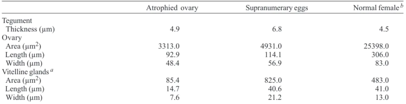
tegu-ment, tubercles with spines and a gynaecophoric canal (Fig. 1). In contrast, the confocal analysis of the corre-sponding body segment from normal adult males clearly developed testicular lobes and a seminal vesicle (Fig. 2). In females worms the atrophy of the ovary observed by brightfield microscopy in one specimen was also con-firmed by CSLM (Fig. 3), contrasting with the regular ovary of normal females (Fig. 4). The supernumerary uterine eggs seen in another specimen exhibited a miracidium in devel-opmental stage and an egg with a formed shell (Fig. 5). In both female specimens the digestive tube was deformed by large dilatations of the lumen, containing amorphous and vacuolated material (Fig. 6). The comparison between the anomalous females, except for the area of the ventral sucker, showed that the specimen with atrophied ovary showed smaller measurements than the female with supranumerary eggs (Tables I, II).
DISCUSSION
The actual incidence of dysmorphogenetic defects among S. mansoni adult worms is completely unknown. However it has been established that this incidence may
TABLE I
Comparative measurements between anomalous and normal Schistosoma mansoni females isolated from Nectomys squamipes
(GD strain), using brightfield microscopy
Atrophied ovary Supranumerary eggs Normal femalesb Suckers
Area oral sucker (µm2) 1810.0 2621.0 2190.1
Area ventral sucker (µm2) 1911.0 1871.0 2112.4
Distance between suckers (µm) 89.0 161.0 160.0
Egg a
Area (µm 2) - 1923.0 2698.0
Larger diameter (µm) - 70.0 97.0
Smaller diameter (µm) - 33.7 34.0
Perimeter (µm) - 186.3 246.0
Egg spine
Area (µm 2) - 133.3 121.5
Larger diameter (µm) - 17.7 17.0
Smaller diameter (µm) - 8.7 8.0
Perimeter (µm) - 50.3 49.4
a: the mean of three eggs; - not detected; b: mean (n = 15)
TABLE II
Comparative measurements between anomalous and normal Schistosoma mansoni females isolated from Nectomys squamipes, using confocal scanning laser microscopy
Atrophied ovary Supranumerary eggs Normal femaleb Tegument
Thickness (µm) 4.9 6.8 4.5
Ovary
Area (µm2) 3313.0 4931.0 25398.0
Length (µm) 92.9 114.1 306.0
Width (µm) 48.4 56.9 83.0
Vitelline glands a
Area (µm2) 85.4 825.0 483.0
Length (µm) 14.7 40.6 41.0
Width (µm) 7.6 21.2 13.0
363 363363 363363 Mem Inst Oswaldo Cruz, Rio de Janeiro, Vol. 98(3), April 2003
1 T
TB
AO
E
E
PE
VC
M
NO GC
ZLT
LT SV
3
5 6
2 50 mµ
25 mµ
50 mµ
50 mµ
50 mµ
50 mµ 4
364 364 364 364
364 Reproductive Abnormalities in S. mansoni • Renata Heisler Neves et al.
vary according to host, environmental/teratogenic fac-tors, including drugs, polygenic disorders, single gene mutations and others (Thorogood 1997).
S. mansoni is a dioecious trematode, as a result of evolutive pressure, acquired different and complemen-tary functions where the males became oriented to fecun-dation (Basch 1990, Platt & Brooks 1997) and female trans-portation to the site of oviposition, which is facilitated by the complex musculature of the suckers (Mair et al. 2000, Morand & Muller-Graf 2000), while the females invested in the production of eggs (Morand & Muller-Graf 2000). Therefore, any imbalance in this relationship can affect the reproduction of this parasitic flatworm. In female schis-tosomes, an intimate and permanent association with the male is necessary for reproduction to occur (Kunz 2001). The finding of atypical adult worms collected from permissive hosts with alterations in the location of the testicular lobes has already been reported (Najim 1951, Machado-Silva et al. 1994, Neves et al. 1998). In the present article, we described a male specimen without testicular lobes (testicular agenesia), not related to immature stage (below 21-days-old). Although sexual maturation with-out synchronism can occur (Barbosa et al. 1978), in our experiment, the hosts were with nine weeks post infec-tion, time enough for all the specimens of the entire popu-lation to reach the adult stage. Furthermore, the tegument of the male with testicular agenesia had tubercles and its thickness was compatible with that of adult stage (Machado-Silva et al. 1994, 1997, 1998, Neves et al. 1998). Finally, it is worthwhile to point out that young speci-mens (21 days old) already present two testicular lobes (Clegg 1965).
Structural modifications of the female reproductive system can occur during maintenance of the trematode in vitro (Basch 1981) or in unisexual infections (Erasmus 1973). In the present experiment, alterations were evi-denced in mixed infections. One of the females of the GD strain presented atrophy in both ovary and vitelline glands. Other components of the reproductive system (oviduct, ootype, uterus) presented normal aspect, resem-bling those of immature females derived from unisexual infection (Erasmus 1973). After 63 days of infection, it is not expected to find immature females, considering that the immature sexual stages appear only during the mor-phogenetic period (30-38 days) (Popiel 1986). Conversely, the preservation of the reproductive morphogenesis is determined by the permanent pairing of specimens (LoVerde & Chen 1991, Southgate et al. 1998), which de-pends on the sex ratio between them. In the present ar-ticle this ratio was balanced (1:1), which lead us to as-sume that all specimens were paired, even though it has been demonstrated that males are not monogamous (Pica-Mattocia et al. 2000). According to Kunz (2001), the male signal does not differentiate pluripotent stem cells into germ cells. Instead, it regulates the multiplication and dif-ferentiation of germ cells that are already present, located in separate organs and committed to become oocytes or vitellocytes before pairing with a male. Vitelline cells are haploid, suggesting a common origin of the vitelline gland and ovary. In S. mansoni cultured in vitro from cercariae to adults, vitelline cells are essentially normal,
demon-strating that specific host influences are not necessary for their development (Basch & Samuelson 1990).
Another female derived from the same strain was also atypical, presenting supernumerary eggs and intestinal dilatation. This situation is rare and confirms a previous report of a specimen with six supranumerary eggs (Machado Filho 1966). Morphological alterations have not been evidenced in the other components of the repro-ductive system, and the eggs were inside a dilated uterus. This observation, together with the fact that the eggs were mature, with miracidium and configurated shell, and the intestinal and uterus lumens were dilated, suggested that the main reason was egg intrauterine retention by unknown obstructive mechanism, rather than egg pro-duction and or secretion defects. The intestinal and uter-ine lumen dilatation may also have been due to impaired or insufficient female musculature. Female reproductive changes could be partially related to intestinal enzymatic deficiencies, since products of hemoglobin digestion by intestinal hemoglobinases are shuttled into the vitellaria, where they serve as egg yolk for the soon-to-develop miracidium (Newport & Agabian 1990).
In our experiment, even considering that the strains are different, they had also been maintained in the labora-tory and, moreover, other analyzed specimens did not present alterations similar to those presently described. As the same intermediate and definitive hosts were uti-lized for both strains, we consider that these strains were under the same selective pressure.
The scientific communication of these types of rare development anomalies or defects, expressing phenotypic variations will undoubtedly help to improve our under-standing of the molecular, genetic and cellular mechanisms underlying normal development in parasitic helminthes.
REFERENCES
Barbosa MA, Pellegrino J, Celho PM, Sampaio IB 1978. Quan-titative aspects of the migration and evolutive asynchronism of Schistosoma mansoni in mice. Rev Inst Med Trop São Paulo 20: 121-132.
Basch PF 1981. Cultivation of Schistosoma mansoni in vitro. II. Production of infertile eggs by worm pairs cultured from cercariae. J Parasitol 67: 186-190.
Basch PF 1990. Why do schistosomes have separate sexes?
Parasitol Today 6: 160-163.
Basch PF, Samuelson J 1990. Cell biology of schistosomes. I. Ultrastructure and transformations. In Modern Parasite Biology. Cellular, Immunological and Molecular Aspects, WH Freeman and Co., New York, p. 91-106.
Cioli D, Knopf PM, Senft AW 1977. A study of Schistosoma mansoni transferred into permissive and nonpermissive hosts. Int J Parasitol 7: 293-297.
Clegg JA 1965. In vitro cultivation of Schistosoma mansoni.
Exp Parasitol 16:133-147.
Dias LC, Ribeiro OB 1980. Schistosoma mansoni: eggs with two miracidia. Trans R Soc Trop Med Hyg 74: 826. Erasmus DA 1973. A comparative study of the reproductive
system of mature, immature and “ unisexual” female Schis-tosoma mansoni. Parasitology 67: 165-183.
Kunz W 2001. Schistosome male-female interaction: induction of germ-cell differentiation. Trends Parasitol 17: 227-231. LoVerde PT, Chen L 1991. Schistosome female reproductive
365 365365 365365 Mem Inst Oswaldo Cruz, Rio de Janeiro, Vol. 98(3), April 2003
Machado Filho BA 1966. Sobre uma fêmea de Schistosoma mansoni Sambon, 1907 com seis ovos “in útero”. Atas Soc Biol Rio de Janeiro 10: 11-12.
Machado-Silva JR, Galvão C, Presgrave OAF, Rey L, Gomes DC 1994. Host-induced morphological changes of Schisto-soma mansoni Sambon, 1907 male worms. Mem Inst Oswaldo Cruz 89: 411-416.
Machado-Silva JR, Lanfredi RM, Gomes DC 1997. Morpho-logical study of adult male worms of Schistosoma mansoni
by scanning electron microscopy. Mem Inst Oswaldo Cruz 92: 647-653.
Machado-Silva JR, Pelajo-Machado M, Lenzi HL, Gomes DC 1998. Morphological study of adult male worms of Schis-tosoma mansoniSambon, 1907 by confocal laser scanning microscopy.Mem Inst Oswaldo Cruz 93: 303-307. Mair GR, Maule AG, Day TA, Halton DW 2000. A confocal
microscopical study of the musculature of adult Schisto-soma mansoni. Parasitology 121: 163-170.
Morand S, Muller-Graf CD 2000. Muscles or testes? Com-parative evidence for sexual competition among dioecious blood parasites (Schistosomatidae) of vertebrates. Parasi-tology 120: 45-56.
Najim AT 1951. A male Schistosoma mansoni with two sets of testes. J Parasitol 37: 545-546.
Neves RH, Pereira MJS, Gomes DC, Oliveira RMF, Machado-Silva JR 1998. Morphometric differences of adult worms from sympatric samples of Schistosoma mansoniSambon, 1907 isolated from rodents and humans. Mem Inst Oswaldo Cruz 93: 309-312.
Newport GR, Agabian N 1990. Molecular biology of schisto-somes and filariae. In Modern Parasite Biology. Cellular, Immunological and Molecular Aspects, WH Freeman and Co., New York, p. 362-383.
Pica-Mattocia L, Moroni R, Tchuem Tchuente LA, Southgate VR, Cioli D 2000. Changes of mate occur in Schistosoma mansoni. Parasitology 120: 495-500.
Platt TR, Brooks DR 1997. Evolution of the schistosomes (Di-genea: Schistosomatoidea): the origin of dioecy and coloni-zation of the venous system. J Parasitol 83: 1035-1044. Popiel I 1986. Male-stimulated female maturation in
Schisto-soma: a review. J Chem Ecol 12: 1745-1754.
Southgate VR, Jourdane J, Tchuente LA 1998. Recent studies on the reproductive biology of the schistosomes and their relevance to speciation in the Digenea. Int J Parasitol 28: 1159-1172.
366 366 366 366