Abstract
Submitted: March 03, 2017 0RGL¿FDWLRQ0D\ Accepted: June 02, 2017
Intradentinal antimicrobial action and
agitation of epoxy resin-based sealer
in endodontic obturation
antimicrobial effect against Ent erococcus faecalis within dentinal tubules.
selected and divided into 2 groups (n=15): with and without UA of the
and the specimens were sectioned at 2, 4 and 6 mm from the apex for stereomicroscope and confocal laser scanner microscopy (CLSM) analysis. In addition, 30 bovine incisors were contaminated with Ent erococcus faecalis
and divided into 3 groups (n=10). The specimens were obturated by using the single cone technique with (G1) and without (G2) UA of the sealer and G3 as the control group. All were sectioned into 6 mm-long cylinders and stained with LIVE/DEAD to assess bacterial viability by CLSM. Results: The UA of the
in sealer penetration in both canals and isthmuses (p<0.05). As regards
groups (p<0.05). Conclusion: Ultrasonic activation of the AH Plus sealer
penetration of sealer, especially in the isthmus area. Additionally, ultrasonic activation of the sealer increased the intradentinal antimicrobial action against
Ent erococcus faecalis
Ke yw or ds: Endodontics. Bacteria. Ultrasonic.
Murilo Priori ALCALDE1
Clóvis Monteiro BRAMANTE1
Rodrigo Ricci VIVAN1
Pablo Andrés AMORSO-SILVA1
Flaviana Bombarda de ANDRADE1
Marco Antonio Hungaro DUARTE1
1Universidade de São Paulo, Faculdade de Odontologia de Bauru, Departamento de Dentística, Endodontia e Materiais Odontológicos, Bauru, SP, Brasil
Introduction
Adequate obturation of the root canal system is
required after biomechanical procedures, to ensure the long-term success of endodontic treatment20. During
root canal obturation, an important objective of using
reached by gutta-percha6. However, the anatomical
obturation2.
The presence of isthmuses has a negative effect 13 as the obturation
4,5. To
quality in terms of reducing unfilled areas and
gaps, and promoting more effective penetration of
sealer into dentinal tubules, recent studies have
recommended the ultrasonic activation (UA) of sealers before obturation with gutta-percha7,16. However, this
effect has only been studied in single rooted teeth
with single canals, and no studies evaluating these
aspects in teeth with anatomical complexities have been conducted so far.
Within the dentinal tubules, bacteria such as
En t er ococcu s f aecalis ( E. f aecalis) have capacity
to penetrate deeply and offer resistance to the antimicrobial agents used in endodontics29. These
bacteria are the microorganisms most frequently
isolated from persistent endodontic infections19,22.
Although the AH Plus sealer has been studied by several authors, and has shown good antimicrobial
effectagainst E. f aecalis18,28, this factor might be
dependent on the depth of penetration of the sealer.
In addition, sealer penetration into the dentinal
bacterial entombing action10,15. Based on the foregoing
considerations, it would be interesting to assess the
effect of ultrasonic activation of the sealer to enhance the antimicrobial activity against E.faecalis within the
dentinal tubules.
Therefore, the aim of the present study was to
in mandibular molars, and analyse the antimicrobial
effect against E. faecalis within the dentinal tubules. The null hypotheses tested were as follows:
Ultrasonic activation of the endodontic sealer would
There would be no difference in the adaptation
of the sealer/dentine interface and intratubular
penetration when ultrasonic activation was performed in the mesial roots of mandibular molars.
The ultrasonic activation would not improve the
intradentinal antimicrobial activity of AH Plus against
E. faecalis.
Material and methods
Sam ple Select ion
The ethics committee approved this research (CEP
788274). The sample calculation was performed before
the mechanical test by using the G*Power v3.1 for Mac (Heinrich Heine, University of Düsseldorf) by selecting
the Wilcoxon– Mann-Whitney test of the t-test family.
The alpha-type error of 0.05, a beta power of 0.95,
and a ratio N2/N1 of 1 were also stipulated. The test showed a total of 8 samples for each group
However, we used an additional 20% of instruments
to compensate possible atypical values that might induce samples loss. To select mandibular molars
with similar anatomical characteristics, 30 extracted
means of micro-computed tomography (micro-CT) SkyScan model 1174 (SkyScan, Kontich, Belgium)
with an isotropic resolution of 16.82 μm. The images
obtained of each specimen were reconstructed with
Nrecon software (v.1.6.3 NRecon; Bruker-microCT, Belgium), providing images of the internal structures
of axial sections of the canals in BMP format.
Based on the three dimensional models, anatomical
pairing was achieved by selecting teeth of similar lengths and presenting the mesial root with Vertucci’s
26 and type V isthmus
classification according to Hsu and Kim9 (1997),
between the two mesial canals. According to this
randomly divided into two groups (n=15).
Finally, the total volume of each root canal system
was calculated by using the Ctan v.1.12 software
(Bruker-microCT), and the Mann–Whitney test was
Sam ple preparat ion
T h e t o o t h c r o w n s w e r e r e m o v e d a t t h e
cementoenamel junction level by using a 0.3 mm
Buehler, Lake Bluff, Illinois, USA). The apical patency
Maillefer, Ballaigues, Switzerland) until it reached
the apical foramen. Then working length (WL) was established at 1 mm short of the apical foramen.
A single operator prepared all the root canals using
the Mtwo rotary instrumentation system (VDW, Munich,
Germany) until a 35.04 diameter was obtained. Root canals were irrigated with 2.5 mL of 2.5% sodium
hypochlorite (NaOCl) after each instrument by using
a disposable syringe and a 27-G NaviTip needle
(Ultradent, South Jordan, UT). On conclusion of root canal instrumentation, three applications of 2.5 mL of
2.5% NaOCl and 17% EDTA were made by means of
passive ultrasonic activation (PUI) for 20 s each, to
achieve improved isthmus cleaning24. Then, the root
and dried with paper points.
Obt urat ion of t he specim ens
The AH Plus sealer (Dentsply Maillefer, Ballaigues, Switzerland) was manipulated in accordance with
(Sigma-Aldrich, St Louis, USA) at 0.1% concentration
viewing by confocal laser scanning microscopy
(CLSM), as in previous studies7. Equal portions of
approximately 0.1012 g of paste A and B were weighed
in an analytical precision balance AR2140 (Ohaus Corporation, Shanghai, China) for the obturation of
each specimen.
The sealer was inserted into the root canals with a Lentulo spiral #35 at a low speed of 200 rpm, by
using the VDW Silver Reciproc motor (VDW, Munich,
Germany) in rotary function. After sealer insertion
in Group 1 specimens (n=15), the sealer was ultrasonically agitated (UA) for 20 s in each mesial
canal, 2 mm short of the WL, in the bucco-lingual
direction, by using an Irrisonic tip (Helse, São Paulo,
SP, Brazil). For the purpose of UA, an ultrasonic device (Jet Sonic; Gnatus, SP, Brazil) was used at 20% of
the power scale. In Group 2 (n=15), no ultrasound
activation (NUA) was applied after sealer insertion.
Subsequently, a 35.04 gutta-percha cone (VDW, Munich, Germany) was inserted into each canal up to
the WL, and the material was seared off and compacted
The excess sealer was removed and the coronal portion
Coltene, Altstatten, Switzerland). All specimens were
then stored at 37°C in 100% humidity for 7 days to
allow the sealers to set completely.
dentine interface and sealer penetration
The specimens were horizontally sectioned at 2, 4
and 6 mm from the apex with a 0.3 mm Isomet saw
(Buehler, Lake Bluff, IL) at 200 rpm, under continuous
water cooling to prevent frictional heat, resulting in a total of 90 slices. All the slices were polished with a
disc polishing machine under continuous water-cooling
(Arotec, Cotia, São Paulo, Brazil) to produce a highly
A high-resolution stereomicroscope (Stemi
areas. By means of Axiovision software (Carl Zeiss), the corresponding areas of the canals and isthmuses
were divided and analysed separately. The total area
of the canals and isthmuses of each cross section
area of the canal and isthmus were then calculated.
After evaluation by stereomicroscopy, all the
samples were observed under an inverted Leica
TCS-objective (Leica Microsystems GmbH, Mannheim,
Germany) at 10 μm below the dentine surface. The absorption and emission wavelengths for rhodamine-B were set to 540 and 590 nm. All images were recorded
pixels. Adaptation at the sealer/dentine interface and
sealer penetration into the dentinal tubules of the canals and isthmuses were evaluated based on the
methods used in other studies13,14 and use of Image
J V1.46r software (National Institutes of Health, Bethesda, MD, USA). The canals and isthmuses were
analysed separately. The perimeter of the canal walls
was obtained; the areas in segments where the sealer
penetrated into the dentinal tubules, and where gaps appeared at the sealer/dentine interface were obtained
and converted into percentages. The same procedure
was performed for the isthmus, measuring the mesial
and distal walls only.
group, performed all the measurements, and the
measurements were repeated twice to ensure
reproducibility.
Intradentinal antimicrobial activity
Sam ple Preparat ion
The crowns of thirty bovine incisor teeth and
2 apical mm were removed; the samples were
standardized to 6 mm lengths by using an Isomet saw (Isomet 1000, Buehler Ltd, Lake Bluff, IL, USA) with
a diamond disk at 250 rpm, under irrigation. The root
#80. Teeth with a wider root canal were discarded. The inorganic part of the smear layer was removed
with 17% ethylenediaminetetraacetic acid (EDTA)
Brazil) for 5 min, and the canals were washed with
deionized water. Two layers of red nail polish (L’Oréal
Colorama, Rio de Janeiro, RJ, Brazil) were used to
cover the external surface of the samples, and dried for 24 h, before being autoclaved at 121°C.
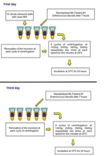
Cont am inat ion prot ocol
E. faecalis (ATCC 29212) was reactivated in Brain
Heart Infusion broth (BHI, Difco, Kansas City, MO, USA)
and maintained at 37°C for 24 h. The bacterial culture
further 24 h in order to achieve exponential growth.
This culture was adjusted to McFarland standard No.
8 CFU/mL) with a spectrophotometer (Bel
Photonics do Brasil Ltda, Osasco, SP, Brazil). The contamination model, proposed by Andrade, et al.1
Sam ple Obt urat ion
After the contamination procedure, the samples were removed from the BHI broth and dried with
sterile paper points size 80 (Dentsply Maillefer, Tulsa,
USA). The AH Plus sealer was manipulated on a sterile plate in accordance with the manufacturer’s
instructions. The sealer was placed in each root canal
by using a K-File size 70 (Dentsply Maillefer, Tulsa,
USA). Then the specimens were divided into 2 groups (n=10) according to ultrasonic activation of the sealer:
ultrasonically agitated (G1) and non-ultrasonically
agitated (G2) groups. In Group G1 activation was
Brasil) adapted to an ultrasonic device (Jet-Sonic Four
Plus; Gnatus, Ribeirão Preto, SP, Brazil) in ‘‘endo’’
mode (20% power). Because the ultrasonic oscillation
occurs in a single plane, the file was activated for 20 seconds in the buccolingual direction as a
standardization procedure. After this, an 80.02
gutta-percha cone (Dentsply Maillefer) was inserted into
the root canal to complete obturation and the excess was removed with a size 15 surgical blade. Group G2
was obturated in the same manner as G1, however,
without previous ultrasonic activation. A third Group (G3) (n=10) did not receive any treatment and served
as positive control. Afterwards, the specimens were
inserted into sterilized microtubules and incubated at
37ºC for one week.
Confocal laser scanning microscopy (CLSM)
analysis
After the above-mentioned time interval, the
specimens were longitudinally sectioned by using an
Isomet saw with a diamond disc, cooled with sterilized
saline solution. The smear layer produced by this sectioning was removed with 2.5 mL of 17% EDTA for
3 min, and the samples were washed with sterilized
saline solution. The halves of the dentinal tubes
® BacLightTM
Bacterial Viability stain (Molecular Probes, Eugene,
OR, USA) for 20 min. This kit contains SYTO 9® dye,
which stains live bacteria with a green pigment, and
propidium iodine dye, which stains dead bacteria with a red pigment, thus enabling viable bacteria to be
blinded calibrated examiners with an inverted Leica
TCS-SPE confocal microscope (Leica Microsystems GmbH, Mannheim, Baden-Württemberg, Germany)
to the root canal surface) and 3 deep (close to the
external root surface) images were acquired and
fragmented using the Leica Application Suite-Advanced Fluorescence software (LAS AF, Leica, Mannheim,
Baden-Württemberg, Germany). For an objective
analysis, the CLSM images were converted into “tiff”
format by the LAS AF software. These images were exported to the bioImageL TM v21 software in order
Statistical analyses
Because of the absence of normal distribution, observed by using the Shapiro–Wilk test, the Mann–
ultrasonic activation by means of stereomicroscopy and
CLSM analysis. Bacterial viability data were evaluated statistically with the Kruskal-Wallis and Dunn tests.
The Prism 6.0 software (GraphPad Software Inc., La
Jolla, USA) was used as the analytical tool, and the
Canal gap 2 mm (%) Canal gap 4 mm (%) Canal gap 6 mm
(%) Isthmus gap 2 mm (%) Isthmus gap 4 mm (%) Isthmus gap 6 mm (%) Canal SP 2 mm (%)
Canal SP 4 mm (%)
Canal SP 6 mm
(%)
Isthmus SP 2 mm
(%)
Isthmus SP 4 mm
(%) Isthmus SP 6 mm (%) UA 0.0 (0.0 -9.49)a 0.0 (0.0 -9.96)a 4.18 (0.0 -16.68)a 0.0 (0.0 -9.31)a 0.0 (0.0 -32.63)a 0.0 (0.0 -40.06)a 83.16 (76.91 -100.0)a 89.03 (67.23 -100.0)a 90.88 (58.58 -100.0)a 87.05 (6.0 -100.0)a 84.36 (24.33 -100.0)a 76.25 (24.4 -100.0)a NUA 4.40 (0.0 -20.46)a 6.43 (0.0 -23.03)a 10.74 (0.0 -28.07)b 0.0 (0.0 - 52.55)b 0.0 (0.0 -30.69)a 2.50 (0.0 -34.43)b 62.73 (35.14 -100.0)b 74.77 (38.80 -100.0)b 71.47 (19.12 -100.0)b 0.0 (0.0 -82.11)b 4.89 (0.0 -100.0)b 36.79 (0.0 -76.22)b
A different letter in each column represents statistical differences between the ultrasonically agitated (UA) and nonultrasonically agitated 18$JURXSVS
Table 2-0HGLDQPLQLPXPDQGPD[LPXPRIJDSVDQGVHDOHUSHQHWUDWLRQVHJPHQWV63LQSHUFHQWDJHVRIWKHFDQDODQGLVWKPXV A different letter in each column represents statistical differences between the ultrasonically agitated (UA) and nonultrasonically agitated 18$JURXSVS Table 1-0HGLDQPLQLPXPDQGPD[LPXPYDOXHVLQSHUFHQWDJHRIXQ¿OOHGDUHDVRIWKHFDQDODQGLVWKPXV Canal Isthmus 8Q¿OOHGDUHDV mm (%) 8Q¿OOHGDUHDV mm (%) 8Q¿OOHGDUHDV mm (%) 8Q¿OOHGDUHDV mm (%) 8Q¿OOHGDUHDV mm (%) 8Q¿OOHGDUHDV mm (%)
0.0 (0.0 – 4.89)a 0.0 (0.0 – 3.28)a 0.0 (0.0 – 4.49)a 0.0 (0.0 – 11.98)a 0.0 (0.0 – 15.66)a 0.0 (0.0 – 11.88)a
Results
areas obtained from the stereomicroscopy analysis are
shown in Table 1. In all sections, ultrasonic activation
(Figure 2A, B).
Results of the CLSM examination are presented
as median and ranges in Table 2. Overall, ultrasonic activation of the sealer showed a significant
improvement in sealer penetration into the dentinal
tubules in both canals and isthmuses (p<0.05) (Figure
2C, D). Gaps at the sealer/dentine interface were
Figure 2-5HSUHVHQWDWLYHVWHUHRPLFURVFRSLF$DQG%DQGFRQIRFDOLPDJHV&DQG'RI¿OOHGPHVLDOURRWVRIPDQGLEXODUPRODUVDWPP IURPWKHDSH[$6DWLVIDFWRU\¿OOLQJZLWKRXWJDSVLQWKHFDQDODQGLVWKPXVLQWKH8$JURXS%*DSVLQVLGHWKHFDQDODQGLVWKPXVLQWKH 18$JURXS&$VLJQL¿FDQWVHDOHUSHQHWUDWLRQLQWRWKHGHQWLQDOWXEXOHVRIWKHFDQDODQGLVWKPXVLVREVHUYHGLQWKH8$JURXSFRQWUDU\WR WKH18$JURXS'ZKHUHIHZHUGHQWLQDOWXEXOHVDUH¿OOHGZLWKVHDOHU
area and at 6 mm only in the canals when the sealer
was ultrasonically agitated (p<0.05). At 4 mm, there
groups (p>0.05).
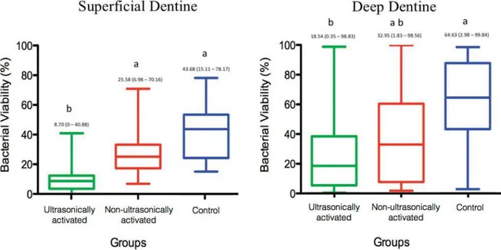
Intradentinal antimicrobial activity
the bacterial viability when compared with the other groups (p<0.05). As regards deep dentine, there was
the ultrasonically agitated group (G1) was compared
with Control group (G3) (p<0.05).
Discussion
Ultrasonic activation of the endodontic sealer
isthmus), adaptation of sealer/dentine interface and intratubular penetration in the UA group. The
antimicrobial activity was also enhanced by UA
in superficial dentine. Therefore, our entire null
hypothesis was rejected. The favourable results
previous studies3,25,27 that showed deficiency in
was capable of leaving tissue remnants, bacteria and debris, increasing the risk of post-treatment apical
periodontitis.
In the present study, PUI for 1 min per canal was
performed in both groups to achieve an improved
isthmus cleaning8 and the sealer was observed to
both groups. However, a low quantity of sealer was
observed inside the isthmus in the NUA group, leading
sections, irrespective of the isthmus width or length (Figure 2B).
activation of the sealer and quantify only the sealer
component inside the isthmus, the single cone
to enable proper differentiation between gutta-percha
and sealer, because when thermoplasticised techniques
are used, a single obturation mass is produced, making
separately. Indeed, Marciano,et al.13 (2011) reported
that the presence of isthmuses increased the incidence
techniques. Moreover, the single cone technique
allowed the insertion of a standard amount of
gutta-percha with reduced compaction forces when compared with other techniques that could push the
sealer and gutta-percha into the isthmus, thereby
interfering with the main study objective. The
above-mentioned factors, previous anatomical pairing, and the similar sealer volume inserted into each specimen
may have provided more reliable results. Therefore,
the results found in the UA group possibly occurred
exclusively as a result of the ultrasonic activation of the sealer.
The improvement in the amount of sealer that
entered inside the isthmus in the UA group was
probably caused by the transmission of acoustic microstreaming energy produced by the ultrasonic tip,
which could have forced the sealer into these areas.
This effect regularly occurs in the irrigating solution
while performing passive ultrasonic irrigation8,24. Although the results of the confocal microscopic
analysis of both the groups showed gaps at the sealer/
dentine interface, ultrasonic activation of the sealer
gaps to minimum values in some canal cross sections.
Furthermore, higher percentages of intratubular sealer
penetration were observed in the canal and isthmus region of the UA group at all the levels evaluated.
Previous studies presented similar results using single
rooted teeth7,16. Macedo, et al.12 (2014) showed that
the ultrasonic activation increased the temperature of irrigants. The same phenomenon can occur during
ultrasonic activation of the sealer, which could promote
higher flowability and reduce the film thickness,
favouring its penetration into dentinal tubules. The medians of sealer penetration of the UA group in all
sections were above 76% when compared with the
NUA group, in which penetration values were below
39%. This result is noteworthy if we consider the antibacterial properties of root canal sealers within
the dentinal tubules29.
This study presented a method for intratubular
contamination of bovine dentine proposed by Andrade, et al.1 (2016). Bovine teeth are commonly used as an
experimental substitute for human teeth because they
are easily available21. Compared with human dentine,
bovine dentine has a higher concentration of dentine tubules per square millimeter, however, this difference
is small. On average, the diameter of dentine tubules
of bovine teeth is larger than that of human teeth,
percentage of intertubular dentine in bovine teeth is
the same as it is in human teeth21.
Vera, et al.25 (2012) showed that microorganisms remain inside the dentinal tubules and survive after
biomechanical preparation and intracanal dressing.
Thus, the sealer penetration into the dentinal tubules
of the antimicrobial action of these materials and their
action of entombing residual microorganism10,15.
Our results showed that ultrasonic activation of the
E. faecalis
other groups (Figure 4A, B and C). As regards deep
E. faecalis viability only in the ultrasonically agitated
group in comparison with the Control group (Figure 4C,
D and F). The AH Plus sealer has antimicrobial action
against Ent er ococcus faecalis18,28. The antimicrobial properties of epoxy resin-based sealers may be related
to the bisphenol-A diglycidyl23 or small quantities of
formaldehyde11 released into the dentinal tubules during the polymerisation process.
The possible explanation of the improvement of
the antimicrobial action of the AH Plus when activated
by ultrasonic tip is due to the transmission of acoustic microstreaming energy produced, which increases the
sealer penetration into the dentine tubules7,16. Thus,
the greater sealer penetration may act as a physical
barrier and may entomb residual microorganisms, thereby separating them from nutrient sources17.
In addition, the greater contact between the sealer
and microorganisms could favour the antimicrobial
action of the AH Plus, consequently reducing the microorganism viability.
Conclusion
The ultrasonic activation of the AH Plus sealer
increased the intratubular penetration of sealer, especially in the isthmus area. Additionally, ultrasonic
activation of the sealer increased the intradentinal
antimicrobial action against Ent er ococcus faecalis,
References
1- Andrade FB, Arias MP, Maliza AG, Duarte MA, Graeff MS,
Amoroso-Silva PA, et al. A new improved protocol for in v it r o intratubular dentinal bacterial contamination for antimicrobial endodontic tests:
standardization and validation by confocal laser scanning microscopy. J Appl Oral Sci. 2015;23(6):591-8.
in mandibular molars after conventional root-canal instrumentation
and after a noninstrumentation technique (NIT). Int Endod J. 2003;36(9):591-8.
3- Carr GB, Schwartz RS, Schaudinn C, Gorur A, Costerton JW. Ultrastructural examination of failed molar retreatment with secondary
Paciomik S. Limited ability of three commonly used thermoplasticized gutta-percha techniques in filling oval-shaped canals. J Endod.
2008;34(11):1401-5.
5- Endal U, Shen Y, Knut A, Gao Y, Haapasalo M. A high-resolution
computed tomographic study of changes in root canal isthmus area by
6- Gatewood RS. Endodontic materials. Dent Clin North Am. 2007;51(3):695-712.
7- Guimaraes BM, Amoroso-Silva PA, Alcalde MP, Marciano MA, Andrade
8- Gutarts R, Nusstein J, Reader A, Beck M. I n vivo
of ultrasonic irrigation following hand-rotary instrumentation in human mandibular molars. J Endod. 2005;31(3):166-70.
9- Hsu YY, Kim S. The resected root surface. The issue of canal isthmuses. Dent Clin North Am. 1997;41(3):529-40.
10- Kokkas AB, Boutsioukis A, Vassiliadis LP, Stavrianos CK. The
by three different root canal sealers: an in v it r o study. J Endod. 2004;30(2):100-2.
11- Leonardo MR, Silva LA, Tanomaru M Filho, Silva RS. Release of formaldehyde by 4 endodontic sealers. Oral Surg Oral Med Oral Pathol
Oral Radiol Endod. 1999;88(2):221-5.
12- Macedo RG, Verhaagen B, Wesselink PR, Versluis M, van der Sluis
on the reaction rate of sodium hypochlorite with bovine dentine during ultrasonic activated irrigation. Int Endod J. 2014;47(2):147-54.
13- Marciano MA, Ordinola-Zapata R, Cunha TV, Duarte MA, Cavenago BC, Garcia RB, et al. Analysis of four gutta-percha techniques
2011;44(4):321-9.
14- Marciano MA, Guimaraes BM, Ordinola-Zapata R, Bramante CM, Cavenago BC, Garcia RB, et al. Physical properties and
interfacial adaptation of three epoxy resin-based sealers. J Endod. 2011;37(10):1417-21.
15- Moon YM, Shon WJ, Baek SH, Bae KS, Kum Ky, Lee W. Effect of
Endod. 2010;36(4):732-6.
16- Nikhil V, Bansal P, Sawani S. Effect of technique of sealer agitation
on percentage and depth of MTA Fillapex sealer penetration: a comparative in- vit ro study. J Conserv Dent. 2015;18(2):119-23.
17- Ordinola-Zapata R, Bramante CM, Graeff MS, del Carpio Perochena
A, Vivan RR, Camargo EJ, et al. Depth and percentage of penetration of endodontic sealers into dentinal tubules after root canal obturation
using a lateral compaction technique: a confocal laser scanning microscopy study. Oral Surg Oral Med Oral Pathol Oral Radiol Endod.
2009;108(3):450-7.
18- Prestegaard H, Portenier I, Orstavik D, Kayaoglu G, Haapasalo M,
Endal U. Antibacterial activity of various root canal sealers and
root-ex vivo with Ent erococcus faecalis. Acta Odontol Scand. 2014;72(8):970-6.
19- Rôças IN, Siqueira JF Jr, Santos KR. Association of Ent erococcus f aecalis with different forms of periradicular diseases. J Endod. 2004;30(5):315-20.
20- Schilder H. Filling root canals in three dimensions. Dent Clin North
Am. 1967 Nov;723-44.
21- Schilke R, Lisson JA, Bauss O, Geurtsen W. Comparison of the
number and diameter of dentinal tubules in human and bovine dentine by scanning electron microscopic investigation. Arch Oral Biol.
2000;45(5):355-61.
22- Siqueira JF Jr, Rôças IN. Polymerase chain reaction-based analysis
of microorganisms associated with failed endodontic treatment. Oral Surg Oral Med Oral Pathol Oral Radiol Endod. 2004;97(1):85-94.
23- Slutzky-Goldberg I, Slutzky H, Solomonov M, Moshonov J, Weiss EI, Matalon S. Antibacterial properties of four endodontic sealers. J
Endod. 2008;34(6):735-8.
24- van der Sluis LW, Versluis M, Wu MK, Wesselink PR. Passive
ultrasonic irrigation of the root canal: a review of the literature. Int Endod J. 2007;40(6):415-26.
25- Vera J, Siqueira Jr JF, Ricucci D, Loghin S, Fernández N, Flores B, et al. One-versus two-visit endodontic treatment of teeth with apical
periodontitis: a histobacteriologic study. J Endod. 2012;38(8):1040-52. 26- Vertucci FJ. Root canal anatomy of the human permanent teeth.
Oral Surg Oral Med Oral Pathol. 1984;58(5):589-99.
detected by endoscopic inspection during periradicular surgery. Int Endod J. 2005;38(3):160-8.
28- Wang Z, Shen Y, Haapasalo M. Dentin extends the antibacterial effect of endodontic sealers against Ent erococcus faecalis
Endod. 2014;40(4):505-8.
29- Zhang C, Du J, Peng Z. Correlation between En t er ococcu s faecalis and persistent intraradicular infection compared with primary intraradicular infection: a systematic review. J Endod.