A SIMPLE AND RELIABLE PCR-RESTRICTION FRAGMENT LENGTH POLYMORPHISM ASSAY TOIDENTIFY
CANDIDA ALBICANS AND ITS CLOSELY RELATED CANDIDA DUBLINIENSIS
Yi Ping Ge, Le Wang, Gui Xia Lu, Yong Nian Shen, Wei Da Liu*
Department of Medical Mycology, Institute of Dermatology, Chinese Academy of Medical Sciences, Nanjing, Jiangsu Province,
China.
Submitted: December 02, 2010; Returned to authors for corrections: May 05, 2011; Approved: June 07, 2012.
ABSTRACT
Candida dubliniensis is an emerging pathogen capable of causing superficial as well as systemic infections. Due to its close similarity to C. albcians, conventional methods based on phenotypic traits are not always reliable in identification of C. dubliniensis. In this study, we developed a PCR-restriction fragment length polymorphism (RFLP) assay to identify and discriminate between the two closely related species. The D1/D2
region of 28S rDNA was amplified by PCR and enzymatically digested by ApaI and BsiEI respectively. PCR products of both species were digested into two fragments by ApaI, but those of other yeast species were undigested. BsiEI cut the PCR products of C. albicans into two fragments but not those of C. dubliniensis. Thus two species were differentiated. We evaluated 10 reference strains representing 10 yeast species, among
which C. albicans and C. dubliniensis were successfully identified. A total of 56 phenotypically characterized clinical isolates (42 C. albicans isolates and 14 C. dubliniensis isolates) were also investigated for intra-species variability. All tested isolates produced identical RFLP patterns to their respective reference strains
except one initially misidentified isolate. Our method offers a simple, rapid and reliable molecular method for
the identification of C. albicans and C. dubliniensis.
Key words: Candida albicans, Candida dubliniensis, PCR-restriction fragment length polymorphism, identification, differentiation
INTRODUCTION
Candida species usually reside as commensals at mucosal membranes in healthy individuals and can be detected in
approximately 50% of the population in this non-virulent form.
However, under conditions when the host’s normal flora is
disrupted or the immunity is impaired, Candida species often become pathogenic. Candida infections have become a
problem of growing significance. The incidence of infections
has increased dramatically over the past a few decades. C. albcians is the most common pathogen in this genus and fourth leading cause of nonsocomial bloodstream infections (4, 21).
However, several non-albicans Candida species, e.g. C. glabrata, C. tropicalis and C. parapsilosis, have emerged as causative agents of candidiasis (20). Another Candida species of growing clinical importance is C. dubliniensis, a novel
opportunistic pathogen first described as a distinct taxon by
Sullivan in 1995 (26). C. dubliniensis was mainly associated with oropharyngeal candidiasis in HIV-infected patients.
Recent evidence, however, indicates that it is also a cause of
superficial and systemic infections in HIV-negative individuals
with an estimated prevalence rate below 5% (7, 10, 27). It is
important to study the epidemiology of C. dubliniensis due to its capability of rapid acquisition of stable fluconazole
resistance, both in vitro and in vivo, after prolonged therapy in HIV-seropositive patients (15, 16, 24).
Identification of C. dubliniensis can be problematic due to its close phenotypic similarity to C. albicans. Both species produces germ tubes, chlamydospores and true hyphae (26).
Several phenotypic assays have been developed to differentiate
C. dubliniensis from C. albicans, including the capacity to grow at 45°C, formation of chlamydospores on selected media,
the ability to assimilate xylose, lactate or α-methyl-D-glucoside
and different colony color produced on CHROMagar Candida
medium (2,5,23,28). These assays can serve as rapid methods
to screen for potential C. dubliniensis isolates. But none of these assays have proved to be efficient and entirely reliable.
At present, the most accurate means of differentiating between
these two closely related species requires the use of molecular
biology-based techniques, such as electrophoretic karyotyping,
DNA fingerprinting analysis with repetitive
sequence-containing DNA probes, randomly amplified polymorphic
DNA analysis, restriction fragment length polymorphism
(RFLP), amplified fragment length polymorphism,
conventional and real-time PCR analysis, or pulsed-field gel
electrophoresis(18,25). These methods proved very effective.
Among them, PCR-RFLP analysis is a simple and reliable one.
In this study we developed and evaluated a PCR-RFLP assay to
identify and discriminate between C. albicans and C. dubliniensis.
MATERIALS AND METHODS
Yeast Strains
C. albicans (SC 5314), C. dubliniensis (CBS 7987), C.
glabrata (ATCC 2001), C. guilliermondii (CBS 6021), C. krusei (ATCC 6258), C. kefyr (CBS 6432), C. lusitaniae (CBS 6936), C. parapsilosis (ATCC 22019), C. tropicalis (CBS 8072) and Trichosporon asahii (CBS 2479) were used as reference yeast strains. Fourteen clinical isolates of
presumptive C. dubliniensis originating from sputum (n=10), vaginal swabs (n=3) and urine (n=1) were studied in
comparison with 42 clinical strains of C. albicans. Presumptive C. dubliniensis isolates were characterized by phenotypic methods including growth on cornmeal Tween-80 agar, growth
at 45°C, characteristic growth on CHROMagar Candida
(CHROMagar, Paris, France) and confirmed by Vitek 2 system
(biomerieux, Marcy l’Etoile, France).
Culture Conditions and Genomic DNA Extraction
Yeast cells were cultured on YPD broth (1% yeast extract,
2% peptone, and 2% dextrose) and were incubated for 24-36
hours at 30°C under shaking conditions (200rpm). The yeast
cells were collected by centrifugation(2ml of the broth culture
at 12000×g for 2 min), suspended in 600μl of 1 M sorbitol-50
mM phosphate buffer (pH 7.5) containing 50U Lyticase
(Sigma-Aldrich, US). After 30 min of incubation at 30°C, the
cells were centrifuged at 1500×g for 10 min. The supernatant
was then discarded and pellet was collected. TianGen Yeast
Genomic DNA Extraction Kit (TianGen Biotech, Beijing,
China) was used to extract genomic DNA from tested isolates
by following the enclosed protocol. DNA obtained was finally
suspended in 100 μl TE buffer and stored at -20°C before use.
Sequence Analyses and Selection of Restriction Enzymes
A total of 30 sequences of the 28S ribosomal DNA
(rDNA) D1/D2 region of 10 tested yeast species were retrieved
from GenBank database (data not shown). Each sequence was
then analyzed for restriction sites using the MapDraw program
of DNA Star Lasergene Version 7.0. Restriction enzymes were
PCR Amplification of the D1/D2 region
Primers NL-1 (5’-GCATATCAATAAGCGGAGGAAA
AG-3’) and NL-4 (5’-GGTCCGTGTTTCAAGACGG-3’) were
used to amplify D1/D2 region of the 28S rDNA genes. PCR
amplifications were carried out in 50-μl volumes containing
1.5μl of each 10μmol/l primer, 25μl of GoTaq Green Master
Mix (Promega, Madison, WI, USA), 3μl DNA template and
corresponding amount of ultra-pure distilled water. PCR was
performed in a PTC-200 DNA Engine thermal cycler
(Bio-Rad) with following parameters: 94°C for 3 min; 94° for 1 min,
52°C for 30 s and 72°C for 1 min, repeated for a total of 32
cycles; 72°C for 10 min and 4°C hold.
Restriction Digests of PCR Products
RFLP analyses were performed in 20μl volumeswith
100-200 ng of amplified DNA products, 25U ApaI or 5U BsiEI(New England Biolabs, Beverly, MA, USA), 2μl 10×digestion buffer, 0.2 µl bovine serum albumin and
corresponding amount of water. Digestion mixtures were
incubated for 0.5-1 h at 37°C for ApaI or at 60°C for BsiEI.
The resulting fragments were separated on 2% agarosegels and
visualized under UV light after ethidium bromide staining,
with a 100bp DNA ladder for fragment size comparison.
RESULT
Sequence Analyses
An extensive analysis of database entries of yeast species
tested in this study was performed with respect to calculated
fragment lengths of the D1/D2 regions generated by
commercially available restriction enzymes. Minor differences
in calculated lengths of PCR products were observed. Finally,
ApaI and BsiEI were selected to be evaluated in experiment. Predicted fragment lengths of ApaI and BsiEI-digested PCR products are given in Table 1. ApaI was expected to cut the PCR amplicons of C. albicans and C. dubliniensis into two fragments, but leave those of other yeast species intact. BsiEI was selected to digest the amplicon of C. albicans into two fragments and those of C. dubliniensis would remain undigested. Thus two related Candida species can be identified and differentiated by distinctive and specific RFLP patterns.
Table 1. Comparison of PCR-RFLP assays to differentiate between C.albicans and C. dubliniensis Length of PCR
products (bp)
Fragments’ length after enzymatic digestion (bp)
No. of isolates tested Target of PCR amplification
and primers
CA / CD Enzyme: CA / CD CA / CD
Reference
ITS region: ITS5 and NL4 approximately 1200 DdeI: 450, 350, 210, 150 / 450, 350, 210, 110
BfaI and HaeIII: differentiable but not specified
78 / 10 8
ITS region:CA-INT-L(R) approximately 600 DdeI: one fragment / two fragments 8 / 2 13 V3 region: CA25SV3L(R) approximately 500 HaeIII: differentiable but not specified 8 / 2 13 ITS2 region: ITS3 and ITS4 approximately 340 NspBII: approximately 160, 180 / 340
BsmAI: approximately 340 / 100, 240
17 / 8 19
ITS2 region: CTSF and CTSR 345 / 350 MspA1I: 35, 143, 167 / 35, 315 1 / 9 6
ITS region: UNI1 and UNI2 586 / 589 HpyF10VI: 141, 184, 261 / 264, 325 61/ 23 1
ITS region: ITS1 and ITS4 540 / 540 BlnI: 540 / 200,340 146 /12 14
D1-D2 region: NL-1 and NL-4 615 / 614 ApaI: 134 , 481/ 134, 480
BsiEI: 181, 434 / 614
43/ 15 This
study CA : C. albicans, CD : C. dubliniensis
PCR Amplification of D1/D2 Regions
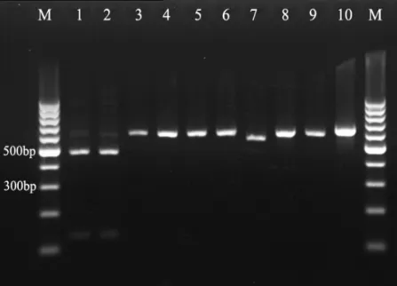
As shown in Fig.1, intended DNA fragments of all
reference strains were successfully amplified with primers
NL-1 and NL-4. PCR products were found to reach 550-600 bp in
length as predicted from sequence analysis. However, most
RFLP Analyses of Reference Strains
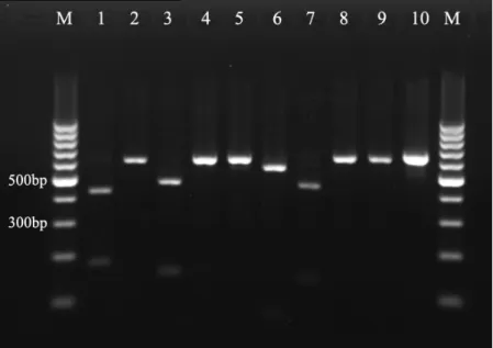
When digested with ApaI, D1/D2 regions of C. albicans and
C. dubliniensis strains shared the same restriction pattern, with
two bands of almost identical sizes (134bp, 481bp for C. albicans
and 134bp, 480bp for C. dubliniensis) (Fig.2). Nevertheless,
amplicons of other yeast strains were undigested by ApaI, which
easily distinguished C. albicans and C. dubliniensis from other
tested species. Digestion of the D1/D2 region with BsiEI generated
distinctive restriction profiles for C. albicans: two fragments of
181bp and 434bp (Fig.3). In addition, BsiEI also cut into two
fragments the PCR-products of C. glabrata (149bp and 475bp), C.
krusei (59bp and 548bp) and C. lusitaniae (126bp and 433bp). As
was seen in Fig.3, the differences in fragment lengths were
sufficient to discriminate C. albicans from C. glabrata, C. krusei
and C. lusitaniae. Other tested species were undigested by BsiEI,
including C. dubliniensis. Therefore, two separate enzymatic
digestions produced species-specific RFLP profiles for C. albicans
and C. dubliniensis.
Figure 1. PCR products from 10 yeast
species: Lane 1: C. albicans (SC 5314) ;
Lane 2: C. dubliniensis (CBS 7987);
Lane 3: C. glabrata (ATCC 2001); Lane
4: C. guilliermondii (CBS 6021); Lane
5: C. kefyr (CBS 6432); Lane 6: C.
krusei (ATCC 6258); Lane 7: C.
lusitaniae (CBS 6936); Lane 8: C.
parapsilosis (ATCC 22019); Lane 9: C.
tropicalis (CBS 8072); Lane 10: T.
asahii (CBS 2479) ; Lane M: 100-bp
ladder.
Figure 2. Restriction digestion of PCR
products of reference yeast strains with
ApaI : Lane 1: C. albicans (SC 5314) ;
Lane 2: C. dubliniensis (CBS 7987);
Lane 3: C. glabrata (ATCC 2001);
Lane 4: C. guilliermondii (CBS 6021);
Lane 5: C. kefyr (CBS 6432); Lane 6:
C. krusei (ATCC 6258); Lane 7: C.
lusitaniae (CBS 6936); Lane 8: C.
parapsilosis (ATCC 22019); Lane 9:
C. tropicalis (CBS 8072); Lane 10: T.
asahii (CBS 2479) ; Lane M: 100-bp
Evaluation for Intra-species Variation of Restriction Sites
To assess intra-species variability of the two restriction
sites, a total of 56 clinical isolates of C. albicans and C. dubliniensis were also investigated. Most tested isolates, after digestion with ApaI and BsiEI respectively, showed identical and consistent RFLP patterns to their respective reference
strains (data not shown). However, one of the 14 presumptive
C. dubliniensis isolates showed RFLP pattern indicative of C. albicans. Further sequencing of the D1/D2 region of this isolate confirmed the identification of C. albicans.
DISCUSSION
Rapid and accurate identification of C. dubliniensis is crucial for the study of epidemiology and clinical management
of infections caused by this opportunistic pathogen. However,
this is hampered by lack of easy and reliable methods for
definite identification. Most phenotypic methods are
presumptive and often subject to error (28). Although several
commercial identification systems such as Vitek 2 system,
have demonstrated useful in separation of C. albicans and C. dubliniensis, results are not always reliable as was seen in our study (3, 12, 23). Confirmatory identification of C. dubliniensis
always requires the molecular methods.
PCR-RFLP assays have been successfully applied to the
identification of Candida species (17, 22, 29). Compared with other molecular methods, PCR-RFLP analysis is generally easy
and rapid to perform. Although more complex RFLP methods
have previously been used for the identification of C. dubliniensis, their use may be limited since they were time-consuming or the results were difficult to interpret (18).Thus in
this study, simpler but more advantageous PCR-RFLP is
preferred due to its increased applicability in clinical
laboratories.
Ribosomal regions, such as the internal transcribed spacer
(ITS) region and 28S rDNA, exhibit a low intraspecific
polymorphism and a high interspecific variability, making
them ideal targets for species identification purposes (9). In this
study, we selected the D1/D2 variable region at the 5’ end of
the 28S rDNA gene as target for PCR amplification.
Sequencing of this region has been demonstrated to be
sufficient for accurate identification of most yeast species (11).
Between C. albicans and C. dubliniensis, there are 13 nucleotide differences in the region D1/D2, sufficiently
variable for reliable differentiation. Two enzymes, ApaI and BsiEI, were selected based on the restriction profiles generated
Figure 3. Restriction digestion of PCR
products of reference yeast strains with
BsiEI : Lane 1: C. albicans (SC 5314) ;
Lane 2: C. dubliniensis (CBS 7987);
Lane 3: C. glabrata (ATCC 2001);
Lane 4: C. guilliermondii (CBS 6021);
Lane 5: C. kefyr (CBS 6432); Lane 6:
C. krusei (ATCC 6258); Lane 7: C.
lusitaniae (CBS 6936); Lane 8: C.
parapsilosis (ATCC 22019); Lane 9:
C. tropicalis (CBS 8072); Lane 10: T.
asahii (CBS 2479) ; Lane M: 100-bp
by these nucleotide differences. Despite that only a limited
number of C. albicans and C. dubliniensis isolates were investigated, those restriction sites for ApaI and BsiEI seemed to be well conserved in these two species.
Several PCR-RFLP assays have been described so far to
discriminate between C. albicans and C. dubliniensis (Table 1). Irobi et al amplified the ITS regions (ITS1, 5.8S, ITS2) of
several medically important Candida species, including C. dubliniensis (8). Further RFLP analysis with BfaI, DdeI or HaeIII revealed distinct differences between the two species. McCullough et al amplified the ITS regions and restricted them
with DdeI. C. albicans produced one fragment while C. dubliniensis produced two fragments (13). In the same study, McCullough and his group also targeted the V3 region of
25S/28S rDNA and cut the PCR amplicons with HaeIII; the discrimination could be made based on fragments of different
sizes. Park and his co-authors amplified a conserved part of the
5.8S rDNA, the adjacent ITS2 region and a part of 28S rDNA.
The differentiation was achieved by analysis of the PCR
products with BsmAI (C. dubliniensis-specific) and NspBIII (C. albicans-specific) (19). Three other PCR-RFLP assays also used a similar strategy by targeting a part or whole of the ITS
region (1, 6, 14). All these methods proved effective for
accurate identification. In comparison, our study used a slightly
different strategy. We first identified a restriction site for ApaI which was specific to both species, instantly separating them
from other yeast species. Subsequently BsiEI was found to produce C. albicans specific pattern and C. dubliniensis was identified by absence of the restriction site.
An advantage of the method described here is the stable
and easy-to-read RFLP patterns. Unlike previous reports, this
method involves only one or two DNA fragments. Besides, it is
a simple and rapid method to perform. With the aid of
time-saving restriction enzymes, the whole process can be
accomplished in less than 6h, requiring no sophisticated
equipments except a conventional thermal cycler. Considering
that DNA sequencer may not be readily available to most
clinical laboratories, this molecular method is applicable for
unequivocal identification and differentiation of C. albicans and C. dubliniensis.
ACKNOWLEDGEMENTS
The authors report no conflicts of interest.
REFERENCES
1. Ahmad, S.; Khan, Z.; Mokaddas, E.; Khan, Z.A. (2004). Isolation and molecular identification of Candida dubliniensis from non-human immunodeficiency virus-infected patients in Kuwait. J Med Microbiol. 53(Pt7): 633–637.
2. Akgül, O.;Cerikçioğlu, N. (2009). Hypertonic sabouraud dextrose agar as a substrate for differentiation of Candida dubliniensis.
Mycopathologia. 167(6):357-359.
3. Cárdenes-Perera, C.D.; Torres-Lana, A.; Alonso-Vargas, R.; Moragues-Tosantas, M.D.; Pontón-San Emeterio, J.;Quindós-Andrés, G.; Arévalo-Morales, M.P.(2004). Evaluation of API ID 32C and Vitek-2 to identify
Candida dubliniensis. Diagn Microbiol Infect Dis.50(3): 219–221. 4. Edmond, M.B.; Wallace, S.E.; McClish, D.K.; Pfaller, M.A.; Jones,
R.N.; Wenzel, R.P. (1999). Nosocomial bloodstream infections in United States hospitals: a three-year analysis. Clin Infec Dis. 29(2): 239-244. 5. Gales, A.C.; Pfaller, M.A.; Houston, A.K.; Joly, S.; Sullivan, D.J.;
Coleman, D.C.; Soll, D.R. (1999). Identification of Candida dubliniensis
based on temperature and utilization of xylose and α-methyl-D-glucoside as determined with the API 20C AUX and Vitek YBC systems. J Clin Microbiol. 37(12): 3804–3808.
6. Graf, B.; Trost, A.; Eucker, J.; Göbel, U.B.; Adam, T. (2004). Rapid and simple differentiation of C. dubliniensis from C. albicans. Diagn Microbiol Infect Dis. 48(2): 149–151.
7. Gutierrez, J.; Morales, P.; Gonzalez, M.A.; Quindos, G. (2002). Candida dubliniensis, a new fungal pathogen. J Basic Microbiol. 42(3): 207–227. 8. Irobi, J.; Schoofs, A.; Goossens, H. (1999). Genetic identification of
Candida species in HIV-positive patients using the polymerase chain reaction and restriction fragment polymorphism analysis of its DNA.
Mol Cell Probes. 13(6): 401–406.
9. Iwen, P.C.; Hinrichs, S.H.; Rupp, M.E. (2002). Utilization of the internal transcribed spacer regions as molecular targets to detect and identify human fungal pathogens. Med Mycol, 40(1): 87-109.
in immunocompetent subjects in Argentina. Oral Microbiol Immunol. 23(6): 505–509.
11. Kurtzman, B.P.; Robnett, C.J. (1997). Identification of clinically important ascomycetous yeasts based on nucleotide divergence in the 50 end of the large-subunit (26S) ribosomal DNA gene. J Clin Microbiol. 35(5): 1216–1223.
12. Mähnss, B.; Stehr, F.; Scäfer, W.; Neuber, K. (2005). Comparison of standard phenotypic assays with a PCR method to discriminate Candida albicans and C. dubliniensis. Mycoses. 48(1): 55–61.
13. McCullough, M.J.; Clemons, K.V.; Stevens, D.A. (1999). Molecular and phenotypic characterization of genotypic Candida albicans subgroups and comparison with Candida dubliniensis and Candida stellatoidea. J Clin Microbiol. 37(2): 417–421.
14. Mirhendi, H.; Makimura, K.; Khoramizadeh, M.; Yamaguchi, H. (2006). A one-enzyme PCR-RFLP assay for identification of six medically important Candida Species. Nippon Ishinkin Gakkai Zasshi. 47(3): 225-229.
15. Moran, G.P.; Sanglard, D.; Donnelly, S.M.; Shanley, D.B.; Sullivan, D.J.; Coleman, D.C. (1998). Identification and expression of multidrug transporters responsible for fluconazole resistance in Candida dubliniensis. Antimicrob Agents Chemother. 42(7): 1819-1830.
16. Moran, G.P.; Sullivan, D.J.; Henman, M.C.; McCreary, C.E.; Harrington, B.J.; Shanley, D.B.; Coleman, D.C. (1997). Antifungal drug susceptibilities of oral Candida dubliniensis isolates from human immunodeficiency virus (HIV)-infected and non-HIV-infected subjects and generation of stable fluconazole-resistant derivatives in vitro.
Antimicrob Agents Chemother. 41(3): 617-623.
17. Mousavi, S.A.A.; Khalesi, E.; Shahidi Bonjar, G.H.; Aghighi, S.; Shaifi, F.; Aram, F. (2007). Rapid molecular diagnosis for Candida species using PCR-RFLP. Biotechnology. 6(4):583-587.
18. Neppelenbroek, K.H.; Campanha, N.H.; Spolidorio, D.M.P.; Spolidorio, L.C.; Seó, R.S.; Pavarina, A.C. (2006). Molecular fingerprinting methods for the discrimination between C. albicans and C. dubliniensis.
Oral Disease.12 (3): 242–253.
19. Park, S.; Wong, M.; Marras, S.A.E.; Cross E.W.; Kiehn T.E.; Chaturvedi V.; Tyagi, S.; Perlin D.S. (2000). Rapid identification of Candida dubliniensis using a species-specific molecular probe. J Clin Microbiol.
38(8): 2829–2836.
20. Pfaller, M.A.; Diekema, D.J. (2007). Epidemiology of invasive candidiasis: a persistent public health problem. Clin Micobio Rev. 20(1): 133-163.
21. Pfaller, M.A.; Jones, R.N.; Messer, S.A.; Edmond, M.B.; Wenzel, R.P. (1998) National surveillance of nosocomial blood stream infection due to
Candida albicans: frequency of occurrence and antifungal susceptibility in the SCOPE Program. Diagn Microbiol Infect Dis. 31(1): 327-332. 22. Pinto, P.M.; Resende, M.A.; Koga-Ito C.Y.; Ferreira, J.A.; Tendler, M.
(2004). rDNA-RFLP identification of Candida species in immunocompromised and seriously diseased patients. Can J Microbiol. 50(7):514-520.
23. Pincus, D.H.; Coleman, D.C.; Pruitt, W.R.; Padhye, A.A.; Salkin, I.F.; Geimer, M.;Bassel, A.; Sullivan, D.J.; Clarke, M.; Hearn, V. (1999). Rapid identification of Candida dubliniensis with commercial yeast identification systems. J Clin Microbiol. 37(11):3533–3539.
24. Ruhnke, M.; Schmidt-Westhausen, A.; Morschhauser, J. (2000). Development of simultaneous resistance to fluconazole in Candida albicans and Candida dubliniensis in a patient with AIDS. J Antimicrob Chemother. 46(2): 291-295.
25. Rocha, B.A.; Barbaro Del Negro G.M.; Yamamoto, L.; de Souza, M.V.B.; Precioso, A.R.; Okay, T.S. (2008). Identification and differentiation of Candida species from paediatric patients by random amplified polymorphic DNA. Rev Soc Bras Med Trop. 41(1): 1–5. 26. Sullivan, D.J.; Westerneng, T.J.; Haynes, K.A.; Bennett, D.E.; Coleman,
D.C. (1995). Candida dubliniensis sp. nov.: phenotypic and molecular characterisation of a novel species associated with oral candidosis in HIV infected individuals. Microbiology. 141(7): 1507–1521.
27. Sullivan, D.J.; Coleman, D.C. (1998). Candida dubliniensis: characteristics and identification. J Clin Microbiol. 36(2): 329–334. 28. Tintelnot, K.; Haase, G.; Seibold, M.; Bergmann, F.; Staemmler, M.;
Franz, T.; Naumann, D. (2000). Evaluation of phenotypic markers for selection and identification of C. dubliniensis. J Clin Microbiol. 38(4):1599–1608.
29. Trost, A.; Graf, B; Eucker, J.; Sezer, O.; Possinger, K.; Göbel, U.; Adam, T. (2004). Identification of clinically relevant yeasts by PCR/RFLP. J Microbio Methods. 56(2): 201-211.