online | memorias.ioc.fiocruz.br
A non-hepatotropic parasite infection increases mortality
in the acetaminophen-induced acute liver failure murine model:
possible roles for IL-5 and IL-6
Marco A de León-Nava1, Carolina Álvarez-Delgado1, Luis Donis-Maturano1, Joselin Hernández-Ruiz2, Aaron N Manjarrez-Reyna2, Edgar Cruz-Avilés2, Sonia Leon-Cabrera3, Jorge Morales-Montor4, José M Fragoso5, Galileo Escobedo2/+
1Centro de Investigación Científica y de Educación Superior de Ensenada, Departamento de Innovación Biomédica, Baja California, México 2Universidad Nacional Autónoma de México, Facultad de Medicina, Unidad de Investigación en Medicina Experimental, Hospital General de México Dr Eduardo Liceaga, Laboratorio de Hígado, Páncreas y Motilidad, Ciudad de México, México 3Universidad Nacional Autónoma de México, Facultad de Estudios Superiores-Iztacala, Unidad de Biomedicina, Carrera de Médico Cirujano,
Los Reyes Iztacala, México 4Universidad Nacional Autónoma de México, Instituto de Investigaciones Biomédicas, Departamento de Inmunología, Ciudad de México, México 5Instituto Nacional de Cardiología Ignacio Chávez,
Departamento de Biología Molecular, Ciudad de México, México
We evaluated the effects of a non-hepatotropic parasite infection (Taenia crassiceps) on the outcome of acetamin-ophen-induced acute liver failure in mice. Uninfected and T. crassiceps infected mice orally received either 300 mg/kg acetaminophen or water as vehicle (n = 5 per group). Survival analysis, hepatocyte necrosis, alanine aminotransferase (ALT) levels, CYP2E1 protein, interleukin (IL-) 5, and IL-6 were assessed for all groups. All infected mice died within 16 h after exposure to acetaminophen (Tc+APAP group), whereas only one-third of uninfected animals exposed to acetaminophen (APAP group) died. Uninfected (Control group) and infected (Tcgroup) mice that received the vehicle showed no liver damage. Tc+APAP mice exhibited massive liver necrosis characterised by marked balloning degenera-tion of hepatocytes and higher serum ALT compared to Control, Tc, and APAP animals. Liver tissue from Tc+APAP mice also displayed increased expression of CYP2E1 protein and higher mRNA and protein levels of IL-5 and IL-6 compared to the other groups. These findings suggest that non-hepatotropic parasite infections may increase mortality following acute liver failure by promoting hepatocyte necrosis via IL-5 and IL-6-dependent CYP2E1 overproduction. This study identifies new potential risk factors associated with severe acute liver failure in patients.
Key words: acute liver failure - acetaminophen - parasite infection - Taenia crassiceps - liver disease - interleukin
The liver carries out more than 500 functions includ-ing regulation of carbohydrate and lipid metabolism, protein synthesis, immune response orchestration, and detoxification processes (Bhatia et al. 2014). Pathologi-cal alterations of the liver are considered serious and expensive public health problems, with high mortality rates when relating to either chronic disease or liver fail-ure (Kandiah et al. 2016). Liver failfail-ure can be classified into four categories: acute, sub-acute, acute-on-chronic, and chronic liver failure (Bernal et al. 2015). Acute liver failure is the clinical manifestation of sudden and severe hepatic damage in patients having no pre-existing liver disease, and it is frequently related to acetaminophen (APAP) overdose (Chalhoub et al. 2014). Acute liver fail-ure is characterised by massive necrosis of hepatocytes, a sudden increase in alanine aminotransferase (ALT) levels, rapid loss of hepatic function, jaundice, encephalopathy,
cerebral edema, multi-organ failure, and extremely high mortality rates ranging from 65-80% (Stravitz & Kramer 2009). Although improved clinical management of acute liver failure has been achieved in the last 20 years, there is still little known regarding the risk factors that increase mortality associated with hepatic disorders.
Acute liver failure is a highly prevalent condition among human populations (Ichai & Samuel 2011). Like-wise, parasite infections are common in developing countries, and they even constitute an emerging health problem in developed nations (Chomicz et al. 2016). It is therefore likely that both of these conditions exhibit comorbidity in thousands of people worldwide. In fact, a growing body of evidence has suggested a relationship between parasite infections and the occurrence of liver pathologies. A previous study reported hepatomegaly and altered albumin synthesis in patients with visceral leishmaniasis (Ural et al. 2015). Additionally, patients with fatal falciparum malaria displayed hepatomegaly, jaundice, and ALT elevation (Prommano et al. 2005). An increased number of eosinophils - a common symptom associated with parasite infections - was also observed in the livers of these malaria patients (dos Santos et al. 2009). IL-5 and IL-6 are cytokines with prominent roles during parasite infections, and they have been shown to increase the severity of experimentally induced
hepati-doi: 10.1590/0074-02760160311
Financial support: Consejo Nacional de Ciencia y Tecnología - CONACYT (grant nº 129316-M), CICESE (grant nº 685-107). + Corresponding author: [email protected]
tis (Louis et al. 2002, dos Santos et al. 2009). Never-theless, the vast majority of reports have only described the relationship between hepatotropic parasites and liver damage. It is still unknown whether non-hepatotropic parasite infections increase liver damage and mortality during an episode of acute liver failure.
To this end, we examined the effects of a non-hep-atotropic parasite infection on the outcome of acute liver failure by inducing an acetaminophen overdose in Taenia crassiceps-infected mice. We also explored the potential molecular mechanisms underpinning the observed pathologies.
MATERIALS AND METHODS
Mice - Pathogen-free female BALB/c mice (6-8 weeks old) were obtained from the breeding facilities of the School of Medicine of the National Autonomous University of Mexico. Animals were fed Purine Diet 5015 (Purine, St. Louis, MO, USA) and water ad libi-tum. All experimental procedures were approved by the Ethical Committees of the General Hospital of Mexico and the School of Medicine of the National Autonomous University of Mexico, according to the University Ani-mal Care and Use Committee.
T. crassiceps inoculation - T. crassiceps larvae were kindly donated by Dr J Morales-Montor of the Biomedi-cal Research Institute of the National Autonomous Uni-versity of Mexico. Briefly, T. crassiceps metacestodes were obtained from a female BALB/c AnN mouse and placed in tubes containing sterile 1x phosphate buffered saline (PBS) supplemented with 100 U/mL penicillin-streptomycin-fungizone (Gibco, Grand Island). Para-sites were washed twice with non-supplemented sterile 1x PBS and centrifuged for 10 min at 290 g at 4ºC. The supernatant was discarded, and T. crassiceps larvae were then resuspended in fresh sterile 1x PBS. Ten viable non-budding T. crassiceps larvae approximately 2 mm in di-ameter were selected for inoculation into the peritoneal cavity of different female mice using a 21 G x 32 mm needle (DL Medica, Mexico). Non-infected control mice were injected with 400 mL sterile 1x PBS instead.
Acetaminophen overdose and experimental groups - Eight weeks after infection, both non-infected (n = 10) and infected (n = 10) female mice were subjected to the following treatments: (a) five intraperitoneally inocu-lated with 400 mL sterile 1x PBS orally received water as a vehicle control (Control group); (b) five animals in-traperitoneally inoculated with 400 mL sterile 1x PBS received a single oral dose of 300 mg/kg acetaminophen (APAP group); (c) five animals intraperitoneally inocu-lated with ten T. crassiceps larvae orally received water (Tc group); (d) five animals intraperitoneally inoculated with ten T. crassiceps larvae received a single oral dose of 300 mg/kg acetaminophen (Tc+APAP group). The ac-etaminophen dose was chosen based on previous reports (Mohar et al. 2014). In one set of experiments, all groups were followed for 24 h and mortality rates were recorded (n = 5 mice per group). In a second set of experiments, all groups were euthanised 12 h after treatment with ac-etaminophen or the vehicle using a CO2-saturated
cham-ber, and liver and blood samples were collected imme-diately (n = 5 mice per group). All sets of experiments were independently repeated twice using five animals per group, as described above.
Liver histology - Liver samples were collected from all experimental groups and fixed/stored in 4% paraformal-dehyde (JT Baker, Mexico) for two weeks. Briefly, tissues were washed twice with 1x PBS (Sigma-Aldrich, USA) and dehydrated with serial incubations in 70%, 80%, 95%, and 100% ethanol (JT Baker, Mexico) for 15 min each. Tissues were placed into xylene (JT Baker, Mexico) for 30 min. They were washed with 1x PBS and equili-brated in liquid paraffin at 60ºC for 30 min. Liquid paraf-fin was replaced with fresh 60ºC parafparaf-fin, and samples were oriented in embedding blocks. Once the blocks were solidified after 24 h at room temperature, liver tissue was sectioned transversely at 4 mM using a microtome (Mi-crotome Olympus Cut 4060, USA). Liver sections were stained with haematoxylin and eosin to assess hepatocyte necrosis and immune infiltration. Microphotographs were acquired at 40x and 100x magnification using a Nikon Microphot-FXA microscope coupled to a Nikon Digital Camera DXM1200F. Hepatocyte necrosis was estimated by quantifying the number of ballooned hepatocytes in 10 high-powered image fields for each mouse.
ALT serum levels - Blood samples were collected from all experimental groups and centrifuged at 454 g
for 30 min. Serum samples were isolated and stored at -86ºC until use. Serum levels of ALT were determined by a conventional colorimetric/fluorometric assay that results in pyruvate formation proportional to ALT activ-ity (Sigma-Aldrich, Mexico).
CYP2E1 detection by western blot - Hepatic tissue from each experimental animal was disrupted in a pro-tein extraction buffer containing a protease inhibitor cocktail (Calbiochem, Darmstadt, Germany) in 500 mM Tris-HCl (1 mL/0.1 g tissue). After 15 min of centrifuga-tion at 20800 g, the supernatant was recovered. Protein was quantified by absorbance at 595 nm using the Brad-ford and Lowry methods (Seevaratnam et al. 2009). In each case, 20 mg of total protein extract was boiled in Laemmli sample buffer, separated by SDS-PAGE (10% acrylamide), and transferred to PVDF membranes. The PVDF membranes were blocked overnight in 1x PBS supplemented with 0.2% Tween 20 and 1% BSA. Mem-branes were washed five times in 1x PBS-Tween 20 and separately incubated for 1 h at room temperature with rabbit anti-mouse CYP2E1 antibody (Santa Cruz Bio-tech, CA, USA). Membranes were then washed three times in 1x PBS-Tween 20 and incubated for 1 h with goat anti-rabbit IgG-HRP antibody (Santa Cruz Bio-tech, CA, USA). Protein bands were visualised by the peroxidase-diaminobenzidine reaction according to the manufacturer’s instructions (Sigma-Aldrich, Mexico). CYP2E1 protein bands were quantified by optical den-sity (OD) analysis using a-TUBULIN as a control.
reagent (1 mL/0.1 g tissue) at 4ºC. Subsequently, 0.2 mL cold chloroform was added to each sample for every 1 mL of TRIzol used. After a 10 min incubation at 4ºC, samples were centrifuged at 20800 g for 15 min at 4ºC. The aque-ous phase of each sample was recovered and separately placed into 1.6 mL Eppendorf tubes. Total RNA was then precipitated with isopropyl alcohol overnight at 4ºC. Af-ter this, RNA samples were centrifuged at 20800 g for 15 min at 4ºC and the supernatant was discarded. The RNA pellet was washed twice with 100% ethanol and dissolved in RNAse-free water treated with DEPC (Sigma-Aldrich, USA). The RNA concentration was determined by ab-sorbance at 260/280 nm, and purity was verified by elec-trophoresis on a 1.0% denaturing agarose gel (Promega, Uniparts, Mexico) in the presence of 2.2 M formaldehyde.
Quantification of IL-5 and IL-6 expression by re-verse transcriptase polymerase chain reaction (RT-PCR) - To quantify the mRNA expression of IL-5 and
IL-6, total RNA samples from hepatic tissues were re-verse-transcribed using the M-MLV Retrotranscriptase system and oligo(dT) primer (Invitrogen, USA). The resulting cDNAs were employed for semi-quantitative PCR using TaqDNA polymerase (Biotecnologías Uni-versitarias, UNAM, México) and gene-specific primers
for IL-5 (forward:
5ˊ-ACATGCTGGGCCTTACTTCT-3ˊ; reverse:
5ˊ-TGGAGTAAACTGGGGGAGGC-3ˊ; product length: 151 bp) and IL-6 (forward:
5ˊ-GCCTTCTTGGGACTGATGCT-3ˊ; reverse: 5ˊ-CTGCAAGTGCATCATCGTTGT-3ˊ; product length:
2 1 7 b p ) . 1 8 S - r i b o s o m a l R N A ( f o r w a r d :
5ˊ- CGCGGTTCTATTTTGTTGGT-3ˊ; reverse: 5ˊ-AGTCGGCATCGTTTATGGTC-3ˊ; product length:
219 bp) was used as a constitutively expressed housekeep-ing gene for normalisation. Briefly, the 25-µL PCR reaction contained 2 µL cDNA, 5 µL 10x PCR-buffer (Perkin-El-mer, USA), 1 mM MgCl2, 0.2 mM of each dNTP, 0.05 µM of each gene-specific primer, 0.5 µL TaqDNA polymerase (Biotecnologías Universitarias, Mexico), and DNAse-free water. After an initial denaturation step at 94ºC for 5 min, the following PCR program was performed for either 28 or 30 cycles (depending on the primer sequence): denaturation at 95ºC for 30 s, annealing at temperatures ranging from 58ºC to 62ºC (depending on the primer sequence) for 30 s, and extension at 72ºC for 45 s. A final extension step was completed at 72ºC for 5 min for each experiment. The 25-µL PCR reactions were then separated by electrophoresis on a 2% agarose gel (Promega, Uniparts, Mexico). The cor-responding bands were visualised by staining with ethid-ium bromide and compared to a 100 bp ladder as a molecu-lar weight marker (Fermentas, Mexico). Relative expression of each amplified gene was determined by OD analysis and normalised to the expression of 18S-ribosomal RNA.
IL-5 and IL-6 protein quantification by enzyme-linked immunosorbent assay (ELISA) - Liver samples were collected and stored in protein extraction buffer at -70ºC until use. Subsequently, 2 g of liver tissue from each animal was individually disrupted in a protein extraction buffer (Calbiochem, Darmstadt, Germany). After centrifugation at 20800 g for 15 min at 4ºC, the supernatant was recovered. Protein concentrations were
determined by absorbance at 595 nm using the Bradford and Lowry methods (Seevaratnam et al. 2009). We used 1 mg total protein to determine IL-5 and IL-6 concen-trations in triplicate using the ELISA according to the manufacturer’s instructions (Peprotech, Mexico). All ELISA measurements were performed at the same time to avoid procedural variations.
Statistics - Survival analysis was performed using Ka-plan-Meier curves followed by the Log-rank Mantel-Cox test for comparison among curves. In Figs 3-6, data are presented as mean ± standard deviation (SD), and anal-ysed using the Shapiro-Wilk test for determination of nor-mality. Data were analysed by one-way ANOVA followed by the post-hoc Tukey test using the GraphPad Prism 5 software. Both experiments performed at 12 h and 24 h were repeated twice under independent conditions using five animals per group in each individual replicate. Dif-ferences were considered significant if p < 0.05.
RESULTS
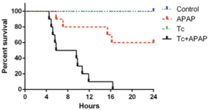
T. crassiceps infection increases mortality in the acetaminophen-induced acute liver failure murine mod-el - The APAP group showed 15% mortality 12 h after acetaminophen administration (Fig. 1). The maximum mortality rate in this group was observed by 17 h, at which time 35% of the animals had died. In contrast, the Tc+APAP group exhibited 85% mortality during the first 12 h of the experiment, reaching 100% by 16 h (Fig. 1). The control and Tc groups showed 0% mor-tality throughout the experiment (Fig. 1). Interestingly, the number of peritoneal parasites did not change in re-sponse to acetaminophen overdose (Tc+APAP group: 437 ± 129, Tc group: 412 ± 156).
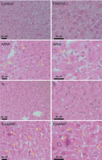
T. crassiceps infection is associated with increased hepatocyte necrosis in the acetaminophen-induced acute liver failure murine model - Ballooning degen-eration of hepatocytes is a well-established indica-tor of hepatic necrosis. Liver histology revealed that Tc+APAP mice displayed higher numbers of ballooned hepatocytes than APAP animals (Fig. 2). Hepatocytes from control and Tc groups did not exhibit ballooning degeneration as that observed in Tc+APAP and APAP animals (Fig. 2). Quantification of the number of bal-looned cells confirmed that acetaminophen exposure induced a 9-fold increase in the amount of necrotic he-patocytes in APAP mice (26.5 ± 10.93) when compared to control and Tc animals (3.16 ± 2.13 and 2.83 ± 1.16,
respectively). However, acetaminophen exposure tripled the number of ballooned hepatocytes when compared to the parasite-infected Tc+APAP group (68.5 ± 15.6) (Fig. 3). Furthermore, little to no leukocyte infiltration was found in the livers of Tc+APAP and APAP mice. This was similar to the control and Tc animals in which no immune cell infiltration was observed as well.
Fig. 2: ballooning degeneration of hepatocytes in Taenia crassiceps -infected mice following an overdose of acetaminophen (APAP). Sig-nificantly more ballooned hepatocytes were observed in Tc+APAP mice compared to APAP animals. Liver tissue from control and Tc mice did not show ballooning degeneration. Left panels show rep-resentative microphotographs at 40x magnification and right panels show the same images enlarged at 100x. Hepatocyte damage can be seen in more detail. Scale bars: 20 mM; yellow arrows: ballooned he-patocytes. Liver samples were collected in experimental sets repeated twice under independent conditions using five animals per group in each individual experiment. Control: uninfected animals receiving sterile water; APAP: uninfected animals receiving 300 mg/kg acet-aminophen; Tc: infected animals receiving sterile water; Tc+APAP: infected animals receiving 300 mg/kg acetaminophen.
Fig. 3: number of ballooned hepatocytes in Taenia crassiceps-infected mice following acetaminophen (APAP) overdose. Hepatocyte necro-sis was estimated by quantifying the number of ballooned hepatocytes in 10 high-powered imaging fields from each mouse. Acetaminophen exposure induced a 9-fold increase in the amount of necrotic hepato-cytes in APAP mice compared to control and Tc animals. Acetamino-phen exposure tripled the number of ballooned hepatocytes following non-hepatotropic parasite infection. Experimental sets were repeated twice under independent conditions using five animals per group in each individual experiment. Data are presented as the mean ± stan-dard deviation. (a) Significant differences compared to the control group; (b) significant differences compared to the APAP group; (c) significant differences compared to the Tc group; (d) significant dif-ferences compared to the Tc+APAP group.
As expected based upon the amount of ballooned hepa-tocytes, the Tc+APAP animals exhibited the most signifi-cant elevation in ALT serum levels compared to the control, Tc, and APAP groups (Fig. 4). Specifically, Tc+APAP mice showed 2-fold higher values for ALT serum than APAP animals (865.2 ± 162.9 U/L and 411.9 ± 238.4 U/L, respec-tively) (Fig. 4). In contrast, serum ALT levels were signifi-cantly lower in control (51.67 ± 8.87 U/L) and Tc (52.33 ± 10.61 U/L) animals compared to APAP and Tc+APAP mice (Fig. 4). It is worth mentioning that ALT serum concentra-tions fell within normal ranges for control and Tc animals, which suggested that the presence of the parasites them-selves had no impact on liver function.
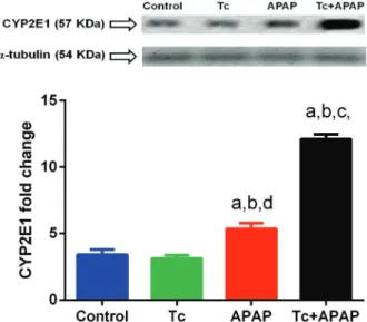
T. crassiceps infection increases CYP2E1 expression in the acetaminophen-induced acute liver failure murine model - In hepatocytes, acetaminophen is metabolised by the cytochrome P450 family 2 subfamily E member 1 (CYP2E1). Native CYP2E1 protein (57 kDa) showed a significant 4-fold increase in the liver tissue of Tc+APAP mice when compared to control and Tc animals (12.1 ± 0.35 versus 3.4 ± 0.38 and 3.1 ± 0.24, respectively) (Fig. 5). CYP2E1 protein levels also exhibited a 2.3-fold in-crease in the liver tissue of Tc+APAP animals compared to those in APAP mice (12.1 ± 0.35 and 5.3 ± 0.41, re-spectively), although CYP2E1 production was 1.8-fold
higher in hepatic specimens of APAP animals compared to that in control and Tc mice (Fig. 5). No differences in the levels of a-TUBULIN (54 KDa) were observed among the experimental groups, indicating that changes in CYP2E1 expression were not related to variations in protein quantification (Fig. 5, upper panel).
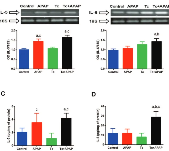
T. crassiceps infection differentially induces IL-5 and IL-6 expression in the acetaminophen-induced acute liver failure murine model - The hepatic expression of
IL-5 was significantly higher in APAP and Tc+APAP animals compared to that in control and Tc mice (Fig. 6A). In contrast, no differences in IL-5 expression lev-els were detected between the APAP and Tc+APAP ani-mal groups (Fig. 6A). Moreover, mRNA levels of IL-6
were significantly higher in the liver tissue of Tc+APAP mice compared to those in control and APAP animals (Fig. 6B). Although the hepatic expression of IL-6 was elevated in Tc mice, this was not a significant increase compared to expression levels in control and APAP ani-mals (Fig. 6B). Consistent with these RT-PCR results, Tc+APAP and APAP mice displayed a 2-fold higher lev-el of hepatic IL-5 protein when compared to control and Tc animals (Fig. 6C). However, no significant differenc-es were observed in Tc+APAP mice versus APAP mice (Fig. 6C). Similarly, the level of IL-6 protein was 3-fold higher in the livers of Tc+APAP animals compared to that in control, Tc, and APAP mice (Fig. 6D). These re-sults cumulatively suggested that IL-5 was mainly pro-duced in response to liver damage, whereas IL-6 played a synergistic role when liver damage occurred in con-junction with a parasite infection.
DISCUSSION
In this study, we showed that the comorbidity of acet-aminophen-induced hepatotoxicity and non-hepatotropic parasite infections increased mortality associated with acute liver failure. Both chronic and acute liver complica-tions are common causes of mortality worldwide (Kan-diah et al. 2016). Furthermore, parasite infections are a public health problem in developing countries and they constitute an emerging concern even in developed nations (Fleury et al. 2012, Fabiani & Bruschi 2013, Chomicz et al. 2016). Since liver disease and parasite infections are prevalent conditions in many countries, their comorbidity is highly likely. Therefore, it is of great clinical relevance to examine whether the presence of parasites causes more severe liver injury, poorer prognoses, and/or increased mortality in patients with acute hepatic disease.
In order to study the possible relationship between acute liver failure and parasite infection, we used two es-tablished experimental paradigms to induce acute liver injury in parasitised mice. Specifically, we inoculated mice with T. crassiceps parasitic larvae and adminis-tered an overdose of acetaminophen to cause liver dam-age (Vargas-Villavicencio et al. 2007, Xie et al. 2015). The acetaminophen-induced acute liver failure in mice resembles many aspects of acetaminophen overdose in humans, including hepatocyte necrosis, ALT serum ele-vation, and massive liver injury (Mohar et al. 2014, Maes et al. 2016). Likewise, inoculation of T. crassiceps larvae
into the peritoneal cavities of mice has been useful for studying the interactions between steroid hormones and immune cells in the context of a chronic infection (Mo-rales-Montor et al. 2008). Here, we found that mortality associated with acute liver failure increased by 100% as a result of the comorbidity of a parasitic infection. Despite recent studies, there is very little information concerning the potential complications of having liver disease and a parasite infection at the same time. A few reports have described hepatomegaly, jaundice, hepatic
malfunction, and poor prognoses in patients infected with parasites (Prommano et al. 2005, Ural et al. 2015). However, the aforementioned studies only examined clinical cases of parasites that already show an affinity for infiltrating the liver, such as Plasmodium falciparum
and Fasciola hepatica (Nacher et al. 2001, Haseeb et al. 2003). This limitation has made it difficult to eluci-date whether non-hepatotropic parasites are capable of directly enhancing liver injury, or whether liver injury is simply the consequence of hepatic decompensation.
For this reason, our experiments involved a liver injury in the presence of a non-hepatotropic parasite infection. Specifically, this parasite proliferated in the mouse peri-toneal cavity without invading the liver. This strategy demonstrated that non-hepatotropic parasites are indeed able to increase liver damage directly, providing some insights into the molecular mechanisms involved.
Acute liver failure results from necrosis of hepato-cytes and a sudden release of ALT into the blood stream. Usually, this involves little to no immune cell infiltration of the liver (Bernal et al. 2015, Yoon et al. 2016). In this study, we found that ballooning degeneration of hepato-cytes (a well-established indicator of hepatic necrosis) (Dias et al. 2007) was significantly increased, following simultaneous acetaminophen overdose and T. crassiceps
infection. Interestingly, levels of serum ALT were higher in parasitised mice that received acetaminophen than in non-parasitised mice that received acetaminophen. In parallel, we observed a mild-to-absent leukocyte infiltra-tion in hepatic specimens of mice that received acetamin-ophen independent of parasite infection, suggesting that the parasite itself was unable to promote a generalised immune cell response that might exacerbate liver injury. These findings support the idea that the severity of acute liver failure may be related to multiple conditions, such as non-hepatotropic parasite infections. Therefore, it is necessary to further investigate risk factors that worsen the prognoses of patients with acute liver injury.
One possible explanation for the relationship be-tween acute liver injury and non-hepatotropic parasite infections might be an interaction between CYP2E1 and IL-6. CYP2E1 is a member of the cytochrome C super-family, and it has prominent roles in metabolising a wide variety of substances like acetaminophen (Kessova & Cederbaum 2003). When acetaminophen accumulates, CYP2E1 is activated and it produces N-acetyl-p-benzo-quinone imine (NAPQI), a reactive metabolite capable of binding to mitochondrial proteins. This leads to per-oxynitrite formation and mitochondrial oxidative stress (Cover et al. 2005). Increased oxidative stress causes the mitochondrial permeability transition pore to open, resulting in a disruption of mitochondrial membrane potential, inhibition of ATP synthesis, mitochondrial dysfunction, DNA fragmentation, and necrosis of hepa-tocytes (Maes et al. 2016). Similarly, our data demon-strated that acetaminophen overdose increased CYP2E1 production in the livers of APAP mice. We also found that CYP2E1 expression increased following liver injury in parasitised mice. This suggests a role for T. crassiceps
in modulating CYP2E1 expression, which might promote the formation of NAPQI and induce hepatic necrosis. A seminal study previously demonstrated that T. crassiceps
increases expression of CYP19A1 (a member of the cyto-chrome P450 superfamily) in the testicles of parasitised male mice (Morales-Montor et al. 1999). Interestingly, investigations revealed that the parasite achieves this, in part, through upregulation of IL-6 (Morales-Montor et al. 2001). IL-6 expression has been shown to increase in re-sponse to T. crassiceps infection (Morales-Montor et al. 2001, 2008). Furthermore, IL-6 upregulates the expres-sion of several members of the cytochrome C
superfam-ily by activating distal intragenic enhancers (Zhao et al. 1997). In our study, mRNA and protein levels of IL-6 were elevated in the livers of parasitised mice that expe-rienced an overdose of acetaminophen. Interestingly, the mRNA levels of IL-6 were higher in the livers of infected animals not exposed to the hepatotoxic agent, although this was not statistically significant. Cumulatively, this suggests that T. crassiceps upregulates CYP2E1 by in-ducing IL-6 expression in liver tissue through a possible enzyme-cytokine mechanism that may be enhanced during acute liver injury. However, additional studies in CYP2E1- and IL-6-deficient mice are needed to under-stand the potential effects of non-hepatotropic parasites on mortality during acute liver failure.
Our study also identified a potential role for IL-5 in the pathology of acetaminophen-induced acute liver failure. IL-5 is a Th2 cytokine that plays pivotal roles in diseases caused by parasitic helminths. IL-5 is also associated with increased liver injury in hepatic dis-ease models, including LPS-induced hepatotoxicity and concanavalin A (ConA)-induced hepatitis (Louis et al. 2002). Furthermore, IL-5 promotes hepatic necrosis in the ConA-induced T cell-mediated hepatitis murine model (Duran et al. 2004). However, to the best of our knowledge, this is the first study reporting an upregula-tion of IL-5 in the livers of mice exposed to acetamino-phen overdose. It is well known that IL-5 increases liver injury by recruiting leukocytes into the hepatic paren-chyma (Jaruga et al. 2003). However, acetaminophen-induced acute liver failure is typically characterised by little to no leukocyte infiltration in the hepatic parenchy-ma, which we likewise observed in this study. Therefore, there are two possible explanations: (a) IL-5 causes liver injury in a leukocyte-independent fashion or (b) IL-5 ex-pression is an indirect consequence of liver damage and has no influence on the pathology. Therefore, we argue that it is of clinical importance for future studies to ex-plore whether IL-5 levels in human liver biopsies or se-rum samples could be a useful predictor of the prognosis of severe acute liver failure in patients.
In conclusion, our results demonstrate that a non-hepatotropic parasite infection is capable of increas-ing the mortality of mice followincreas-ing acute liver injury. The underlying mechanism might involve a synergism among CYP2E1, IL-6, and IL-5 that leads to hepatocyte necrosis. Further clinical research is needed to evaluate the potential benefits of using IL-5 and/or IL-6 as pos-sible biomarkers for identifying patients with a higher risk of developing severe acute liver failure.
REFERENCES
Bernal W, Jalan R, Quaglia A, Simpson K, Wendon J, Burroughs A. Acute-on-chronic liver failure. Lancet. 2015; 386(10003): 1576-87.
Bhatia SN, Underhill GH, Zaret KS, Fox IJ. Cell and tissue engineer-ing for liver disease. Sci Transl Med. 2014; 6(245): 245sr242.
Chalhoub WM, Sliman KD, Arumuganathan M, Lewis JH. Drug-induced liver injury: what was new in 2013? Expert Opin Drug Metab Toxicol. 2014; 10(7): 959-80.
Cover C, Mansouri A, Knight TR, Bajt ML, Lemasters JJ, Pessayre D, et al. Peroxynitrite-induced mitochondrial and endonuclease-mediated nuclear DNA damage in acetaminophen hepatotoxicity. J Pharmacol Exp Ther. 2005; 315(2): 879-87.
Dias Jr LB, Alves VA, Kanamura C, Oikawa RT, Wakamatsu A. Ful-minant hepatic failure in northern Brazil: morphological, immu-nohistochemical and pathogenic aspects of Labrea hepatitis and yellow fever. Trans R Soc Trop Med Hyg. 2007; 101(8): 831-9.
dos Santos DC, Martinho JMSG, Pacheco-Moreira LF, de Araujo CCV, Caroli-Bottino A, Pannain VL, et al. Eosinophils involved in fulminant hepatic failure are associated with high interleu-kin-6 expression and absence of interleukin-5 in liver and periph-eral blood. Liver Int. 2009; 29(4): 544-51.
Duran A, Rodríguez A, Martin P, Serrano M, Flores JM, Leitges M, et al. Crosstalk between PKCzeta and the IL4/Stat6 pathway dur-ing T-cell-mediated hepatitis. EMBO J. 2004; 23(23): 4595-4605.
Fabiani S, Bruschi F. Neurocysticercosis in Europe: still a public health concern not only for imported cases. Acta Trop. 2013; 128(1): 18-26.
Fleury A, Sciutto E, Larralde C. Neurocysticercosis is still prevalent in Mexico. Salud Publica Mex. 2012; 54(6): 632-6.
Haseeb AN, el-Shazly AM, Arafa MA, Morsy AT. Evaluation of ex-cretory/secretory Fasciola (Fhes) antigen in diagnosis of human fascioliasis. J Egypt Soc Parasitol. 2003; 33(1): 123-38.
Ichai P, Samuel D. Epidemiology of liver failure. Clin Res Hepatol Gastroenterol. 2011; 35(10): 610-7.
Jaruga B, Hong F, Sun R, Radaeva S, Gao B. Crucial role of IL-4/ STAT6 in T cell-mediated hepatitis: up-regulating eotaxins and IL-5 and recruiting leukocytes. J Immunol. 2003; 171(6): 3233-44.
Kandiah PA, Olson JC, Subramanian RM. Emerging strategies for the treatment of patients with acute hepatic failure. Curr Opin Crit Care. 2016; 22(2): 142-51.
Kessova I, Cederbaum AI. CYP2E1: biochemistry, toxicology, reg-ulation and function in ethanol-induced liver injury. Curr Mol Med. 2003; 3(6): 509-18.
Louis H, Le Moine A, Flamand V, Nagy N, Quertinmont E, Paulart F, et al. Critical role of interleukin 5 and eosinophils in concanavalin A-induced hepatitis in mice. Gastroenterol. 2002; 122(7): 2001-10.
Maes M, Vinken M, Jaeschke H. Experimental models of hepatotox-icity related to acute liver failure. Toxicol Appl Pharmacol. 2016; 290: 86-97.
Mohar I, Stamper BD, Rademacher PM, White CC, Nelson SD, Kava-nagh TJ. Acetaminophen-induced liver damage in mice is associ-ated with gender-specific adduction of peroxiredoxin-6. Redox Biol. 2014; 20(2): 377-87.
Morales-Montor J, Baig S, Mitchell R, Deway K, Hallal-Calleros C, Damian RT. Immunoendocrine interactions during chronic cysti-cercosis determine male mouse feminization: role of IL-6. J Im-munol. 2001; 167(8): 4527-33.
Morales-Montor J, Escobedo G, Vargas-Villavicencio JA, Larralde C. The neuroimmunoendocrine network in the complex host-parasite relationship during murine cysticercosis. Curr Top Med Chem. 2008; 8(5): 400-07.
Morales-Montor J, Rodríguez-Dorantes M, Cerbon MA. Modified expression of steroid 5 alpha-reductase as well as aromatase, but not cholesterol side-chain cleavage enzyme, in the reproductive system of male mice during (Taenia crassiceps) cysticercosis. Parasitol Res. 1999; 85(5): 393-8.
Nacher M, Treeprasertsuk S, Singhasivanon P, Silachamroon U, Van-naphan S, Gay F, et al. Association of hepatomegaly and jaundice with acute renal failure but not with cerebral malaria in severe falci-parum malaria in Thailand. Am J Trop Med Hyg. 2001; 65(6): 828-33.
Prommano O, Chaisri U, Turner GD, Wilairatana P, Ferguson DJ, Viriyavejakul P, et al. A quantitative ultrastructural study of the liver and the spleen in fatal falciparum malaria. Southeast Asian J Trop Med Public Health. 2005; 36(6): 1359-70.
Seevaratnam R, Patel BP, Hamadeh MJ. Comparison of total protein concentration in skeletal muscle as measured by the Bradford and Lowry assays. J Biochem. 2009; 145(6): 791-7.
Stravitz RT, Kramer DJ. Management of acute liver failure. Nat Rev Gastroenterol Hepatol. 2009; 6(9): 542-53.
Ural S, Kaptan F, Sezak N, El S, Örmen B, Türker N, et al. Evaluation of clinical and laboratory findings of adult visceral leishmaniasis cases. Mikrobiyol Bul. 2015; 49(4): 586-93.
Vargas-Villavicencio JA, Larralde C, De León-Nava MA, Escobedo G, Morales-Montor J. Tamoxifen treatment induces protection in murine cysticercosis. J Parasitol. 2007; 93(6): 1512-7.
Xie J, Liu J, Chen TM, Lan Q, Zhang QY, Liu B, et al. Dihydromyric-etin alleviates carbon tetrachloride-induced acute liver injury via JNK-dependent mechanism in mice. World J Gastroenterol. 2015; 21(18): 5473-81.
Yoon E, Babar A, Choudhary M, Kutner M, Pyrsopoulos N. Acet-aminophen-Induced Hepatotoxicity: a comprehensive update. Journal Clin Transl Hepatol. 2016; 4(2): 131-42.