Contents lists available atScienceDirect
Veterinary Parasitology
j o u r n a l h o m e p a g e :w w w . e l s e v i e r . c o m / l o c a t e / v e t p a r
Cytokine and transcription factor profiles in the skin of dogs naturally
infected by
Leishmania (Leishmania) chagasi
presenting distinct
cutaneous parasite density and clinical status
Daniel Menezes-Souza
a,b,c, Rodrigo Corrêa-Oliveira
b, Renata Guerra-Sá
c, Rodolfo Cordeiro
Giunchetti
a,b, Andréa Teixeira-Carvalho
d, Olindo Assis Martins-Filho
d, Guilherme Corrêa
Oliveira
e, Alexandre Barbosa Reis
a,b,f,∗aLaboratório de Imunopatologia, Núcleo de Pesquisas em Ciências Biológicas, Universidade Federal de Ouro Preto, 35400-000, Ouro Preto, Minas Gerais, Brazil
bLaboratório de Imunologia Celular e Molecular, Centro de Pesquisas René Rachou, Fundac¸ão Oswaldo Cruz, 30190-002, Belo Horizonte, Minas Gerais, Brazil cLaboratório de Bioquímica e Biologia Molecular, Núcleo de Pesquisas em Ciências Biológicas, Universidade Federal de Ouro Preto, 35400-000, Ouro Preto, Minas Gerais, Brazil
dLaboratório de Biomarcadores de Diagnóstico e Monitorac¸ão, Centro de Pesquisas René Rachou, Fundac¸ão Oswaldo Cruz, 30190-002, Belo Horizonte, Minas Gerais, Brazil
eLaboratório de Parasitologia Celular e Molecular, Centro de Pesquisas René Rachou, Fundac¸ão Oswaldo Cruz, 30190-002, Belo Horizonte, Minas Gerais, Brazil
fDepartamento de Análises Clínicas, Escola de Farmácia, Universidade Federal de Ouro Preto, 35400-000, Ouro Preto, Minas Gerais, Brazil
a r t i c l e
i n f o
Article history:
Received 13 October 2009
Received in revised form 7 November 2010 Accepted 10 November 2010
Keywords:
Canine visceral leishmaniasis Cytokines
Transcription factors Real-time PCR Leishmania chagasi Parasitism
a b s t r a c t
The immune response in the skin of dogs infected withLeishmania chagasiand its asso-ciation with distinct levels of tissue parasitism and clinical progression of canine visceral leishmaniasis (CVL) are poorly understood and limited studies are available. A detailed analysis of the profiles of cytokines (IFN-␥, IL-4, IL-5, IL-10, IL-12, IL-13, TGF-1 and TNF-␣) and transcription factors (T-bet, GATA-3 and FOXP3) in the skin of 35 naturally infected dogs was carried out using real-time PCR alongside determinations of skin parasite density and the clinical status of CVL. A mixed cytokine profile with high levels of expression of IFN-␥, TNF-␣and IL-13 was determined in asymptomatic dogs. Additionally, the levels of transcription factors GATA-3 and FOXP3 were correlated with the asymptomatic disease. A mixed cytokine profile was also observed during active CVL. Moreover, high levels of IL-10 and TGF-1, concomitant with the low expression of IL-12, may represent a key condition that allows persistence of parasite replication in the skin. The results obtained indicate that in asymptomatic disease or lower levels of skin parasite density, a mixed inflammatory, regulatory immune response profile may be of major relevance for both the maintenance of the clinical status of the dogs as well as for parasite persistence and replication at low levels.
© 2010 Elsevier B.V. All rights reserved.
∗ Corresponding author at: Universidade Federal de Ouro Preto,
Depar-tamento de Analises Clinicas, Rua Costa Sena, 171, 35400-000, Ouro Preto, Minas Gerais, Brazil. Tel.: +55 21 31 3559 1694; fax: +55 21 31 3559 1680.
E-mail address:alexreis@nupeb.ufop.br(A.B. Reis).
1. Introduction
Visceral leishmaniasis (VL) caused by the protozoan
Leishmania (Leishmania) chagasi[syn.Leishmania (Leishma-nia) infantum], is one of the most important of zoonotic diseases affecting dogs and humans in Europe and Latin America (Desjeux, 2004). Dogs are considered to be
excel-0304-4017/$ – see front matter© 2010 Elsevier B.V. All rights reserved.
lent models for the study of human VL because the natural history of the canine disease is very similar to that observed in human (Moreno and Alvar, 2002). A num-ber of reports are available concerning the parasite load found in different tissues and the immunopathological changes related to the progression of clinical forms of canine visceral leishmaniasis (CVL) (Chamizo et al., 2005; Reis et al., 2006a,b,c; Giunchetti et al., 2006; Lage et al., 2007; Giunchetti et al., 2008a,b; Alves et al., 2009; Carrillo and Moreno, 2009; Guerra et al., 2009; Manna et al., 2009; Reis et al., 2009).
It has been established that the skin is an important reservoir for parasites in asymptomatic and symptomatic
Leishmania-infected dogs, and the high parasite loads found in this organ suggest that the skin may play an impor-tant role in the transmission and epidemiology of the disease (Abranches et al., 1991). Previous investigations have revealed that symptomatic CVL-infected dogs exhibit an intense diffuse dermal inflammatory infiltrate and high parasitic burden in comparison with their asymptomatic counterparts (Giunchetti et al., 2006). On this basis it was proposed that the immunopathological changes in the skin and the levels of cutaneous parasitism are directly related to the clinical severity of the disease.
Earlier evaluations of the immune response pattern in
Leishmania-infected dogs have been based on the analy-sis of cytokines profiles in peripheral blood mononuclear cells (PBMCs), skin, lymph nodes, bone marrow and spleen. Thus,Pinelli et al. (1994)found higher levels of IL-2 and TNF-␣ in supernatants from in vitro-stimulated PBMCs derived from asymptomatic dogs, and proposed that these cytokines could be used as markers of disease progression. Furthermore,Chamizo et al. (2005)reported that PBMCs of asymptomatic CVL-infected dogs exhibited preferential expression of TH1 cytokines (Chamizo et al., 2005). Some
authors have demonstrated the ability of IL-12 to augment the production of IFN-␥by PBMCs derived from dogs with experimental or natural symptomatic CVL, and stressed the importance of these cytokines in the resolution of the dis-ease (Dos-Santos et al., 2004; Strauss-Ayali et al., 2005). In a recent study, both type 1 and 2 immune responses were demonstrated to occur in the spleen during CVL ( Strauss-Ayali et al., 2007), whileLage et al. (2007)suggested that CVL is marked by the balanced splenic production of type 1 and 2 cytokines with the predominant accumulation of IL-10 and IFN-␥as a consequence of increased parasitic load and progression of the disease.
In the present study, the immunopathology of CVL has been further investigated by performing a detailed analy-sis of the expression of type 1 (IL-12, IFN-␥and TNF-␣), type 2 4, IL-5 and IL-13) and immunoregulatory (IL-10 and TGF-1) cytokines in the skin of dogs naturally infected byLeishmania (L.) chagasi. In addition, the lev-els of the transcription factors T-bet, GATA-3 and FOXP3 have been assessed during CVL. Attention was particularly focussed on the possible association between clinical status and skin parasite density, but the key objective of the study was to explore novel biomarkers, including the relationship between type 1 and 2 cytokine patterns and transcription factors that might influence susceptibility and resistance to infection.
2. Materials and methods
2.1. Study population and clinical evaluation
The investigation was approved by the Ethics Commit-tee on Animal Experimentation (CETEA) of the Universi-dade Federal de Minas Gerais, Brazil. The study population comprised 51 adult dogs (aged between 2 and 6 years) of both genders that had been captured by the Center of Zoonosis Control in Belo Horizonte (Minas Gerais, Brazil), a region with a high prevalence of CVL and human VL. The animals were maintained under quarantine at the kennels of the Institute of Biological Sciences (Universidade Fed-eral de Minas Gerais) and treated for intestinal helminthic infections (Endal Plus®; Schering-Plough Coopers, Brazil) and immunised against parvovirosis, leptospirosis, distem-per, parainfluenza and hepatitis (Vanguard®HTLP 5/CV-L vaccine; Pfizer, New York, NY, USA). Experimental ani-mals were categorised on the basis of serological results from an indirect immunofluorescence assay test (IFAT), the “gold standard” immunological test in Brazil for the diag-nosis of CVL. Sixteen dogs presenting negative IFAT assays with serum samples diluted 1:40, and negative parasito-logical examinations forLeishmaniain tissue smears (bone marrow, ear skin, spleen, liver and popliteal lymph node), were considered to be non-infected and were employed as the control group (CD,n= 16). Thirty-five animals with positive IFAT titres≥1:40 were considered CVL-positive and were included in the groups of infected animals.
Leishmania-infected dogs were sub-divided on the basis of the presence or absence of signs of infection accord-ing to Mancianti et al. (1988) as follows: absence of indicative signs of the disease – asymptomatic group (AD,
n= 10); presence of a maximum of three clinical signs of the disease including opaque bristles and/or localised alopecia and/or moderate loss of weight – oligosymp-tomatic group (OD, n= 10); presence of characteristic clinical signs of the disease including cutaneous lesions, onycogryphosis, opaque bristles, severe loss of weight, apa-thy and keratoconjunctivitis – symptomatic group (SD,
n= 15).
2.2. Sample collection and assessment of skin parasite load
Animals were euthanised with sodium thiopental (Abbott Laboratories, Abbott Park, IL, USA; 30 mg/kg body weight) and samples of skin tissue were collected from the ears without lesions. One fragment of the skin was used for tissue imprints on microscopic slides. The samples were fixed in methanol, stained with Giemsa and exam-ined under an optical microscope.Leishmaniaamastigote stages were counted and parasite densities were expressed as Leishman Donovan Units (LDU) as described byStauber (1955)with some modifications. Parasite densities were categorised statistically into tertiles according to Reis et al. (2006a)as absent (LDU = 0; CD group, n= 16), low (LDU = 1–9; LP group, n= 12), medium (LDU = 10–130; MP group,n= 11) and high (LDU = 131–7246; HP group,
177 (2011) 39–49 41
Table 1
Sequences of primers used for quantification of mRNA expression by real-time PCR. F: forward primer, R: reverse primer. GeneBank accession number of the sequence used to design primers and their product length are shown as well as each PCR efficiency andR2.
Gene Primer sequence (5′–3′) Product length (bp) Reaction efficiency (%) R2
GAPDH
F TTCCACGGCACAGTCAAG 115 99.1 0.996
R ACTCAGCACCAGCATCAC
IL-12p40
F CAGCAGAGAGGGTCAGAGTGG 109 96.5 0.989
R ACGACCTCGATGGGTAGGC
IFN-␥
F TCAACCCCTTCTCGCCACT 113 95.4 0.967
R GCTGCCTACTTGGTCCCTGA
TNF-␣
F CGTCCATTCTTGCCCAAAC 94 97.2 0.983
R AGCCCTGAGCCCTTAATTC
IL-4
F CACCTCCCAACTGATTCCAA 123 96.9 0.991
R CTCGCTGTGAGGATGTTCAA
IL-5
F GCCTATGTTTCTGCCTTTGC 106 95.3 0.979
R GGTTCCCATCGCCTATCA
IL-13
F CCTCCTCAGAGCAAAGTG 148 96.7 0.973
R CCCAGCACAAACAAAGAC
IL-10
F AGAACCACGACCCAGACATC 129 97.1 0.993
R CCACCGCCTTGCTCTTATTC
TGF-1
F AGGATCTGGGCTGGAAGTG 134 95.1 0.981
R CGGGTTGTGCTGGTTGTA
T-bet
F GCTTCCAACACACACATC 80 96.0 0.977
R TGAGTGATCTCCGCATTC
GATA3
F ATGACACGCTGGAGGACTTC 106 98.5 0.969
R TGGCTGGAGTGGCTGAAA
FOXP3
F AAACAGCACATTCCCAGAGTTC 102 95.1 0.981
R AGGATGGCCCAGCGGATCAG
2.3. Extraction of total RNA and synthesis of first strand cDNAs
The second fragment of ear skin was stored at−80◦C
until required for RNA analysis. Total RNA was extracted by homogenising approximately 20 mg of skin tissue with 1 mL of TRIzol reagent (Invitrogen Brasil, São Paulo, SP, Brazil) in a rotor stator. The lysate was incubated at room
temperature for 10 min, mixed with chloroform (200L)
by tube inversion, and centrifuged at 12,000×gfor 10 min
at 4◦C. The aqueous phase was collected and RNA
extrac-tion continued using the SV Total RNA Isolaextrac-tion System (Promega, Madison, WI, USA) according to the recom-mendations of the manufacturer, which included a DNase treatment step. The efficiency of DNAse treatment was evaluated by PCR amplification of the cDNA reaction mix without the addition of the Thermoscript enzyme. Finally, each q-PCR run was performed with 2 internal controls assessing both potential genomic DNA contaminations (no reverse transcriptase added) and purity of the reagents used (no cDNA added). Strand cDNAs were synthesised
from 1.0g of total RNA using the ThermoScriptTMRT-PCR
System (Invitrogen Brasil, São Paulo, SP, Brazil) with oligo-dT primers according to the manufacturer’s instructions.
2.4. Design of primers for gene evaluation
Primers were designed with the aid of Gene Runner version 3.05 (copyright Hasting Software Inc. 2004) using specific canine sequences obtained from GenBank with accession numbers GAPDH (AB038240), IL-4 (AF239917), IL-5 (AF331919), IL-10 (U33843), IL-12p40 (U49100), IL-13
(AF244915), IFN-␥(AF126247), TGF-1 (L34956), TNF-␣
(DQ923808), FOXP3 (XM 548996), GATA-3 (XM 844060) and T-bet (XM 548164). The sequences of the primers
employed are listed inTable 1. The primers were
synthe-sised by Eurogentec (Southampton, U.K.) and reconstituted in nuclease free water.
2.5. Real-time PCR, cloning and sequencing of amplicons
Fig. 1.Relationship between clinical status and the expression of mRNAs for cytokines in the skin of dogs naturally infected withLeishmania chagasi. Animals were categorised as asymptomatic (AD), oligosymptomatic (OD) and symptomatic (SD) according to the clinical progression of CVL: the control group (CD) comprised uninfected animals. Box plots show the median value (horizontal line across the box), the interquartile ranges (horizontal ends of the box), and the highest and lowest values (lines extending from the box and terminating in horizontal lines). The log number of messenger RNA relative expression for IFN-␥, IL-4, IL-5, IL-10, IL-12, IL-13, TGF-and TNF-␣are shown. Significant differences (p< 0.05) compared with CD, AD, OD and SD are indicated by the letters ‘a’, ‘b’, ‘c’ and ‘d’, respectively. Spearman’s correlation indexes (randp-values) are shown on the graphs where applicable. The data was also evaluated as mean fold-differences relative to the each messenger RNA expression of the cytokines in the clinical groups in comparison to the values of the control group. Statistically significant increase in the target transcript levels of AD to TNF-␣, IL-13 and IL-10 as compared to SD (p= 0.0491; p= 0.0225 andp< 0.05, respectively) were observed. Moreover, there was an increase in the target transcript levels of OP to IL-10 as compared to SD (p< 0.05).
primer and cDNA diluted at 1:5. The samples were incu-bated at 95◦C for 10 min and then submitted to 40 cycles of 95◦C for 15 s and 60◦C for 1 min, during which time fluorescence data were collected. The efficiency of each pair of primers was evaluated by serial dilution of cDNA according to the protocol developed by PE Applied Biosys-tems. In order to evaluate gene expression, three replicate analyses were performed and the amount of target RNA was normalised with respect to the control (housekeep-ing) gene GAPDH and expressed according to the 2−Ct
method. PCR products were cloning with pGEM®-T Easy Vector (Promega) and sequenced to check specificity using an ABI 3100 Automated Sequencer (PE Applied Biosystems) and a Dye Terminator Kit.
2.6. Statistical analysis
Statistical analyses were performed with the aid of GraphPad Prism software package version 5.0 (GraphPad Software, San Diego, CA, USA). Normality of the data was established using the Kolmogorov–Smirnoff test. In the
parametric data, one-way analysis of variance was used for the comparative study between groups, followed by Tukey’s test. In the nonparametric data, Kruskal–Wallis test was used for between group comparative study, fol-lowed by Dunns’ test for multiple comparisons. Spearman’s rank correlation was also computed in order to investi-gate relationships between the expression of cytokine and transcription factor mRNAs with clinical forms and skin parasite density. In all cases, differences were considered significant when the probabilities of equality, p values, were≤0.05.
3. Results
3.1. Asymptomatic dogs show high expression of IFN-, TNF-˛and IL-13 in the skin
177 (2011) 39–49 43
Fig. 2. Relationship between skin parasite density and the expression of mRNAs for cytokines in the skin of dogs naturally infected withLeishmania chagasi. Animals were categorised with low (LP), medium (MP) and high (HP) parasite densities according toReis et al. (2006a): the control group (CD) comprised uninfected animals. Box plots show the median value (horizontal line across the box), the interquartile ranges (horizontal ends of the box), and the highest and lowest values (lines extending from the box and terminating in horizontal lines). The log number of messenger RNA relative expression for IFN-␥, IL-4, IL-5, IL-10, IL-12, IL-13, TGF-and TNF-␣are shown. Significant differences (p< 0.05) compared with CD, LP, MP and HP are indicated by the letters ‘a’, ‘b’, ‘c’ and ‘d’, respectively. Spearman’s correlation indexes (randp-values) are shown on the graphs where applicable. The data was also evaluated as mean fold-differences relative to the each messenger RNA expression of the cytokines in the clinical groups in comparison to the values of the control group. Increase in the target transcript levels of LP and MP to IL-12,p= 0.0337 andp= 0.0307, respectively as well as MP to IL-13,p= 0.0420 as compared to HP were observed. Moreover, there was an increase in the target transcript levels of HP to IL-10 as compared to LP (p= 0.0311) and MP (0.0070), respectively.
and OD groups when compared with the CD group (p< 0.05). TNF-␣ was highly expressed in AD in rela-tion to CD and SD (p< 0.05). The data revealed that the impaired expression of IFN-␥ and TNF-␣ correlated (r=−0.3988/p= 0.0263 andr=−0.5496/p= 0.0020, respec-tively) with the morbidity of the disease. Interestingly, asymptomatic animals presented increased levels of IL-13 in comparison with all other groups (p< 0.05), and this was significantly negatively correlated with clinical pro-gression (r=−0.6879/p< 0.0001). Additionally, AD showed a significant increase in IL-5 expression in comparison with CD (p< 0.05), while OD exhibited an enhanced expression (p< 0.05) of IL-10 when compared with CD and AD. Analy-sis of TGF-1 expression showed levels were significantly higher in OD than in CD (p< 0.05).
The data was also evaluated as mean fold-differences relative to the each messenger RNA expression of the cytokines according to clinical groups in relation to the val-ues of the control group. Similar findings were found in comparison to those evaluated during the analysis of the expression of cytokine genes with statistically significant increase in the target transcript levels of AD to TNF-␣, IL-13 and IL-10 as compared to SD (p= 0.0491;p= 0.0225 and
p< 0.05, respectively). Moreover, there was an increase in the target transcript levels of OP to IL-10 as compared to SD (p< 0.05).
3.2. Enhanced IL-10 and TGF-ˇ1 cytokines and a concomitant low expression of IL-12 may represent a key condition for parasite replication in the skin of infected dogs
Fig. 3.Correlations between the expression of the mRNAs of mixed cytokines in the skin of dogs presenting CVL (Infected dogs,n= 35). The results are displayed as scatter diagrams of individual values. Spearman’s correlation indexes (randp-values) are shown on the graphs while connecting lines illustrate positive and negative correlation indexes.
Analysis of IL-12 expression indicated that a signifi-cant up-regulation of this cytokine occurred in the LP and MP groups in comparison with the HP group (p< 0.05). Moreover, there was a significant negative correlation (r=−0.5928/p= 0.0002) between the decrease in the rel-ative expression of IL-12 and the increase in parasite load (Fig. 2), and a negative correlation between the levels of IL-12 and those of IL-10 or TGF-(r=−0.5777/p= 0.0005 andr=−0.5013/p= 0.0030, respectively;Fig. 3). Consistent with these observations, a significant increase in the ratio of expression of IL-12 to IL-10 was observed in groups with a lower (p< 0.05) parasite burden (LP: 69.95±85.06; MP: 90.80±97.24; HP: 16.13±31.06). The relationship between inflammatory and regulatory responses was con-firmed by the ratio of expression of IFN-␥/IL-10, which was found to be significantly higher (p< 0.05) in LP and MP when compared with HP (LP: 1845±6138; MP: 1780±4169; HP: 40.58±128.2). The presence of the parasite was associated with an increase in the pro-inflammatory cytokines IFN-␥and TNF-␣(p< 0.05) in all infected groups when compared with the control group, although no correlation could be established between the expression of these cytokines and skin parasite density (Fig. 2).
The data was also evaluated as mean fold-differences relative to the each messenger RNA expression of the cytokines according to parasitism in relation to the val-ues of the control group. Similar findings were found in comparison those evaluated during the analysis of the expression of cytokine genes with statistically significant increase in the target transcript levels of LP and MP to IL-12,p= 0.0337 andp= 0.0307, respectively as well as MP to IL-13 as compared to HP,p= 0.0420. Moreover, there was an increase in the target transcript levels of HP to IL-10 as compared to LP and MP (p= 0.0311 and 0.0070), respec-tively.
3.3. Mixed cytokine profile is a hallmark of active CVL following L. chagasi infection
A detailed analysis of the correlations between of type 1 and type 2 cytokines expressed in the skin of dogs naturally infected by L. chagasi are depicted in Fig. 3. Correlation analyses revealed that the type 1 cytokines IFN-␥, IL-12 and TNF-␣ were positively correlated with IL-13 expression (r= 0.3646/p= 0.0476;
177 (2011) 39–49 45
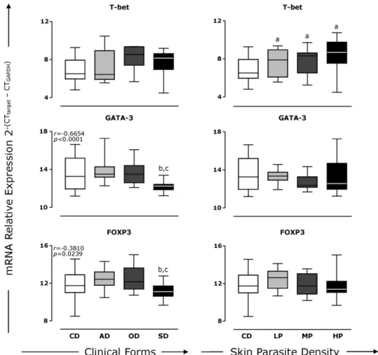
Fig. 4. Analyses of the expression of mRNAs for transcription factors FOXP3, GATA-3 and T-bet in the skin of dogs naturally infected withLeishmania chagasi. In left plate, animals were categorised as asymptomatic (AD), oligosymptomatic (OD) and symptomatic (SD) according to the clinical progression of CVL. In right plate, animals were categorised with low (LP), medium (MP) and high (HP) parasite densities according toReis et al. (2006a). In each case the control group (CD) comprised uninfected animals. Box plots show the median value (horizontal line across the box), the interquartile ranges (horizontal ends of the box), and the highest and lowest values (lines extending from the box and terminating in horizontal lines). The log number of messenger RNA relative expression for T-bet, GATA-3 and FOXP3 are shown. Significant differences (p< 0.05) compared with CD, AD or LP, OD or MP and SD or HP are indicated by the letters ‘a’, ‘b’, ‘c’ and ‘d’, respectively. Spearman’s correlation indexes (randp-values) are shown on the graphs where applicable. The data was also evaluated as mean fold-differences relative to the each messenger RNA expression of the transcription factors in the clinical groups in comparison to the values of the control group. Statistically significant decrease in the target transcript levels of SD to GATA-3 and FOXP3 has been observed as compared to the transcript levels of the AD (p= 0.0188 andp< 0.05) or OD (p= 0.0296 andp= 0.0256), respectively.
IL-13 was observed in AD (Fig. 1). Simultaneous expres-sion of IL-5 with IFN-␥ and TNF-␣(r= 0.3691/p= 0.0447 and r= 0.5673/p= 0.0009, respectively) was found during CVL, and similar situations were observed with respect to IL-4 with TNF-␣(r= 0.5243/p= 0.0012) and IL-4 with IL-12 (r= 0.6643/p< 0.0001) in all infected dogs, independent of clinical status and/or skin parasite burden (Fig. 3).
3.4. The transcription factors GATA-3 and FOXP3 are correlated with less severe clinical forms
In an attempt to determine whether the expression of the transcription factors FOXP3, GATA-3 and T-bet might be reliable biomarkers of clinical status and skin parasite load in CVL, the association between the levels of these variables was investigated. Data analyses revealed signif-icant negative correlations between FOXP3 and GATA-3 with respect to clinical evolution (r=−0.6654/p< 0.0001;
r=−0.3810/p= 0.0239, respectively;Fig. 4, left panel), but no correlation between the levels of the transcription fac-tors and skin parasite load (Fig. 4, right panel). The presence of the parasite was associated with an increase in T-bet in all infected groups in comparison with CD (p< 0.05; Fig. 4, right panel). In this sense, high levels of T-bet were found in OD and SD compared with CD (p< 0.05;Fig. 4, left panel), but no associations could be established between the expression of T-bet and clinical status or dermal para-site burden (Fig. 4).
transcript levels of the AD (p= 0.0188 andp< 0.05) or OD (p= 0.0296 andp= 0.0256), respectively.
4. Discussion
The skin is an important immune compartment that actively participates in host protection at both the early and later phases of infection. A wide variety of cells, includ-ing intra-epithelial T lymphocytes and Langerhans cells, are present in the skin and these provide considerable capacity to generate and maintain local immune reactions. Leish-maniasis is typically transmitted by the bite of sand flies infected with the pathogen and the skin is clearly the first point of contact with the protozoan. Apparently normal skin of dogs naturally infected byL. chagasiis intensely par-asitised by amastigote forms ofL. chagasi(Giunchetti et al., 2006) that reflects a compartmentalized profile of cytokine associated with resistance or susceptibility toLeishmania
infection.
In this context, IL-12 is known to perform a number of key functions including the induction of IFN-␥ produc-tion by PBMC and NK cells, the stimulaproduc-tion of proliferaproduc-tion in pre-activated T-cells and NK cells, production of NO from macrophages and plays in the development of specific type 1 T-cell-mediated immunity (Trinchieri et al., 2003). Additionally, it has been postulated that IL-10 modulates the type 1 immune response inLeishmania-infections by inhibiting IFN-␥production viathe suppression of IL-12 synthesis in antigen presenting cells (Lage et al., 2007; Peruhype-Magalhães et al., 2005). This would imply that the balance between IFN-␥and IL-10 during infection is particularly important in the control of VL as suggested in an earlier study involving functional models (Silvestre et al., 2007). In the present study, we have the unique opportu-nity to perform a compartmentalized characterisation of an immune response in skin from naturallyL. chagasi-infected dogs. Since skin is important site to transmission of the infection, the study of the immune response in the skin of dogs infected withL. chagasiand its association with dis-tinct levels of tissue parasitism and clinical progression of CVL will permit new insights elucidating the progressive or protective mechanisms during the infection (Kemp et al., 1996; Reis et al., 2009).
In the present investigation, dogs showing high skin parasitism exhibited a predominantly immunoregulatory pattern of immune response characterised by increased expression of IL-10 and TGF- in comparison with the CD, LP and MP groups (Fig. 2). These findings are con-sistent with previous reports relating the ability of IL-10 and TGF-to down-regulate T-cell responses and inhibit the leishmanicidal activity of macrophages thus leading to the progression of leishmaniasis and/or prevention of cure (Vouldoukis et al., 1997; Gantt et al., 2003).Alves et al. (2009)observed that the increased of IL-10 and TGF- in lymph nodes are correlated with high parasite bur-den and symptomatic diseases in dogs naturally infected withL. chagasi. Since the experimental animals typically exhibited active CVL, the present results suggest that an increase in IL-10 and TGF-may lead to the maintenance of parasite multiplication and therefore disease status. More-over, the IFN-␥/IL-10 (LP: 1845±6138; MP: 1780±4169;
HP: 40.58±128.2) and IL-12/IL-10 (LP: 69.95±85.06; MP: 90.80±97.24; HP: 16.13±31.06) ratios were lower in the HP group than in the other groups (p< 0.05). Accordingly, LP showed a significant increase in IL-12 expression in comparison HP and a negative correlation was observed between IL-12 levels and increase of skin parasite den-sity (Fig. 2). Additionally, the data revealed that increases in IL-12 were negatively correlated with the levels of the immunoregulatory cytokines IL-10 and TGF-1 (Fig. 3). The results presented here thus re-emphasise the involvement of immunoregulatory cytokines in the suppression of the immune response involved in the control of parasite repli-cation in the skin.
Few studies have been concerned with the involve-ment of transcription factors in the regulation of cytokine gene transcription in canine models.Biller et al. (2007) have, however, demonstrated increased expression of the mRNAs of FOXP3 and of the cytokines IL-10 and TGF-in regulatory T cells (TReg) of dogs with cancer. In the present
study, although specific cell populations such as TReg
lym-phocytes were not assessed, a positive correlation between the expression of FOXP3 and immunoregulatory (IL-10 and TGF-) cytokines was confirmed (r= 0.6764/p< 0.001;
r= 0.3151/p< 0.05, respectively). Additionally, low levels of FOXP3 expression were observed in the SD group com-pared with the AD and OD groups, and this was negatively correlated with clinical progression. Nevertheless, no cor-relation between the up-regulation of IL-10 and TGF-1 by FOXP3 and the clinical development of the disease could be observed. An explanation for this finding could be that FOXP3 is involved in regulating the expression of many genes, as described in recent studies involving murine models (Marson et al., 2007; Zheng et al., 2007), or it might indicate that other cells could be involved in the produc-tion of these cytokines, how macrophages with the purpose of modulation of the immune response. It is, however, imperative that the role of FOXP3 in the development of regulatory cells in CVL be ascertained in future studies.
Analysis of the cytokine profile with respect to par-asite burden revealed increases in the expression of pro-inflammatory cytokines IFN-␥and TNF-␣and in the transcription factor T-bet in all infected groups (LP, MP and HP) in comparison with the CD group (Figs. 2 and 4). T-bet is a key protein in the immune system and has been described as a TH1-specific T-box transcription factor controlling the
development of TH1 cells and the expression of the
hall-mark type 1 cytokine, IFN-␥, in TH1 and NK cells. A number
of studies have established that T-bet plays an essential role in the control of TH1 cell-dependent protozoan infections
177 (2011) 39–49 47
clinical status revealed in AD group increases in IFN-␥, TNF-␣(Fig. 1) and IFN-␥/IL-4 ratios in comparison with the SD and CD groups (CD: 0.32±0.15; AD: 0.77±0.50; OD: 0.80±0.43,p< 0.05). In addition, a negative correla-tion could be established between high levels of IFN-␥and TNF-␣and clinical evolution (Fig. 1). These findings are consistent with previous reports implicating the involve-ment of IFN-␥ and TNF-␣ secretion in optimal parasite clearance through activation of macrophages and, conse-quently, induction of nitric oxide production (Vouldoukis et al., 1997). Furthermore, IL-4 has been proven to have no role in disease progression of the visceralising species (Satoskar et al., 1995). In contrast to the present results, some investigations have found no difference in the expres-sion of IFN-␥and TNF-␣in bone marrow (Quinnell et al., 2001) or spleen cells (Lage et al., 2007) in naturally infected dogs presenting different clinical forms. Moreover, a recent study bySanchez-Robert et al. (2008)demonstrated that higher IFN-␥expression in PBMCs was associated with an increase of clinical signs in CVL. One possible explanation of this observation is a distinct in situimmune response againstL. chagasiin target organs of naturally infected dogs, as previously described inSanchez et al. (2004).
Even though IL-12 plays a major role in determining a type 1 immune response, no difference in expression of the mRNA of this cytokine was detected among the CVL clinical groups evaluated in the present study (Fig. 1). In according to our results,Lage et al. (2007)andAlves et al. (2009)not observed differences in the frequency and expression of this cytokine in dogs presenting different clinical forms of CVL.
High levels of IL-5 in the skin of asymptomatic
Leishmania-infected dogs were observed (Fig. 1). Previous authors had suggest that IL-5 and associated IFN-␥ produc-tion could be involved in the control of infecproduc-tion in such animals or humans, possibly by promoting differentiation and activation of eosinophils and enhancing the gener-ation and activgener-ation of specific cytotoxic T lymphocytes (Nagasawa et al., 1991; Mary et al., 1999; Peruhype-Magalhães et al., 2005).
In murine leishmaniasis, several researchers have observed that IL-13 synthesis promotes initial IFN-␥ pro-duction and influences the assembly and maturation of tissue granuloma. However, such experiments have not addressed the mechanism(s) by which IL-13 regulates the expression of anti-leishmanial type 1 response (Murray et al., 2006). In the present study it was demonstrated that asymptomaticLeishmania-infected animals presented a high expression of IL-13, and a negative correlation of this cytokine with clinical progression in CVL was observed (Fig. 1). In addition, a concomitant high IFN-␥expression (Fig. 1) was found in the AD group and this was positively correlated with the expression of IL-13 (Fig. 3). In a recent longitudinal study,Sanchez-Robert et al. (2008)evaluated the cytokine profile in PBMCs of experimentally infected dogs and observed that most of the animals that devel-oped clinical leishmaniasis expressed IL-13 during the first 4 months after infection, a result that was in complete con-trast to that presented by the asymptomatic group.
A reduction of GATA-3 in the SD group (Fig. 4B) and a high negative correlation of this transcription factor with
clinical evolution were detected in the present study. The level of GATA-3 also showed a positive correlation with the expression of the type 2 cytokines 4, 5 and IL-13 (data not shown) in the skin of infected dogs. However, among these cytokines, only IL-13 presented a concomitant expression with GATA-3 in the AD group that was nega-tively correlated with clinical progression (Figs. 1 and 4) in CVL. This finding is in agreement with that ofKitamura et al. (2005)who evaluated the correlation between the expres-sion of GATA-3 and type 2 cytokines in human helper T-cell clones and demonstrated that only IL-13 was strongly cor-related with the mRNA levels of the transcription factor. It has been reported that GATA-3 plays an important role in IL-13 production in both T cells and mast cells, and also facilitates chromatin remodelling of TH2 cytokine gene loci,
including the IL-13 gene. In addition, a GATA-3 binding site in the proximal IL-13 promoter is necessary for cell type-specific expression of IL-13 (Murray et al., 2006). This interesting correlation found in the dermal compartment may encourage further studies of the role of GATA-3 in the determination of CD4+ T cell phenotype and in the
expression of type 2 cytokines in canine models. Thus, the results presented in this study suggest that high levels of IL-13 and GATA-3 can be considered as good biomarkers of asymptomatic clinical forms in CVL. However, due to high dispersion in the expression of GATA-3 in the groups stud-ied, further investigations should be performed to confirm the importance of this gene as a biomarker in CVL.
Several investigations have demonstrated that a mixed cytokine pattern can be associated with resistance or sus-ceptibility in vaccine models and Leishmania-infections (Raziuddin et al., 1994; D’Andrea et al., 1995; Peruhype-Magalhães et al., 2006). The mixed type 1/type 2 immune profile revealed in the present study demonstrated the ability of naturally infected dogs to respond toL. chagasi
infections independent of clinical status and skin parasite density. The immune profile was characterised by a posi-tive correlation between cytokine levels of type 1 (IFN-␥, IL-12 and TNF-␣) and of type 2 (IL-4, IL-5 and IL-13) (Fig. 3). In agreement with these results,Raziuddin et al. (1994) reported enhanced production of IL-4 and TNF-␣in both VL and in cutaneous leishmaniasis. Furthermore, a study of the immune response to lipopolysaccharide or Staphylococ-cus aureusin PBMCs pre-treated with IL-4 or IL-13 revealed a significant increase in the production and accumulation of IL-12 and TNF-␣in such cells, and this could be inhibited by anti-IL-4 neutralising antibodies (D’Andrea et al., 1995). These findings were confirmed in the present study by the demonstration of positive correlations between 4 or IL-13 and IL-12 or TNF-␣(Fig. 3). Even though only IL-13 was directly correlated with IFN-␥, the concomitant increase in IL-12 and IL-4 suggests an up-regulation of expression of IL-13 cytokine, reflecting a complex regulatory role of the mixed cytokine profile that is conducive to a protective response inLeishmania-infected dogs (Fig. 3).
enhanced expression of the GATA-3 transcription factor suggest that these genes could be biomarkers for asymp-tomatic clinical forms in CVL. Moreover, IL-12 could play a protective role against parasite replication. On the other hand, in order to guarantee the survival and persistence of amastigotes in the skin compartment, the establish-ment of a regulatory profile, triggered by an increase in the immunoregulatory cytokines IL-10 and TGF-, is cru-cial. The results indicate that a concomitant expression of mixed cytokines, without the necessity for an absolute polarised profile, can tilt the immune system toward either a progressive or protective response in CVL. An advance in our knowledge of the mechanism that determines the protective immune response toL. chagasiinfection in dogs will permit the establishment of a rational strategy for the development of vaccines and immunological therapies against CVL.
Acknowledgments
The study was supported by the Fundac¸ão de Amparo à Pesquisa do Estado de Minas Gerais, Brazil (PRONEX 2007). RCO, GCO, ABR, ATC and OAMF thank CNPq for fellowships. The authors wish to express their appreciation of the hard work carried out by the staff of the Fundac¸ão Nacional da Saúde during the execution of this project. The authors are also grateful for the use of facilities at CEBIO, Universidade Federal de Minas Gerais and Rede Mineira de Bioterismo (FAPEMIG), and for support with the provision of experi-mental animals.
References
Abranches, P., Silva-Pereira, M.C., Conceic¸ão-Silva, F.M., Santos-Gomes, G.M., Janz, J.G., 1991. Canine leishmaniasis: pathological and eco-logical factors influencing transmission of infection. J. Parasitol. 77, 557–561.
Alves, C.F., de Amorim, I.F., Moura, E.P., Ribeiro, R.R., Michalick, M.S., Kalapothakis, E., Bruna-Romero, O., Tafuri, W.L., Teixeira, M.M., Melo, M.N., 2009. Expression of IFN-gamma, TNF-alpha, IL-10 and TGF-beta in lymph nodes associates with parasite load and clinical form of dis-ease in dogs naturally infected withLeishmania (Leishmania) chagasi. Vet. Immunol. Immunopathol. 128 (4), 349–358.
Biller, B.J., Elmslie, R.E., Burnett, R.C., Avery, A.C., Dow, S.W., 2007. Use of FoxP3 expression to identify regulatory T cells in healthy dogs and dogs with cancer. Vet. Immunol. Immunopathol. 116, 69–78. Carrillo, E., Moreno, J., 2009. Cytokine profiles in canine visceral
leishma-niasis. Vet. Immunol. Immunopathol. 128, 67–70.
Chamizo, C., Moreno, J., Alvar, J., 2005. Semi-quantitative analysis of cytokine expression in asymptomatic canine leishmaniasis. Vet. Immunol. Immunopathol. 103, 67–75.
D’Andrea, A., Ma, X., Aste-Amezaga, M., Paganin, C., Trinchieri, G., 1995. Stimulatory and inhibitory effects of interleukin (IL)-4 and IL-13 on the production of cytokines by human peripheral blood mononuclear cells: priming for IL-12 and tumor necrosis factor-␣production. J. Exp. Med. 181, 537–546.
Desjeux, P., 2004. Leishmaniasis: current situation and new perspectives. Comp. Immunol. Microbiol. Infect. Dis. 27, 305–318.
Dos-Santos, L.R., Barrouin-Melo, S.M., Chang, Y.F., Olsen, J., McDonough, S.P., Quimby, F., dos Santos, W.L., Pontes-de-Carvalho, L.C., Oliveira, G.G., 2004. Recombinant single-chain canine interleukin 12 induces interferon gamma mRNA expression in peripheral blood mononu-clear cells of dogs with visceral leishmaniasis. Vet. Immunol. Immunopathol. 98, 43–48.
Gantt, K.R., Schultz-Cherry, S., Rodriguez, N., Jeronimo, S.M., Nascimento, E.T., Goldman, T.L., Recker, T.J., Miller, M.A., Wilson, M.E., 2003. Acti-vation of TGF-beta byLeishmania chagasi: importance for parasite survival in macrophages. J. Immunol. 170, 2613–2620.
Giunchetti, R.C., Mayrink, W., Genaro, O., Carneiro, C.M., Corrêa-Oliveira, R., Martins-Filho, O.A., Marques, M.J., Tafuri, W.L., Reis, A.B., 2006. Relationship between canine visceral leishmaniasis and the Leishma-nia (LeishmaLeishma-nia) chagasiburden in dermal inflammatory foci. J. Comp. Pathol. 135, 100–107.
Giunchetti, R.C., Martins-Filho, O.A., Carneiro, C.M., Mayrink, W., Marques, M.J., Tafuri, W.L., Corrêa-Oliveira, R., Reis, A.B., 2008a. Histopathol-ogy, parasite density and cell phenotypes of the popliteal lymph node in canine visceral leishmaniasis. Vet. Immunol. Immunopathol. 121, 23–33.
Giunchetti, R.C., Mayrink, W., Carneiro, C.M., Corrêa-Oliveira, R., Martins-Filho, O.A., Marques, M.J., Tafuri, W.L., Reis, A.B., 2008b. Histopathological and immunohistochemical investigations of the hepatic compartment associated with parasitism and serum biochem-ical changes in canine visceral leishmaniasis. Res. Vet. Sci. 84, 269– 277.
Guerra, L.L., Teixeira-Carvalho, A., Giunchetti, R.C., Martins-Filho, O.A., Reis, A.B., Corrêa-Oliveira, R., 2009. Evaluation of the influence of tissue parasite density on hematological and phenotypic cellular parameters of circulating leukocytes and splenocytes during ongoing canine visceral leishmaniasis. Parasitol Res. 104, 611–622. Kemp, M., Theander, T.G., Kharazmi, A., 1996. The contrasting roles of
CD4+ T cells in intracellular infections in humans: leishmaniasis as an example. Immunol. Today 17, 13–16.
Kitamura, N., Kaminuma, O., Mori, A., Hashimoto, T., Kitamura, F., Miyag-ishi, M., Taira, K., Miyatake, S., 2005. Correlation between mRNA expression of Th1/Th2 cytokines and their specific transcription fac-tors in human helper T-cells clones. Immunol. Cell Biol. 83, 536– 541.
Lage, R.S., Oliveira, G.C., Busek, S.U., Guerra, L.L., Giunchetti, R.C., Corrêa-Oliveira, R., Reis, A.B., 2007. Analysis of the cytokine profile in spleen cells from dogs naturally infected byLeishmania chagasi. Vet. Immunol. Immunopathol. 115, 135–145.
Manna, L., Reale, S., Vitale, F., Gravino, A.E., 2009. Evidence for a relation-ship between Leishmania load and clinical manifestations. Res. Vet. Sci. 87, 76–78.
Mancianti, F., Gramiccia, M., Gradoni, L., Pieri, S., 1988. Studies on canine leishmaniasis control. I. Evolution of infection of different clinical forms of canine leishmaniasis following antimonial treatment. Trans. R. Soc. Trop. Med. Hyg. 82, 566–567.
Marson, A., Kretschmer, K., Frampton, G.M., Jacobsen, E.S., Polansky, J.K., MacIsaac, K.D., Levine, S.S., Fraenkel, E., von Boehmer, H., Young, R.A., 2007. Foxp3 occupancy and regulation of key target genes during T-cell stimulation. Nature 445, 931–935.
Mary, C., Auriault, V., Faugere, B., Dessein, A., 1999. Control of Leishma-nia infantuninfection is associated with CD8+and gamma interferon and interleukin-5-production CD4+ antigen-specific T-cells. Infect. Immun. 67, 5559–5566.
Moreno, J., Alvar, J., 2002. Canine leishmaniasis: epidemiological risk and the experimental model. Trends Parasitol. 18, 399–405.
Murray, H.W., Tsai, C.W., Liu, J., Ma, X., 2006. VisceralLeishmania donovani infection in interleukin-13−/−mice. Infect. Immun. 74, 2487–2490. Nagasawa, M., Ohshiba, A., Yata, J., 1991. Effect of recombinant
inter-leukin 5 on the generation of cytotoxic T cells (CTL). Cell Immunol. 133, 317–326.
Peruhype-Magalhães, V., Martins-Filho, O.A., Prata, A., Silva, L.De-A., Rabello, A., Teixeira-Carvalho, A., Figueiredo, R.M., Guimarães-Carvalho, S.F., Ferrari, T.C., Corrêa-Oliveira, R., 2005. Immune response in human visceral leishmaniasis: analysis of the correlation between innate immunity cytokine profile and disease outcome. Scand. J. Immunol. 62, 487–495.
Peruhype-Magalhães, V., Martins-Filho, O.A., Prata, A., Silva, L.De-A., Rabello, A., Teixeira-Carvalho, A., Figueiredo, R.M., Guimarães-Carvalho, S.F., Ferrari, T.C., Van Weyenbergh, J., Correa-Oliveira, R., 2006. Mixed inflammatory/regulatory cytokine profile marked by simultaneous raise of interferon-gamma and interleukin-10 and low frequency of tumour necrosis factor-alpha(+) monocytes are hall-marks of active human visceral Leishmaniasis due toLeishmania chagasiinfection. Clin. Exp. Immunol. 146, 124–132.
Pinelli, E., Killick-Kendrick, R., Wagenaar, J., Bernadina, W., del Real, G., Ruitenberg, J., 1994. Cellular and humoral immune responses in dogs experimentally and naturally infected withLeishmania infantum. Infect. Immun. 62, 229–235.
Quinnell, R.J., Courtenay, O., Shaw, M.A., Day, M.J., Garcez, L.M., Dye, C., Kaye, P.M., 2001. Tissue cytokine responses in canine visceral leish-maniasis. J. Infect. Dis. 183, 1421–1424.
177 (2011) 39–49 49
Reis, A.B., Martins-Filho, O.A., Teixeira-Carvalho, A., Carvalho, M.G., Mayrink, W., Franca-Silva, J.C., Giunchetti, R.C., Genaro, O., Corrêa-Oliveira, R., 2006a. Parasite density and impaired bio-chemical/hematological status are associated with severe clinical aspects of canine visceral leishmaniasis. Res. Vet. Sci. 81, 68– 75.
Reis, A.B., Teixeira-Carvalho, A., Vale, A.M., Marques, M.J., Giunchetti, R.C., Mayrink, W., Guerra, L.L., Andrade, R.A., Correa-Oliveira, R., Martins-Filho, O.A., 2006b. Isotype patterns of immunoglobulins: hallmarks for clinical status and tissue parasite density in Brazilian dogs nat-urally infected byLeishmania (Leishmania) chagasi. Vet. Immunol. Immunopathol. 112, 102–116.
Reis, A.B., Teixeira-Carvalho, A., Giunchetti, R.C., Guerra, L.L., Carvalho, M.G., Mayrink, W., Genaro, O., Corrêa-Oliveira, R., Martins-Filho, O.A., 2006c. Phenotypic features of circulating leucocytes as immunolog-ical markers for clinimmunolog-ical status and bone marrow parasite density in dogs naturally infected byLeishmania chagasi. Clin. Exp. Immunol. 146, 303–311.
Reis, A.B., Martins-Filho, O.A., Teixeira-Carvalho, A., Giunchetti, R.C., Carneiro, C.M., Mayrink, W., Tafuri, W.L., Corrêa-Oliveira, R., 2009. Sys-temic and compartmentalized immune response in canine visceral leishmaniasis. Vet. Immunol. Immunopathol. 128, 87–95.
Rosas, L.E., Snider, H.M., Barbi, J., Satoskar, A.A., Lugo-Villarino, G., Keiser, T., Papenfuss, T., Durbin, J.E., Radzioch, D., Glimcher, L.H., Satoskar, A.R., 2006. Cutting Edge: STAT1 and T-bet play distinct roles in determining outcome of visceral leishmaniasis caused byLeishmania donovani. J. Immunol. 177, 22–25.
Sanchez, M.A., Diaz, N.L., Zerpa, O., Negron, E., Convit, J., Tapia, F.J., 2004. Organ-specific immunity in canine visceral leishmaniasis: analysis of symptomatic and asymptomatic dogs naturally infected with Leish-mania chagasi. Am. J. Trop. Med. Hyg. 70, 618–624.
Sanchez-Robert, E., Altet, L., Alberola, J., Rodriguez-Cortés, A., Ojeda, A., López-Fuertes, L., Timon, M., Sanchez, A., Francino, O., 2008. Longitu-dinal analysis of cytokine gene expression and parasite load in PBMC
inLeishmania infantumexperimentally infected dogs. Vet. Immunol. Immunopathol. 15, 168–175.
Satoskar, A., Bluethmann, H., Alexander, J., 1995. Disruption of the murine interleukin-4 gene inhibits disease progression duringLeishmania mexicanainfection but does not increase control ofLeishmania dono-vaniinfection. Infect. Immun. 63, 4894–4899.
Silvestre, R., Cordeiro-Da-Silva, A., Santarém, N., Vergnes, B., Sereno, D., Ouaissi, A., 2007. SIR2-deficientLeishmania infantuminduces a defined IFN-gamma/IL-10 pattern that correlates with protection. J. Immunol. 179, 3161–3170.
Stauber, L.A., 1955. Leishmaniasis in the hamster. In: Cole, W.H. (Ed.), Some Physiological Aspects and Consequences of Parasitism. Rugers University Press, New Brunswick, pp. 77–90.
Strauss-Ayali, D., Baneth, G., Shor, S., Okano, F., Jaffe, C.L., 2005. Interleukin-12 augments a Th1-type immune response manifested as lymphocyte proliferation and interferon gamma production in Leish-mania infantum-infected dogs. Int. J. Parasitol. 35, 63–73.
Strauss-Ayali, D., Baneth, G., Jaffe, C., 2007. Splenic immune responses during canine visceral leishmaniasis. Vet. Res. 38, 547–564. Szabo, S.J., Sullivan, B.M., Stemmann, C., Satoskar, A.R., Sleckman, B.P.,
Glimcher, L.H., 2002. Distinct effects of T-bet in TH1 lineage com-mitment and IFN-␥production in CD4 and CD8 T cells. Science 295, 338–342.
Trinchieri, G., Pflanz, S., Kastelein, R.A., 2003. The IL-12 family of heterodimeric cytokines: new players in the regulation of T cell responses. Immunity 19, 641–644.
Vouldoukis, I., Bécherel, P.A., Riveros-Moreno, V., Arock, M., da Silva, O., Debré, P., Mazier, D., Mossalayi, M.D., 1997. Interleukin-10 and interleukin-4 inhibit intracellular killing ofLeishmania infantumand Leishmania majorby human macrophages by decreasing nitric oxide generation. Eur. J. Immunol. 27, 860–865.