online | memorias.ioc.fiocruz.br
The role of platelet and plasma markers of antioxidant status and
oxidative stress in thrombocytopenia among patients with vivax malaria
Claudio F Araujo, Marcus VG Lacerda1, Dulcineia SP Abdalla2, Emerson S Lima/+
Faculdade de Ciências Farmacêuticas, Universidade Federal do Amazonas, Manaus, AM, Brasil 1Laboratório de Malária, Fundação de
Medicina Tropical do Amazonas, Manaus, AM, Brasil 2Departmento de Análises Clínicas e Toxicológicas, Faculdade de Ciências
Farmacêuticas, Universidade de São Paulo, São Paulo, SP, Brasil
Malaria remains an important health problem in tropical countries like Brazil. Thrombocytopenia is the most common hematological disturbance seen in malarial infection. Oxidative stress (OS) has been implicated as a pos-sible mediator of thrombocytopenia in patients with malaria. This study aimed to investigate the role of OS in the thrombocytopenia of Plasmodium vivax malaria through the measurement of oxidant and antioxidant biochemical markers in plasma and in isolated platelets. Eighty-six patients with P. vivax malaria were enrolled. Blood samples were analyzed for total antioxidant and oxidant status, albumin, total protein, uric acid, zinc, magnesium, bilirubin, total thiols, glutathione peroxidase (GPx), malondialdehyde (MDA), antibodies against mildly oxidized low-density lipoproteins (LDL-/nLDL ratio) and nitrite/nitrate levels in blood plasma and GPx and MDA in isolated platelets. Plasma MDA levels were higher in thrombocytopenic (TCP) (median 3.47; range 1.55-12.90 µmol/L) compared with the non-thrombocytopenic (NTCP) patients (median 2.57; range 1.95-8.60 µmol/L). Moreover, the LDL-/nLDL au-toantibody ratio was lower in TCP (median 3.0; range 1.5-14.8) than in NTCP patients (median 4.0; range 1.9-35.5). Finally, GPx and MDA were higher in the platelets of TPC patients. These results suggest that oxidative damage of platelets might be important in the pathogenesis of thrombocytopenia found in P. vivax malaria as indicated by alterations of GPx and MDA.
Key words:malaria - oxidative stress - platelet - thrombocytopenia - malondialdehyde - glutathione peroxidase
Reactive oxygen (ROS) or nitrogen species (RNS) are considered to play diverse roles in many aspects of physiological and pathological events (Akaike & Maeda 2000). When pro-oxidants increase or antioxidants fall, oxidative stress (OS) ensues that leads to excessive mo-lecular damage and tissue injury (Januel et al. 2006). However, OS has been defined as an imbalance of in-creased oxidants and dein-creased antioxidants. During metabolism, aerobic organisms form ROS, such as anion radical superoxide (O2•‾), hydrogen peroxide (H2O2), hy-pochlorous acid, hydroxyl (•OH), hydroperoxyl or RNS such nitric oxide (NO•), nitrogen dioxide radical and anion peroxynitrite. These radicals are constantly pro-duced during normal aerobic metabolism and are safe-ly removed by a variety of biological endogenous and exogenous antioxidants (Gutteridge 1995). OS can be measured in biological fluids by analysis of endogenous products of lipid peroxidation such as malondialdehyde (MDA) or by measurement of enzymes involved in an-tioxidant mechanisms. MDA is an endogenous aldehyde produced by fatty acid oxidation and has been used as
Financial support: CNPq (Millennium Group, Redoxoma) + Corresponding author: [email protected]
Received 1 October 2007 Accepted 8 September 2008
marker of OS. Glutathione peroxidase (GPx) is a sele-nium-dependent and lipid peroxide-scavenging enzyme that effectively reduces lipid peroxides with the concomi-tant oxidation of glutathione. Its activity can be altered under OS conditions (Gutteridge 1995).
Activated phagocytes produce ROS and RNS that help to kill some types of microorganisms. However, the method by which these species destroy microorgan-isms remains unclear; both direct oxidative damage and indirect damage (whereby reactive species promote the actions of other antibacterial agents) are involved, and no single mechanism is likely to account for the killing of all microorganisms. Neutrophils, monocytes, eosino-phils and macrophages respond to appropriate stimuli by a marked increase in O2 uptake, termed the respiratory burst (Halliwell 2006).
The presence of OS during malaria infection is al-ready known, although not totally understood. Either a protective or deleterious role of this OS seems to occur in patients with malaria (Pabón et al. 2003). Malarial in-fection induces the generation of •OH radical in the liver, which may be responsible for the induction of OS and apoptosis (Guba et al. 2006). NO• is a molecule that has been proposed to have a crucial role in malaria patho-genesis (Sobolewscki et al. 2005). The malaria parasite itself is reported to generate large quantities of H2O2 and O2 (Hunt & Stocker 1990, Mishra et al. 1994).
al. 2001). To characterize the role of OS in thrombocy-topenia mediated by P. vivax infection, markers of OS and antioxidant status were analyzed in blood plasma and platelets of patients with vivax malaria.
SUBJECTS, MATERIALS AND METHODS
Subjects - The study population was constituted of outpatients with acute febrile syndrome attending the Fundação de Medicina Tropical do Amazonas (FMT-AM), from March-October 2006. Exclusion criteria were pregnancy and current use of antimalarials. A total of 86 patients (60 male and 26 female) between the ages of 18-60 years with P. vivax malaria were randomly enrolled in the study. Randomization was performed through the selection of patients with the diagnosis of malaria by thick blood smear arriving at the FMT-AM, using a random-digit table.The diagnosis was made initially by a routine thick blood smear and confirmed a posteriori
by polymerase chain reaction (Snounou et al. 1993). All the individuals completed a written informed consent form. The study was approved by the Ethics Committee Board of the FMT-AM.
Blood and platelet samples - Venous blood was col-lected in K2EDTA Vacutainer® tubes for automatized
platelet count. Thrombocytopenia was defined as plate-let count under 150,000/µL. Venous blood samples were also collected in Vacutainer® tubes containing sodium
citrate (0.15%). Blood was centrifuged at 500 rpm for 5 min at rt to obtain platelet-rich plasma (PRP). The plate-lets were washed three times at 2,000 rpm for 10 min. After each centrifugation, the supernatant was decanted and discarded, and the platelet pellet was resuspended into 500 µL of a sodium chloride solution (0.89%) and immediately refrigerated at 4ºC until the biochemical assays were performed. Platelet-poor plasma was sepa-rated from the remaining blood after the PRP separation by centrifugation at 3,500 rpm by 10 min.
Biochemical assays - The concentrations of total pro-tein, albumin (ALB), uric acid (UA), total bilirubin (TB), conjugated bilirubin (CB) and magnesium were mea-sured in blood plasma by using commercial kits (Labtest Diagnóstica®, Minas Gerais, Brazil). Zinc concentration
and GPx activity in plasma and platelets were measured with commercially available reagent kits (Randox Labo-ratories LTDA®). All measurements were made with an
automatic chemical analyzer (Cobas Mira Plus®, Roche
Diagnostic Systems®, Inc, Branchburg, NJ).
Markers of OS and antioxidant status - The concen-tration of total sulfhydryl (SH) groups was measured according to Ellman (1959) and Hu (1994) adapted to an autoanalyzer (Cobas Mira Plus®) as described by
Costa et al. (2006). The total antioxidant status (TOS) of blood plasma was measured with 2, 2-azinobis 3-eth-ylbenzothiazoline-6-sulfonate (Erel 2004). The TOS was measured as described by Erel (2005). MDA lev-els were determined by the TBARs spectrophotometric test (Esterbauer & Cheeseman 1990). Antibodies against mildly oxidized low-density lipoproteins (LDL-) and
na-tive LDL (nLDL) were determined by enzyme-linked immunosorbent assay according to Oliveira et al. (2006) and expressed as LDL-/nLDL autoantibody ratios. Nitrite/
Nitrate concentrations were measured by chemilumines-cencein the gas phase (NOA 280®, Sievers, Boulder, CO,
USA) after reduction with acidic vanadium (III) chloride.
Statistical analysis - Parameters are shown as mean ± standard deviation or median and range. Student’s t test was used to estimate differences of means. Mann-Whitney rank sum test was used when the data were not normally distributed. Correlations were examined by the Pearson correlation test when the data were normally distributed. Analyses were performed with Epi Info 3.3 (CDC/Atlan-ta). Statistical significance was assumed when p < 0.05.
TABLE I
Markers of oxidative stress and antioxidant status in nonthrombocytopenic (NTCP) and thrombocytopenic (TCP) patients with P.vivax malaria. Results are expressed as mean ± SD
Parameters NTCP (n = 24) TCP (n = 62) p-valuea
Uric acid (mg/dL) 4.05 ± 1.2 3.68 ± 1.0 0.331
Protein (g/dL) 6.74 ± 0.8 6.66 ± 0.5 0.698
Albumin (g/dL) 3.74 ± 0.4 3.56 ± 0.4 0.143
Magnesium (mg/dL) 2.58 ± 0.4 2.57 ± 0.3 0.959
Total antioxidant status (mEqTrolox/L) 1.28 ± 0.3 1.28 ± 0.3 0.971
Thiols (μmol/L) 0.27 ± 0.1 0.24 ± 0.1 0.179
Total Bilirubin (mg/dL) 0.64 ± 0.2 1.13 ± 0.7 0.024b
Conjugated Bilirubin (mg/dL) 0.12 (0.03 - 0.22) 0.25 (0.07 -1.53) 0.040c
Zinc (μmol/L) 4.35 ± 1.3 4.52 ± 1.3 0.701
LDL-/LDLn autoantibodies ratio 4.0 (1.9 - 35.5)d 3.0 (1.5 - 14.8) 0.019c
NOx (μmol/L) 14.83 ± 20.1 12.55 ± 12.9 0.915
Malondialdehyde (μmol/L) 2.57 (1.95 - 8.6) 3.47 (1.55 - 12.9) 0.033c
Total oxidant status (μmolH202/L) 23.97 ± 8.9 23.14 ± 11.6 0.785
Glutatione peroxidase (U/L) 246.81 ± 100.5 221.43 ± 62.3 0.516
RESULTS
Thrombocytopenia (platelet count < 150,000/µL) was found in 72% of the malaria patients. The patients were classified as non-thrombocytopenic (NTCP) or orthrombocytopenic (TCP). The comparison between plasma biochemical markers in the two groups is shown in Table. In the TCP group, TB, CB and MDA concen-trations were higher than in NTCP subjects (p < 0.05). Moreover, the LDL-/nLDL autoantibody ratio was lower
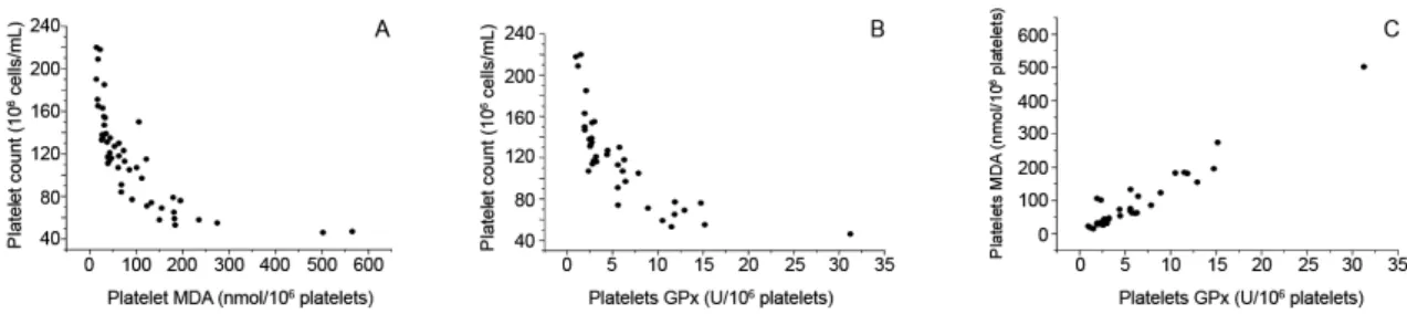
in TCP compared to NTCP patients. There was a negative correlation between platelet count and time of infection, parasitaemia, TB, platelet GPx and platelet MDA level (p > 0.05). MDA concentration (Fig. 1) and GPx activity (Fig. 2) were higher in the platelets of TCP patients. Fig. 3 shows the negative correlations between platelet number and platelet MDA concentrations (r = -0.701; p = < 0.001) (Fig. 3A); platelet number and platelet GPx levels (r = -0.737; p = < 0.001) (Fig. 3B); and platelet GPx activity and plate-let MDA concentration (r = 0.961; p = < 0.001) (Fig. 3C).
DISCUSSION
The aim of this study was to investigate the role of OS and antioxidant status in thrombocytopenia mediated by P. vivax infection. In order to achieve this objective, markers of OS and antioxidant status were analyzed in plasma and platelets of TCP and NTCP patients. Plasma MDA levels were higher in TCP when compared with the NTCP patients. Moreover, the LDL-/nLDL
auto-an-tibody ratio was lower in TCP than in NTCP patients. Also, importantly, GPx and MDA were higher in plate-lets of TCP patients.
Thrombocytopenia has long been observed in human and animal malaria infection (Osim et al. 1991). Several mechanisms have been suggested for this thrombocy-topenia, including disseminated intravascular coagula-tion, immune mechanisms due to absorption of soluble malaria antigen by platelets and subsequent attachment of antibodies to such antigens. Other factors suggested are defective platelet formation and hypersplenism and OS. However, the exact mechanism has not been eluci-dated (Abdalla 1990, Kumar & Shashirekha 2006).
Human plasma protection against free radical injury is offered by a wide spectrum of antioxidants with syn-ergic action; individual measurements of antioxidant concentrations in blood do not always reflect the level of antioxidant status. We showed that the total plasmatic antioxidant capacity was not decreased in TCP patients, despite the low levels of ALB and thiol groups, which demonstrates an adequate capacity of plasma to protect its environment from free radical aggression. As the TOS activity of human plasma is mainly attributable to UA, protein thiol groups, and bilirubin (Halliwell & Gutteridge 1999), the low plasma levels of thiols and the increase in UA concentrations found in plasma of pa-tients with malaria might account for the maintenance of the overall redox network in plasma of these patients. In the present study, although not statistically different, in
Fig 1: malondialdehyde (MDA) levels in platelets of nonthrombocy-topenic (NTCP) and thrombocynonthrombocy-topenic (TCP) malaria vivax patients. The results are expressed in nmol/106 platelets (p < 0.001;
Mann-Whitney rank sum test).
Fig 2: glutathione peroxidase (GPx) in platelets of nonthrombocy-topenic (NTCP) and thrombocynonthrombocy-topenic (TCP) malaria vivax patients. The results are expressed in U/106 platelets (p < 0.001; Mann-Whitney
rank sum test).
the TCP group, total plasma thiols, a parameter of non-oxidation of SH groups, were lower than in the NTCP patients. This could be suggestive of a decrease in anti-oxidant protection in these subjects.
In the present study, we found that plasma MDA levels of TCP were higher than those of NTCP. The in-creased plasma MDA concentration in TCP suggests the role of free radicals in the pathogenesis of this disease. Despite the possible interference of bilirubin in the MDA measurement, no correlation was observed between total plasmatic bilirubin and MDA in our sample (Pearson´s test; r = 0.188; p = 0.211). This means that in the present study bilirubin did not interfere in MDA results.
The electronegative LDL (LDL-) is a
pro-inflamma-tory LDL subfraction that has been related to OS (Da-masceno et al. 2006). In the present study, the auto-anti-bodies reactive to LDL-, expressed as the ratio between
antibodies anti-LDL- and anti-native LDL, were lower in
TCP than in NTCP. However, the amount of anti-native LDL was 2.5 times higher in TCP, which is probably due to the polyclonal B cell activation observed in chronic malaria infection (Donati et al. 2006).
Platelet membranes are less resistant to OS and the membranes of platelets are thinner than those of erythro-cytes. While some erythrocytes are being lysed, the lysis of platelets will also be unavoidable. It is expected that increased OS may lead to increase in platelet lysis. Erel et al. (2001) found that platelet count, platelet superox-ide dismutase (SOD) and GPx activities of patients with vivax malaria were lower and platelet lipid peroxidation levels were higher than normal controls, thus suggesting OS as a possible cause of thrombocytopenia. Ohyashiki et al. (1991) showed that platelet lipid peroxidation increase when rat platelets were exposed to free oxygen radicals, what parallels the decrease of platelet aggregation capac-ity. Sohail et al. (2007) found higher lipid peroxidation levels in P. vivax infected patients than in healthy sub-jects. It is feasible that a significant amount of MDA gen-erated during malaria infection can be due to activation of the immune response (Pabón et al. 2003). The increase in lipid peroxidation is probably due to the production of ROS species by the immune cells and also due to the synchronized release of O
-2 during hemoglobin
degrada-tion by the malarial parasite. It has been shown that in-tact Plasmodium falciparum trophozoite infected human red cells produce H2O2 and OH- radical about twice as
much as the normal erythrocyte (Sohail et al. 2007). The deficiency of selenium may result in an ineffec-tive antioxidant system, e.g., low levels of GPx (Akaike & Maeda 2000). In order to maintain a redox equilib-rium, malaria parasites are equipped with a range of low weight antioxidants, the most prominent being the tripeptide glutathione, as well as with antioxidant enzymes (Becker et al. 2004). In the present work, in-creased GPx was found in the platelets of thrombocy-topenic patients that is in accordance with previously reported data. Pabón et al. (2003) showing increased activities of GPx, SOD and total antioxidant status (CAT) in malaria patients. This suggests that the in-creased GPx levels found here may represent a compen-satory response to increased OS in thrombocytopenic
patients as indicated by their high amount of platelet MDA. We also demonstrated that levels of platelet GPx, as well as platelet MDA, are negatively correlated with platelet number in P. vivax malaria infection. GPx, CAT and SOD are the primary intracellular antioxidant de-fense mechanism against OS. Both GPx and CAT have the ability to inactivate the intracellular H2O2. GPx has been considered the preferential pathway for elimina-tion of low concentraelimina-tions of H2O2 (Jakob & Jandl 1966). Pabón et al (2003) reported similar data showing incre-ment of MDA/GPx ratio (caused by increase of both MDA and GPx) in patients with malaria, suggesting that this may be a consequence of an augmentation of lipid peroxidation (high MDA levels) followed by increased GPx synthesis and activity. The increase of GPx could also be due to the fact that Plasmodium produces GPx in response to ROS species formed during hemoglobin degradation (Ginsburg & Atamna 1997, Gamain et al. 1999). It is clear that MDA/GPx ratios are affected in malaria patients; this supports the concept that a great amount of free radicals are generated during malaria in-fection, which are responsible for changes in the activity of antioxidant enzymes.
Thus, among the variety of mechanisms postulated as the cause of thrombocytopenia in malaria, none have been unequivocally proven. It is possible that several of these factors are responsible acting together. OS has been proposed as an underlying mechanism that contrib-utes to endothelial dysfunction associated with malaria. The clinical significance of this pathogenic pathway re-mains to be substantiated, because no general consensus on the existence of systemic oxidant stress in malaria has yet been attained. Free oxygen radicals may play an important role in structural and functional damages of platelets and in the mechanism of thrombocytopenia.
In conclusion, our results suggest that MDA and GPx are important markers of platelet OS in malaria caused by P. vivax and could be implicated in the mechanisms of malaria-induced thrombocytopenia. Further studies are needed in order to clarify the differences of the as-sociation of OS and thrombocytopenia between patients with P. falciparum and P. vivax malaria.
ACKNOWLEDGEMENTS
To Miss Carolina Marinho da Costa, for her technical as-sistance, to the patients and the personnel of the Fundação de Medicina Tropical do Amazonas and to the group of the Labo-ratório de Bioquímica do Departmento de Análises Clínicas Universidade de São Paulo.
REFERENCES
Abadalla SH 1990. Hematopoiesis in human malaria. Blood Cells 16: 401-416.
Akaike T, Maeda H 2000. Nitric oxide and virus infection. Immunol 100: 300-308.
Becker K, Tilley L, Vennerstrom JL, Roberts D, Rogerson S, Gins-burg H 2004. Oxidative stress in malaria parasite-infected eryth-rocytes: host-parasite interactions. Int J Parasitol34: 163-189.
Damasceno NR, Sevanian A, Apolinário E, Oliveira JM, Fernandes I, Abdalla DSP 2006. Detection of electronegative low density lipo-protein (LDL-) in plasma and atherosclerotic lesions by monoclo-nal antibody-based immunoassays. Clin Biochem 39: 28-38.
Donati D, Mok B, Chêne A, Xu H, Thangarajh M, Glas R, Chen R, Wahlgren M, Bejarano MT 2006. Increased B cell survival and preferential activation of the memory compartment by a malaria polyclonal B cell activator.J Immunol177:3035-3044.
Ellman GL 1959. Tissue sulf hydryl groups. Arch Biochem Biophys 82: 70-77.
Erel O 2004. A novel automated direct measurement method for total antioxidant capacity using a new generation, more stable ABTS radical cation. Clin Biochem37: 277-285.
Erel O 2005. A new automated colorimetric method for measuring total antioxidant status. Clin Biochem38: 1103-1111.
Erel O, Vural H, Aksoy N, Aslan G, Ulukanligil M 2001. Oxidative stress of platelets and thrombocytopenia in patients with vivax malaria. Clin Biochem34: 341-344.
Esterbauer H, Cheeseman KH 1990. Determination of aldehydic lipid peroxidation products: malonaldehyde and 4-hydroxynonenal. Methods Enzymol186: 407-421.
Guba M, Kumar S, Choubey V, Maity P, Bandyopadhyay U 2006. Apoptosis in liver during malaria: role of oxidative stress and im-plication of mi tochondrial pathway. FASEB J20: 1224-1226.
Gutteridge JMC 1995. Lipid peroxidation and antioxidants as bio-markers of tissue damage. Clin Chem41: 1819-1828.
Halliwell B 2006. Phagocyte-derived reactive species: salvation or suicide? Trends Biochem Sci31: 509-515.
Hu ML 1994. Measurement of protein thiol groups and glutathione in plasma. Methods Enzymol233: 380-385.
Hunt NH, Stocker R 1990. Oxidative stress and the redox status of malaria-infected erythrocytes. Blood Cells16: 499-526.
Jacob HS, Jandl JH 1966. Effects of sulphydryl inhibition on red blood cells. II Glutathione in the regulation of the hexoses mono-phosphate pathway. J Biol Chem241: 4243-4250.
Januel C, El Hentati FZ, Carreras M, Arthur JR, Calzada C, Lagarde M, Vericel E 2006. Phospholipid-hydroperoxide glutathione (GPx-4) localization in resting platelets, and compartmental change during platelet activation. Biochim Biophys Acta 1761: 1228-1234.
Kumar A, Shashirekha 2006. Thrombocytopenia - an indicator of acute vivax malaria. Indian J Pathol Microbiol49: 505-508.
Mishra NC, Kabilan L, Sharma A 1994. Oxidative stress and malaria-infected erythrocytes. Indian J Malariol 31: 77-87.
Ohyashiki T, Kobayashi M, Matsui K 1991. Oxygen radical mediated lipid peroxidation and inhibition of ADP-induced platelet aggre-gation. Arch Biochem Biophys288: 282-286.
Oliveira JA, Sevanian A, Rodrigues RJ, Apolinario E, Abdalla DSP 2006. Minimally modified electronegative LDL and its autoanti-bodies in acute and chronic coronary syndromes. Clin Biochem 39: 708-714.
Osim EE, Adegunloye BJ, Emeribe AO 1991. In vivo platelet aggrega-tion in acute malaria. Acta Tropica49: 227-232.
Pabón A, Carmona J, Burgos LC, Blair S 2003. Oxidative stress in patients with non-complicated malaria. Clin Biochem36: 71-78.
Snounou G, Viriyakosol S, Zhu XP, Jarra W, Pinheiro L, Rosario VE, Thaithong S, Brown KN 1993. High sensitivity of detection of human malaria parasites by the use of nested polymerase chain reaction. Mol Biochem Parasitol61: 315-320.
Sobolewscki P, Gramaglia I, Frangos J, Intaglietta M, van der Heyde HC 2005. Trends Parasitol21: 415-422.