ISSN 2221-1896 (PRINT)
www.roavs.com
ISSN 2223-0343 (ONLINE)
Acute toxicity of the methanolic extracts of
Terminalia brownii
bark in rats
Thoria, O. O1, Galal, M. A2, Ashour, N. A2, Hussain, A. M. and Samia H. Abdelrahman
1
Veterinary Research Institute, Soba, P.O. Box 8067 (Elamarat) Khartoum, Sudan; 2National Centre for Research;
3
Department of Pathology, Faculty of Veterinary Medicine University of Khartoum, P.O. Box 32 Khartoum North North, Sudan; 4El Razi College of Medicine and Technology, Khartoum, Sudan.
Abstract
This study was designed to reveal the toxicopathological effects of the methanolic extract of the bark of Terminalia brownie in Swiss albino rats. The methanolic extracts of the bark were given at doses rate of 100, 500 and 1000 mg/kg (group 1, 2 and 3 respectively) body weight to Swiss albino rats. Oral administration of the extracts caused symptoms such as dullness, inappetence and decreased activity in group 2 and 3 rats. The mean values of haemoglobin (Hb) concentration, red blood cell counts (RBC) and packed cell volume (PCV) decreased in the three treated groups. Serum analysis indicated no changes in the activity of the enzyme alanine amino transferase (ALT) or the concentration of urea, creatinine, bilirubin, total protein, and total albumin in the sera of all treated rats during the course of the experiment. However, there was an increase in the activity of the enzyme aspartate amino transferase (AST) in the sera of group 3 and 4 rats. The main lesions found were congestion and hemorrhage in the liver, kidney, lung, stomach and small intestine.
Keywords: Bark of Terminalia brownii, Rats, Toxicity
Introduction
Terminalia brownii (Combertacaea) is a traditional Sudanese medicinal plant known locally as shagarat elsobag. It is widely spread tree throughout the Sudan. Details of its botanical description were given by Elgaszali et al. (1997) and Omer and Elnima (1999). The maceration of the bark of the plant has been used in traditional medicine for the treatment of cough and bronchitis in the west and Souteast Sudan (Elgazali et al., 1997) and for the treatment of diarrhoea and gonorrhoea (Zakaria et al., 2007).
Screening of active principles in the bark revealed the abundance of tannins, saponins, appreciable amounts of flavonoids and traces of other constituents (Omer and ElNima, 1999). Whereas, in vitro studies revealed a high antimicrobial activity residing in the extracts of bark and leaves of the plant against bacterial pathogens (Omer and Elnima, 1999; Zakaria et al., 2007).
The objective of this research was to find the effect of the methanolic extract of the bark of Terminalia brownii in albino rats and to correlate the clinical and
pathological changes with possible changes in the concentration of certain serum constituents of these animals.
Materials and Methods
Twenty four Wister albino rats weighed 150-200 gm were used in this experiment. They were housed in laboratory cages, fed with pellets and fresh vegetables and were watered ad libidum throughout the experimental period. The green bark of the plant was collected, dried in the shade and then coarsely ground using a morter and pestle. About 500 g from the coarsely ground bark was put in soxhlet apparatus (Quick Fex 5183). One liter of 70% methanol was added to the plant and left for 24 hours in the apparatus. The methanolic extract was then filtered. The filtrate was evaporated under vacuum till dryness at 40oC using a rotary evaporators. The obtained solid extract was removed, weight and kept as a stock solution for use.
The rats were allowed one week for adaptation. After the adaptation period, the rats were divided into four groups randomly. One group was kept as control.
The other groups were given the methanolic extract 100, 500 and 1000 mg/kg body weight respectively for 6 days. All the rats were bled before dosing and then after every two days. Serum was analyzed for the activities of the alanine amino transferase (ALT) and aspartate aminotransferase (AST) by the method described by Reitman et al. (1967). The concentrations of total proteins were detected using the methods of Weichselbaum (1946). For total albumin, the methods of Doumas et al. (1971) and Webster et al. (1974) were used. Total bilirubin was estimated by the method described by Jendarassik and Groph (1938). Creatinine levels were measured by the method of Falling and Hausen (1971) and urea by the method of Varley (1967). For histopathological studies, tissues were fixed in 10% formal saline, and 6-µm paraffin sections were stained with haematoxylin and Eosin (H and E).
Statistical analysis
All data were subjected to the statistical package for social sciences (SPSS). Mean was tested using two way analysis of variance (ANOVA) procedure, according to Mead and Gurnow (1983) and then separate using Duncan's multiple range test (DMRT).
Results
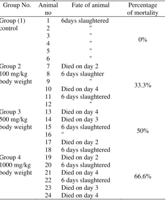
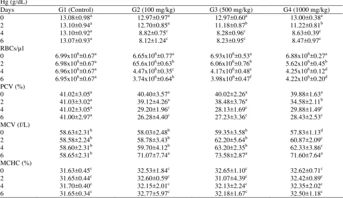
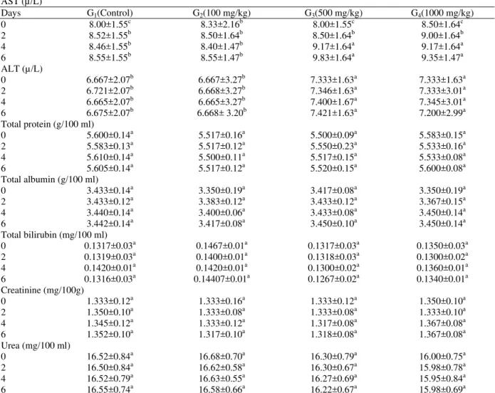
The dosing schedule and time of expiry of rats dosed with Terminalia brownii bark methanolic extract are shown in Table 1. Rats of group 2 showed no clinical signs throughout the course of dosing. Rats of group 3 and 4 showed dullness, inappetence and decreased activity after the 4th dose. The post mortem findings of rats of the four groups are summarized in Table 2. The lesions were similar in all treated rats. However, there were variations in severity of lesions between the different doses used. The predominant lesions were congestion and haemorrhages in the lung, liver, kidney, stomach and small intestines. The mean values of haemoglobin concentration (Hb), RBCs and PCV percentages decreased significantly in the three treated groups. The mean values of MCV increased significantly in all treated groups (P<0.05), while there was no significant changes in the values of MCHC in all group (Table 3). The biochemical changes associated with the bark extract are shown in Table 4. There are no significant changes in the activity of the enzyme ALT in the serum of any rat of all treated groups, but there was a significant increases in the concentration of the enzyme AST in the sera of groups 3 and 4 rats (P<0.05). There were no significant changes in the concentration of total protein, total albumin, urea, creatinine and bilirubin in the sera of all treated rats during the course of the experiment. Histopathological changes were congestion and
intestine. In group 4 rats there are local areas of necrosis and infiltration of mononuclear cells in the portal areas of the liver.
Table 1: Dosing schedule and fate of rates given
methanolic extract of Terminalia brownii bark
for 6 days
Group No. Animal no
Fate of animal Percentage of mortality Group (1) control 1 2 3 4 5 6 6days slaughtered " " " " " 0% Group 2 100 mg/kg body weight 7 8 9 10 11 12
Died on day 2 6 days slaughter
" Died on day 4 6 days slaughtered
" 33.3% Group 3 500 mg/kg body weight 13 14 15 16 17 18
Died on day 4 Died on day 3 6 days slaughtered "
Died on day 2 6 days slaughtered
50% Group 4 1000 mg/kg body weight 19 20 21 22 23 24
Died on day 2 6 days slaughtered Died on day 4 6 days slaughtered Died on day 3 Died on day 4
66.6%
Discussion
In the present study, it was shown that the methanolic extract of the bark of the plant was fatal to many rats of all groups dosed orally with different concentrations of the extract. In the animals congestions and haemorrhages of the liver, lungs, kidneys, stomach and small intestine were the main gross and microscopic lesions observed. This is probably due to the toxic substance present in the plant extract (Adam, 1974; Adam and Magzoub, 1975; Thoria et al., 2001). Similarly, congestion in the internal organs was also reported in experimental animals fed ginger and garlic (Ali and Mohammed, 1986).
The decreased values of RBCs and Hb could be attributed to the liver and renal damage which usually lead to lack of erythropoietin (Erselve, 1995). Similarly, decreased values of RBCs and Hb occurred in intramuscularly treated rats with the methanolic extract of Ambrosia maritima (Fatih-ElRahman, 2003).
Table 2: Necropsy findings in Albino rats dosed with methanolic extract Terminalia brownii bark for 6 days
Site Lesion
Group 1 (Control)
Group 2 (100 mg/kg body
weight)
Group 3 (100 mg/kg body
weight)
Group 4 (100 mg/kg body
weight) Liver Congestion
necrosis fatty changes
- - -
+ - -
++ + -
+++ + - Kidneys Haemorrhages congestion
atrophy of the fat in the renal pelvis
- - -
- + -
+ ++
-
+ +++
- Heart Congestion
Serous atrophy of the cardiac fat
- -
+ -
+ -
+ -
Lungs Pulmonary congestion and haemorrhage Pulmonary oedema
- ++ -
+++ +
+++ +
Stomach Gasterities - + + ++
Small intestine Enterilies - + + ++
+ +++, increasing severity of lesions; (-), Absence of lesions
Table 3: Mean haematological values in blood of rats dosed with methanolic extract Terminalia brownii bark for 6 days
Hg (g/dL)
Days G1 (Control) G2 (100 mg/kg) G3 (500 mg/kg) G4 (1000 mg/kg)
0 13.08±0.98a 12.97±0.97a 12.97±0.60a 13.00±0.38a
2 13.10±0.94a 12.70±0.85a 11.18±0.87b 11.22±0.81b
4 13.10±0.92a 8.82±0.75c 8.28±0.96c 8.63±0.39c
6 13.07±0.93a 8.12±1.24c 8.23±0.95c 8.47±0.97c
RBCs/µl
0 6.99x10⁶±0.67a 6.65x10⁶±0.77a 6.93x10⁶±0.53a 6.88x10⁶±0.27a
2 6.98x10⁶±0.67a 65.6x10⁶±0.63b 6.06x10⁶±0.76b 5.62x10⁶±0.45b
4 6.96x10⁶±0.67a 4.47x10⁶±0.35c 4.17x10⁶±0.48e 4.25x10⁶±0.12d
6 6.95x10⁶±0.67a 3.74x10⁶±0.64h 3.98x10⁶±0.47f 4.22x10⁶±0.20d
PCV (%)
0 41.02±3.05a 40.40±3.57a 40.02±2.26a 39.88±1.63a
2 41.03±3.02a 39.12±4.26a 38.48±3.76a 34.58±2.11b
4 41.02±3.05a 29.20±1.96c 28.13±1.69c 29.88±1.49c
6 41.00±2.97a 26.28±4.40c 27.23±3.36c 28.43±2.53c
MCV (f/L)
0 58.63±2.31b 58.03±2.48b 59.35±3.58b 57.83±1.13d
2 58.58±2.24b 58.78±3.43b 62.20±5.64b 60.87±2.09c
4 58.60±2.31b 59.70±4.12b 63.20±2.35b 62.33±3.86c
6 58.65±2.31b 71.07±7.74a 73.58±2.87a 71.60±7.64a
MCHC (%)
0 31.63±0.45c 32.53±1.84c 32.65±1.10c 32.62±0.71c
2 31.65±0.44c 32.60±0.59c 31.07±4.39c 32.42±0.89c
4 31.70±0.40c 32.15±2.01c 32.13±2.24c 32.35±2.02c
6 31.65±0.34c 32.77±5.97c 32.18±1.67c 32.50±1.18c
Mean values (±SD) having different superscript letters in columns and rows (between groups and days) are significantly different (P<0.05)
of liver injury caused by poisonous plants and by trematode parasites in domestic animals (Gopinath and Ford, 1969).
In our study, there were no significant changes in total protein or albumin concentrations. This may indicate that the ability of the liver to synthesize protein was not affected. It is documented that over 90% of serum protein including albumin, are synthesized by the liver (Mohamoud, 1977). The absence of bilirubinaemia and splenic haemosiderosis in these experimental
animals suggested that the animals did not suffer from haemolytic anaemia (Ford et al., 1968; Adam and Magzoub, 1975). Dosing of the bark extract to albino rats caused no significant changes in the non protein, nitrogen constituents of serum, urea and creatinine, which usually increase when severe glomerular damage occurs (Mahmoud, 1977).
Table 4: Mean concentration of serum constituents in rats given methanolic extract of Terminalia brownii bark for 6 days
AST (µ/L)
Days G1(Control) G2(100 mg/kg) G3(500 mg/kg) G4(1000 mg/kg)
0 8.00±1.55c 8.33±2.16b 8.00±1.55c 8.50±1.64c
2 8.52±1.55b 8.50±1.64b 8.50±1.64b 9.00±1.64b
4 8.46±1.55b 8.40±1.47b 9.17±1.64a 9.17±1.64a
6 8.55±1.55b 8.55±1.47b 9.83±1.64a 9.35±1.47a
ALT (µ/L)
0 6.667±2.07b 6.667±3.27b 7.333±1.63a 7.333±1.63a
2 6.721±2.07b 6.668±3.27b 7.346±1.63a 7.333±3.01a
4 6.665±2.07b 6.665±3.27b 7.400±1.67a 7.345±3.01a
6 6.675±2.07b 6.668± 3.20b 7.421±1.63a 7.200±2.99a
Total protein (g/100 ml)
0 5.600±0.14a 5.517±0.16a 5.500±0.09a 5.583±0.15a
2 5.583±0.13a 5.517±0.12a 5.550±0.23a 5.533±0.16a
4 5.610±0.14a 5.500±0.11a 5.517±0.15a 5.533±0.08a
6 5.605±0.14a 5.517±0.12a 5.520±0.15a 5.600±0.08a
Total albumin (g/100 ml)
0 3.433±0.14a 3.350±0.19a 3.417±0.08a 3.350±0.19a
2 3.433±0.12a 3.383±0.12a 3.433±0.12a 3.367±0.15a
4 3.440±0.14a 3.400±0.06a 3.433±0.08a 3.450±0.14a
6 3.442±0.14a 3.417±0.08a 3.450±0.10a 3.450±0.14a
Total bilirubin (mg/100 ml)
0 0.1317±0.03a 0.1467±0.01a 0.1317±0.03a 0.1350±0.03a
2 0.1319±0.03a 0.1400±0.01a 0.1318±0.03a 0.1300±0.02a
4 0.1420±0.01a 0.1420±0.01a 0.1300±0.02a 0.1360±0.01a
6 0.1316±0.03a 0.14407±0.01a 0.1267±0.02a 0.1340±0.01a
Creatinine (mg/100g)
0 1.333±0.12a 1.333±0.16a 1.333±0.12a 1.350±0.10a
2 1.350±0.10a 1.333±0.08a 1.333±0.08a 1.333±0.10a
4 1.345±0.12a 1.333±0.12a 1.317±0.08a 1.367±0.08a
6 1.352±0.10a 1.317±0.10a 1.318±0.08a 1.367±0.08a
Urea (mg/100 ml)
0 16.52±0.84a 16.68±0.70a 16.30±0.79a 16.00±0.75a
2 16.50±0.84a 16.62±0.58a 16.30±0.67a 15.98±0.78a
4 16.52±0.79a 16.63±0.55a 16.27±0.69a 15.95±0.84a
6 16.55±0.74a 16.58±0.66a 16.22±0.67a 15.98±0.69a
Mean values (±SD) having different superscript in columns and rows (between groups and days) are significantly different (P<0.05)
References
Adam, S.E.I. 1974. Hepatotoxic activity of plant poisons and mycotoxins in domestic animals. Veterinary Bulletin, 44: 767-776.
Adam, S.E.I. and Magzoub, M. 1975. Toxicity of Jatropha curcas for goats. Toxicology, 4: 347-354. Ali, M. and Mohammed, S.Y. 1986. Selective
suppression of platelet, thromboxan formation with sparing of vascular prostacyclin by aqueous extract of garlic in rabbits. Prostaglandin. Lukota Medicine, 25: 139-145.
Doumas, B. and Watson, W. 1971. Clinica Chimica Acta, 31: 87-82.
Elgazali, G.E.B., Tohami, M.S., Elegami, A.B., Abdalla, W. I. and Galal, M.A. 1997. Medicinal plants of the Sudan: Medicinal plants of northern Kordofan. Part IV. Omdurman Islamic University
Fabing, D.L. and Ethinghausen, G. 1971. The kinetic method without deprotenisation (Jaffreaction) for determination of serum creatinine. Clinical Chemistry, 17: 391.
Fatih-Elrahman, A.O. 2003. Toxicopathological effects of Ambrosia martima and Guiera sengalensis in rats. M.Sc. Thesis, University of Khartoum, Sudan. Ford, E.J.H., Richie, H.E. and Thorpe, E. 1968. Serum changes following the feeding of ragmorth (Senecio jacoboea) to calves. Journal of Comparative Pathology, 78: 207-218.
Gopinath, C. and Ford, E.J.H. 1969. The effect of Lantana Camara on the liver of sheep. Journal of Pathology, 99: 75-85.
Mahmoud, O.M. 1977. Toxicity of Calotropis Procera to sheep and goats M.Sc. Thesis, University of Khartoum, Sudan.
Reitman, S., Frankel, A. and Amer, S. 1967. A colormetric method for the determination of serum glutamic oxaloacetic and glutamic pyruvic transaminase. American Journal of Clinical Pathology, 28: 56-63.
Thoria, O.O., Adam, S.E.I., Sahar, M.A.N. and Hussain, A.M. 2001. Toxicity of Jatropha curcas in goats. The Sudan Journal of Veterinary Research, 17:109-115.
Varley, H. 1967. Practical clinical biochemistry, 4th (ed.), William Heinemann Medical Books Ltd., and
Interscience Book Inc. New York. P: 407.
Weichselbaum, T.E. 1946. An accurate and rapid method for the determination of proteins in small amount of blood serum and plasma. American Journal of Clinical Pathology, 16:-40-43.