The role of the miR-200 family in the
epithelial-to-mesenchymal transition (EMT)
Epithelial genes Snail1 Zeb2 Smad2/5 Ajuba Prmt5 E-box E-box SBE 14 -3 -3 β / γ Nucleus TGFβ/
BMP rER CM Crtap CyPB P3H1 Collagen Decorin TGFβ Collagen
miR-200
Epithelial genes Snail1 Snail1 Zeb2 Zeb2 Smad2/5 Smad2/5 Ajuba Ajuba Prmt5 Prmt5 E-box E-box SBE 14 -3 -3 β / γ 14 -3 -3 β / γ Nucleus TGFβ/BMP TGFβ/
BMP rER CM Crtap CyPB P3H1 Crtap Crtap CyPB CyPB P3H1 P3H1 Collagen Decorin TGFβ Collagen
miR-200
Ricardo Perdigão Henriques
Dissertation presented to obtain a Ph.D. degree in
Biochemistry, Molecular Biology at the Instituto de
Tecnologia Química e Biológica, Universidade
Nova de Lisboa
epithelial-to-mesenchymal transition (EMT)
Ricardo Perdigão Henriques
Dissertation presented to obtain a Ph.D. degree in Biochemistry,
Molecular Biology at the Instituto de Tecnologia Química e Biológica,
Universidade Nova de Lisboa
Supervisors:
Prof. Judy Lieberman and Prof. Manuel Carrondo
Instituto de Tecnologia Química e Biológica Universidade Nova de Lisboa
The role of the miR-200 family in the epithelial-to-mesenchymal transition (EMT)
by Ricardo Perdigão Henriques
First edition: August 2015
Copyright number:
Prof. Judy Lieberman’s Laboratory Program in Cellular and Molecular Medicine Boston Children’s Hospital
Harvard Medical School 200 Longwood Ave, WAB 255 Boston MA 02115
http://labs.idi.harvard.edu/lieberman/
ITQB-UNL/IBET Animal Cell Technology Unit
Instituto de Tecnologia Química e Biológica-Universidade Nova de Lisboa/ Instituto de Biologia Experimental e Tecnológica
Av. da República EAN, 2780-157 Oeiras, Portugal Fax: +351 21 442 11 61; Phone: +351 21 446 91 00 http://www.itqb.unl.pt
http://www.ibet.pt
Prof. Dr. Judy Lieberman, Chair in Cellular and Molecular Medicine, Boston Children's Hospital, Boston, USA and Professor of Pediatrics, Harvard Medical School, Boston, MA, USA.
Prof. Dr. Manuel Carrondo, Cathedratic Professor of Chemical and Biochemical Engineering at Faculdade de Ciências e Tecnologia, Universidade Nova de Lisboa, Caparica, Portugal and IBET Executive Director, Oeiras, Portugal.
Jury
Dr. Luís Moita, Principal Investigator and Head of the Innate Immunity and Inflammation laboratory at Instituto Gulbenkian de Ciência, Oeiras, Portugal.
Prof. Dr. Paula Alves, Principal Investigator and Head of the Animal Cell Technology laboratory at ITQB, Oeiras, Portugal and IBET CEO, Oeiras, Portugal.
Dr. Philippe Couttet, Head of the Pathway Integration laboratory at Novartis Pharma AG, Basel, Switzerland.
Foreword
The present thesis dissertation is the result of six years of research at Prof. Judy Lieberman’s laboratory at the Harvard Medical School.
I would like to express my deepest gratitude to everyone that supported me during my doctoral studies. Innumerous people contributed directly or indirectly to the work within this dissertation.
I would like to thank my advisor Prof. Manuel Carrondo for accepting me as his doctoral student and for all his guidance and moral support during all these years. He was critical to my move to the USA, as he offered me the opportunity to work at MIT in a collaboration with Prof. Daniel Wang. This was an enormous step in my career that subsequently opened many opportunities. For this I will always be grateful and will never forget his support.
I would also like to thank my advisor Prof. Judy Lieberman for accepting me as her student in her world-renowned research group, for her mentorship and financial support over many years. Her support and guidance allowed me to pursue interesting research questions with great scientific rigor. I thank her for always being willing to take time out of her busy schedule to meet with me and offer her scientific insights.
I also want to thank Prof. Paula Alves for always supporting my career and offering me her guidance from the time I was an intern student at her laboratory. She has been a great mentor and friend.
I am also grateful to all Lieberman lab members for years of advice, support and friendship. Specially, I would like to thank Dr. Fabio Petrocca, who taught me all basic molecular biology techniques and nurtured my scientific interests. His patience and knowledge allowed me to rapidly transition from the bioengineering field to the molecular oncology field. I will always remember his support during difficult times, and it was an honor to work with him in this project. Thank you for being a remarkable scientist and person!
I also thank Prof. Guilherme Ferreira (UALG) and Prof. João Crespo (UNL) for accepting to be part of my thesis committee.
A special thanks to my thesis defense jury members, Dr. Luís Moita, Prof. Paula Alves, Dr. Philippe Couttet and Dr. Sónia Melo for taking the time to evaluate my work.
I would also like to thank current and previous members of the Lieberman laboratory Radiana Trifonova, Zhan Xu, Michael Walch, Ashish Lal, Nan Yan, Anders Wittrup, Adi Gilboa-Geoffen, Elizabeth O'Day, Gregory Idos, Emre Basar, Jerome Thiery, Danielle Rajani, Dennis Martinvalet, Laura Maliszewski, Dipanjan Chowdhury, Farokh Dotiwala, Changying Guo, Sachin Mulkin, Brooke Bollman and Xing Liu for all their support and friendship during my six year stay in the lab.
I also thank my friends at MIT Raga Markely, David McClain, Sohan Patel and Andrés Abin for their support during my work there.
I also express my deepest gratitude to my friends in Boston that helped me survive the cold and long New England winters, Pedro Caetano, Jonathan Soto, Elvira Rocha, Francisco Queiró, Jorge Fradinho, Pedro Pinto, Marcus Dahlem, Priyadarshi Panda, Sigurd Østrem and Luca Pinto.
I would also like to thank current and previous members of the Animal Cell Technology laboratory that have supported me since I met them in 2006: António Roldão, Marcos Sousa, Tiago Ferreira, Margarida Serra, Nuno Carinhas, Ana Teixeira, Ana Coroadinha, Catarina Brito, Cristina Peixoto, Carina Silva and Pedro Cruz.
I also thank my longtime friends in Portugal, that even at a great distance always gave me their support and never forgot me: Bruno Vilares, Dr. Adelino Cunha, João Carreiro, Hugo Botelho and Rita Vaz.
I also thank the Portuguese Ministry of Science and Technology (FCT) for my Ph.D. fellowship SFRH/BD/37188/2007.
Um sentido agradecimento ao meu grande amigo Bruno Vilares, foi crítico o teu apoio durantes todos estes anos de intensa amizade!
Um especial agradecimento para ti Adiari, companheira de vida e de luta há 8 anos! O teu Amor fez-me Sonhar mais Alto e alcançar muito Mais... Obrigado por tudo!
Agradeço também ao Quim Perdigão, Tina, Juka, Teresa Sampaio e Família Vázquez Rodríguez todo o apoio que não esquecerei!
Perdigão-Henriques R, Petrocca F, Altschuler G, Thomas MP, Le MT, Tan SM, Hide W, Lieberman J. miR-200 promotes the mesenchymal to epithelial transition by suppressing multiple members of the Zeb2 and Snail1 transcriptional repressor complexes. Oncogene. Epub Mar 23, 2015.
Le MT, Hamar P, Guo C, Basar E, Perdigão-Henriques R, Balaj L, Lieberman J. miR-200-containing extracellular vesicles promote breast cancer cell metastasis. J Clin Invest. 2014; 124(12):5109-28.
Publications in conference proceedings
Perdigão-Henriques R, et al. (2009, 2010, 2011, 2012 and 2013). The role of miR-200 in breast cancer metastasis, Annual Harvard Medical School-Immune Disease Institute Retreat, Boston, USA.
Perdigão-Henriques R, et al. (2011). The role of miR-200 in breast cancer metastasis, 6th Microsymposium on small RNAs, Vienna, Austria.
Dissertation Abstract
Over the last decade, microRNAs (miRNAs) have earned a lot of attention due to their critical roles in several biological processes and human diseases. However, progress in this field has been limited by the difficulty of discovering miRNA target genes, as each miRNA can potentially bind to hundreds of different mRNAs. Therefore, the main focus of this thesis was to develop an integrated framework that could be used to discover novel genes and pathways regulated by a given miRNA. We applied this framework to study the miR-200 family, which was previously shown to be involved in embryonic development and tumor metastasis but only a few target genes were known.
In Chapter 1, miRNA biogenesis and action mechanism are described. miRNA target prediction tools, both computational and experimental, are also reviewed. Also, a basic overview of the epithelial to mesenchymal transition (EMT) and TGF-β signaling is given as both are regulated by miR-200. We also provide an overview of the miR-200 family and a detailed list of published miR-200 target genes.
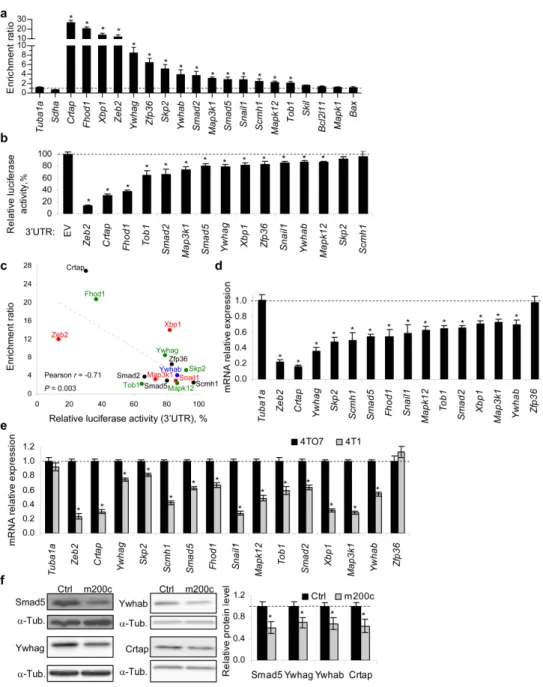
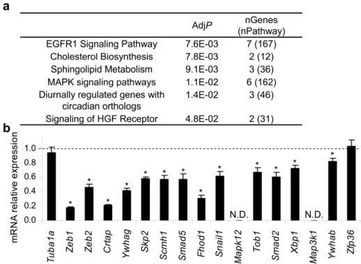
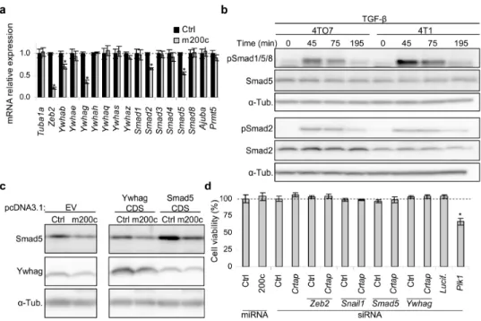
beta (TGF-β) signaling pathway, among other pathways. Accordingly, we experimentally showed that miR-200c counteracts the suppressive effects of TGF-β and bone morphogenetic protein-2 (BMP-2) on epithelial gene expression. Additionally, we showed that miR-200c regulates the 3’-untranslated regions (3’UTRs) of 12 (Crtap, Fhod1, Smad2, Map3k1, Tob1, Ywhag/14-3-3γ, Ywhab/14-3-3β, Smad5, Zfp36, Xbp1, Mapk12, Snail1) of 14 putative miR-200c-binding mRNAs tested. These 12 genes were experimentally validated as miR-200c targets by identifying their 3’UTR miR-200c recognition elements. We also experimentally showed that for these 12 genes the extent of mRNA binding to miR-200c strongly correlated with gene suppression.
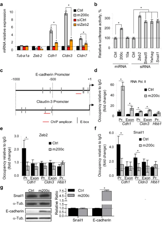
Smad2 and Smad5 form a complex with Zeb2 and Ywhab/14-3-3β and Ywhag/14-3-3γ form a complex with Snail1. These complexes that repress transcription assemble on epithelial gene promoters. We showed that miR-200c over-expression increases RNA polymerase II binding to epithelial gene promoters, while reducing binding of Zeb2 and Snail1 complexes to these promoters. Expression of miR-200c-resistant Smad5 modestly, but significantly, reduced epithelial gene induction by miR-200c.
miR-200 expression or Zeb2 knockdown are known to inhibit cell invasion in in vitro assays. Knockdown of each of 3 novel miR-200 target genes identified here, Smad5, Ywhag and Crtap, also profoundly suppressed cell
invasion. Thus miR-200 suppresses TGF-β/BMP signaling, promotes epithelial gene expression and suppresses cell invasion by regulating a network of genes.
Durante a última década os microRNAs (miRNAs) têm recebido muita atenção devido ao seu relevante papel em vários processos biológicos e em várias doenças humanas. Contudo, o progresso nesta área do conhecimento tem sido limitado pela grande dificuldade em descobrir os genes que são regulados por cada miRNA, já que cada miRNA pode potencialmente ligar-se a centenas de mRNAs diferentes. Por isso, o principal objectivo desta tese consistiu em desenvolver um framework
integrado que pudesse ser usado para descobrir os genes e vias de sinalização que são regulados por um dado miRNA. Este framework foi aplicado ao estudo da família miR-200, a qual já se sabia que participava no desenvolvimento embrionário e na formação de metástases, mas para a qual apenas alguns mRNAs alvo eram conhecidos.
No Capítulo 1, a biogénese dos miRNAs e o seu mecanismo de acção são abordados. As ferramentas computacionais e experimentais que existem para descobrir que genes são regulados pelos miRNAs são também descritas. Além disso, a transição epitelial-mesenquimal (EMT) e a via de sinalização TGF-β são também revistos pois ambos estes processos são regulados pela família miR-200. Também é feita uma revisão da família miR-200, incluindo uma lista detalhada dos genes que foram publicados como sendo regulados por esta família.
complementares à sequência do miR-200c e para a grande maioria deles a sua expressão é negativamente regulada pelo miR-200c. Além disso, mostrámos que este framework permite descobrir novos mRNAs regulados pelo miR-200c com um reduzido número de falsos positivos. Os nossos resultados mostram que o miR-200c principalmente regula 13 vias de sinalização celular. As duas vias de sinalização mais significativamente reguladas pelo miR-200c são a do receptor do factor de crescimento epidérmico (EGFR) e a da MAP cinase (MAPK). Adicionalmente, os resultados mostram que o miR-200c também regula a via de sinalização do factor beta de transformação do crescimento (TGF-β), entre outras vias. De acordo com estes resultados, mostrámos experimentalmente que o miR-200c contraria o efeito suppressor que o TGF-β e a proteína morfogenética do osso tipo 2 (BMP-2) têm na expressão de genes epiteliais. Adicionalmente, mostrámos que o miR-200c regula a região não traduzida da extremidade 3’ (3’UTR) de 12 (Crtap, Fhod1, Smad2, Map3k1, Tob1, Ywhag/14-3-3γ, Ywhab/14-3-3β, Smad5, Zfp36, Xbp1, Mapk12,
Snail1) de 14 mRNAs que foram escolhidos para serem validados como sendo regulados pelo miR-200c. Esta validação foi feita através da identificação precisa dos elementos que são reconhecidos pelo miR-200c no 3’UTR de cada um destes 12 mRNAs. Também mostrámos experimentalmente que o grau de interacção entre cada um destes 12 mRNAs e o miR-200c está fortemente correlacionado com o grau que cada um dos mRNAs é reprimido.
modestamente, a indução de genes epiteliais que se verifica quando o miR-200c é sobre-expressado.
É sabido que a expressão da família miR-200 ou a supressão de Zeb2
inibem a invasão celular em ensaios in vitro. Supressão de cada um dos três mRNAs que nós indentificámos como sendo regulados pelo miR-200c,
Smad5, Ywhag and Crtap, também inibiram fortemente a invasão celular. Em conclusão, mostrámos experimentalmente que a família miR-200 suprime a via de sinalização do TGF-β/BMP, promove a expressão de genes epiteliais e inibe a invasão celular através da regulação de uma rede de genes.
No Capítulo 3, são discutidas as conclusões principais e as suas limitações assim como possíveis futuras linhas de investigação.
Table of Contents
CHAPTER 1:Introduction
1. microRNAs ...2
1.1. Function ...2
1.2. Biogenesis and action mechanism...2
1.3. Computational prediction of miRNA targets ...6
1.3.2. Experimental identification of miRNA targets ...8
1.3.2.1. Reporter assays ...8
1.3.2.2. mRNA and protein expression profiling ...10
1.3.2.3. Immunoprecipitation of miRISC components...11
1.3.2.4. Pull-down of biotinylated miRNAs ...16
2. Epithelial to mesenchymal transition...17
2.1. TGF-β superfamily signaling ...20
3. miR-200 family ...22
3.1. Published miR-200 family target genes ...24
3.2. miR-200 family role in EMT and metastasis...29
4. Thesis Scope ...30
5. References ...32
CHAPTER 2:The role of the miR-200 family in EMT 1. Introduction...51
2. Materials and methods ...54
3. Results...66
3.1. miR-200c pull-down ... 66
3.2. miR-200 bound genes are enriched for genes involved in EGFR, MAPK and TGF-β signaling and metabolic pathways implicated in metastasis and oncogenic transformation ... 69
3.3. miR-200-bound genes form a dense interaction network ... 70
3.4. Experimental validation of the predictions of the Bi-miR-200c pull-down ... 72
3.5. miR-200 targets Snail1 and Zeb2 transcription complexes ... 77
3.8. The miR-200c target Crtap promotes cell invasion ... 86
3.9. CRTAP and miR-200 expression are inversely correlated in NCI-60 tumors and primary human breast cancers ... 87
4. Discussion... 89
5. Note added in proof... 95
6. Conflict of Interest ... 97
7. Acknowledgements ... 97
8. Supplemental figures ... 98
10. Supplemental tables... 102
9. References ... 135
CHAPTER 3:Discussion and conclusions 1. Discussion... 149
1.1. Bi-miR-200c pull-down false-positive rate... 149
1.2. miR-200c-regulated pathways ... 150
1.3. miR-200c effect on Snail1 protein ... 151
1.4. Regulation of E-cadherin expression by Ywhag and Smad5 ... 152
2. Conclusions ... 153
3. Future work ... 154
Abbreviations
Abbreviation Full form
4SU 4-Thiouridine
Ago Argonaute protein
Ago2 Eukaryotic translation initiation factor 2C, 2, Argonaute 2
BMP Bone morphogenetic protein
bps Base pairs
BSA Bovine serum albumin
cDNA Complimentary DNA
CDS Coding sequence
ChIP chromatin-immunoprecipitation
co-IP Co-immunoprecipitation
DDX6 DEAD (Asp-Glu-Ala-Asp) box helicase 6
DMEM Delbecco's modified essential medium
DNA Deoxyribonucleic acid
ECM Extracellular matrix
EDTA Ethylenediaminetetraacetic acid
EGF Epidermal growth factor
EGFR Epidermal growth factor receptor
EMT Epithelial to mesenchymal transition
ERK Extracellular regulated kinase
GTP Guanosine 5′-triphosphate
HGF Hepatocyte growth factor
IP Immunoprecipitation
JNK c-Jun N-terminal kinase
MAPK Mitogen activated protein kinase
MET Mesenchymal to epithelial transition
MIS Mullerian inhibitory substance
MRE miRNA-recognition element
miRISC miRNA-induced silencing complex
miR/miRNA microRNA
PD Pull-down
PDGF Platelet-derived growth factor
PI3K Phosphoinositide 3-kinase
pri-miRNA Primary miRNA
qRT-PCR Quantitative real-time PCR
RhoB Ras homolog gene family, member B
RNA Ribonucleic acid
RNAse Ribonuclease
shRNA Short-hairpin RNA
siRNA Small-interfering RNA
S1P Sphingosine-1-phosphate
Smad Small mothers against decapentaplegic
Smurf SMAD-ubiquitination-regulatory factor
snoRNA Small-nucleolar RNA
TALEN Transcription activator-like effector nucleases
TGF-β Transforming growth factor-beta
tRNA Transfer RNA
UTR Untranslated region
Zeb1 Zinc finger E-box binding homeobox 1
List of Figures
Figure 1. Mechanisms that regulate miRNA biogenesis and RISC loading...3
Figure 2. Methods for identifying miRNA targets. ...9
Figure 3. Illustration of PAR-CLIP.. ...14
Figure 4. Schematic representation of the iCLIP protocol. ...15
Figure 5. Overview of the pulldown method. ...17
Figure 6. Epithelial to mesenchymal transition (EMT) ...18
Figure 7. EMT contribution to cancer progression...20
Figure 8. General mechanism of TGF-β superfamily signaling and Smad activation. ...21
Figure 9. miR-200 family. ...23
Figure 10. miR-200c targets several genes regulating numerous processes involved in cancer development and progression. ...29
Figure 11. Bi-miR-200c PD identifies 520 putative miR-200c target genes...67
Figure 12. Interactome of directly-interacting pulled-down genes. ...71
Figure 13. Experimental validation of miR-200c target genes identified by the PD. ...73
Figure 14. Identification of miR-200c microRNA recognition elements (MREs) in the 3’UTRs of 12 new target genes...76
Figure 15. miR-200 induces epithelial gene expression by repressing Zeb2 and Snail1 complexes ...78
Figure 16. Snail1, Smad5, Ywhag and Crtap are physiologically important miR-200c targets. ...83
Figure 17. Schematic model of how miR-200c induces epithelial gene expression . ...88
Figure S1. ...98
Figure S2. ...99
Figure S3. ...100
Table 1. Characteristic features of algorithms providing information beyond
classical miRNA target gene interaction ... 7
Table 2. EMT-promoting Smad complexes and their target genes... 22
Table 3. Published miR-200 target genes. ... 24
Table S1. Published miR-200 target genes and their overlap with the PD gene list.. ... 102
Table S2. Set of genes enriched in the Bi-miR-200c PD with an enrichment ratio R>2 and P<0.01 ... 107
Table S3. Bi-miR-200c PD genes (R>2, P<0.01) in each of the most enriched canonical pathways (Wikipathways) (AdjP<0.01) ... 124
Table S4. Bi-miR-200c PD genes in each of the most enriched canonical pathways. .... 127
Table S5. Predicted miR-200c MREs in the 3’UTRs of target genes ... 128
Chapter 1
CONTENTS
1. microRNAs ...2
1.1. Function ... 2 1.2. Biogenesis and action mechanism ... 2 1.3. Computational prediction of miRNA targets ... 6 1.3.2. Experimental identification of miRNA targets ... 8
1.3.2.1. Reporter assays... 8
1.3.2.2. mRNA and protein expression profiling ... 10
1.3.2.3. Immunoprecipitation of miRISC components ... 11
1.3.2.4. Pull-down of biotinylated miRNAs... 16
2. Epithelial to mesenchymal transition ...17
2.1. TGF-β superfamily signaling ... 20 3. miR-200 family ...22
3.1. Published miR-200 family target genes... 24 3.2. miR-200 family role in EMT and metastasis ... 29 4. Thesis Scope...30
1. microRNAs
1.1. Function
microRNAs (miRNAs) are short (~22-nucleotide (nt)-long), noncoding single-stranded RNA molecules that regulate gene expression by binding to partially complementary sequences in target mRNAs to induce mRNA decay, translational repression or both1. Each miRNA can potentially bind to hundreds of mRNAs, sometimes having a strong effect but more often having a weak fine-tuning effect. miRNAs participate in a variety of biological processes such as development, differentiation, proliferation, antiviral defense, metabolism and cellular stress signaling1-2. At least 60% of human protein-coding genes are regulated by miRNAs (mostly downregulated). Dysregulation of miRNA expression levels has been associated with several pathologies such as cancer, neurodevelopmental, autoimmune, liver, muscle, skin and cardiovascular diseases3-4. Also, mutations in miRNA genes have been shown to cause genetic diseases such as nonsyndromic autosomal dominant progressive hearing loss which is induced by point mutations in the seed sequence of miR-96. Likewise, mutations in miR-196a-2 elevate the risk of developing lung, breast, liver and gastric cancer.
1.2. Biogenesis and action mechanism
Figure 1. Mechanisms that regulate miRNA biogenesis and RISC loading. (A) Biogenesis of a miRNA is a stepwise process involving (i) transcription of a primary transcript (pri-miRNA), (ii) processing (“cropping”) by the Drosha/DGCR8 microprocessor complex to produce a pre-miRNA, (iii) export from the nucleus into the cytoplasm, mediated by the exportin XPO5 and the guanosine 5′-triphosphate (GTP)–binding protein Ran, and (iv) additional processing (“dicing”) by a complex containing the RNase Dicer, a catalytic component, Argonaute (Ago), and the RNA-binding protein TRBP (transactivation response RNA binding protein) to produce the mature miRNA. The resulting complex of Ago and the mature, single-stranded miRNA is called the RISC. The miRNA binds complementary sequences in mRNAs; Ago either inhibits the translation of the mRNA or cleaves it to cause its degradation. Figure and legend reproduced from Hata A, Lieberman J. Sci Signal 20156.
transcribed from individual, non-protein coding genes which have their own promoters8.
These pri-miRNA transcripts contain one or more stem-loop structures (also called hairpin structures) in which miRNAs are embedded2. Still in the nucleus, these stem-loop structures are recognized by the double-stranded RNA-binding protein DGCR8 (DiGeorge syndrome critical region 8), also known as Pasha in invertebrates. DGCR8 associates with Drosha, a ribonuclease (RNase) III family enzyme, and together they form the microprocessor complex. Drosha then cleaves the pri-miRNA into a precursor miRNA (pre-miRNA). The pre-miRNA (~70-nt long), is then transported to the cytoplasm by a nuclear complex formed by Exportin-5 together with the guanosine 5′-triphosphate (GTP)–binding protein Ran. Once in the cytoplasm, the pre-miRNA is cleaved by Dicer (another RNase III enzyme) together with the RNA-binding protein TRBP (transactivation-response RNA-binding protein) to generate an RNA duplex. This duplex consists of the mature miRNA (depicted in red in Figure 1), and the miRNA star (miRNA*, RNA strand partially complementary to the mature miRNA). While the mature miRNA is loaded into an Argonaute (Ago) protein, the miRNA* is usually released and degraded. Ago proteins also bind to members of the GW182 protein family (in humans there are three GW182 paralogs known as TNRC6A-C) and this complex bound to a miRNA forms the miRNA-induced silencing complex (miRISC)6. Ago proteins are the core components of the miRISC complex as they bind to the miRNA and mediate recognition of its target mRNAs.
1.3. Computational prediction of miRNA targets
suggesting that as many as 25% of functional MREs are in mRNA coding regions23.
Table 1. Characteristic features of algorithms providing information beyond classical miRNA target gene interaction. Table reproduced from Tarang S, Weston MD RNA Biology 201428.
Prediction
method Characteristic features Resource
Diana Micro-T Target prediction made with miRNA or mRNA sequences as input diana.cslab.ece.ntua.gr/ microT/ FAME Experimentally verified miRNA pathways infer biological process affected by miRNAs acgt.cs.tau.ac.il/ fame/ Hoctar Information on host genes regulating expression of its embedded microRNA hoctar.tigem.it Magia Query of miRNA target prediction, analysis of expression profiles, and post-transcription regulatory network gencomp.bio.unipd.it/ magia/start/
MaMi
miRNA target prediction based on hybridization energies and secondary structures for the miRNA-mRNA hybrid where
parameters can be modified by the user
mami.med.harvard.edu
Microinspector
Identification of potential miRNA binding sites in user-submitted sequences, searching against databases of known
miRNA binding sites
bioinfo.uni-plovdiv.bg/ microinspector/ miR2disease Information on miRNA association to a disease process www.mir2disease.org
miRBase Complete repository of miRNA sequences and targets www.mirbase.org mirDIP mirDIP integrates 12 microRNA prediction data sets from six microRNA prediction databases ophid.utoronto.ca/ mirDIP/index.jsp miRecords
Two main modules, experimentally validated targets, and integrated information across 11 independent prediction
softwares
mirecords.umn.edu/ miRecords/index.php
miRGator Integrates miRNA expression data with mRNA and protein to interpret the biological functions of miRNAs genome.ewha.ac.kr/ miRGator/miRGator.html miRNAmap Information on statistics of miRNA sequences and target genes mirnamap.mbc.nctu.edu.tw
MirPath Identification of altered molecular pathways by the expression of specific miRNAs 83.212.96.7/DianaTools New/index.php?r=mirpath miRTar pairs and on miRNA sites on the alternative spliced transcriptsInformation on the biological function of miRNA-target gene mirtar.mbc.nctu.edu.tw/ human/ MiRTarBase
Information on experimentally verified miRNA targets by data mining and manually surveying pertinent literature related to
functional studies on miRNAs
mirtarbase.mbc.nctu.edu.tw
miRWalk
miRNA-target information on the complete sequence (promoter, 5′-UTR, CDS, and 3′-UTR) and target interaction
information across eight other types of prediction software; information on experimentally validated targets
www.umm.uni-heidelberg.de/
apps/zmf/mirwalk/index.html Patrocles Polymorphisms on miRNA sequences and target genes www.patrocles.org
PITA Secondary structure of the miRNA-mRNA hybrid for target gene prediction genie.weizmann.ac.il/ pubs/mir07/index.html Tarbase Experimentally validated miRNA targets diana.cslab.ece.ntua.gr/
tarbase/
Also, computational analyses need to take into account cell-specific mRNA isoforms. In biological processes such as proliferation29, differentiation30 and cancer31 some mRNAs are alternatively polyadenylated and generate transcripts with unique 3′UTRs, which may have different MREs. Therefore, high-throughput mRNA sequencing of the biological system of interest combined with computational prediction of miRNA targets can more accurately identify functional MREs32.
1.3.2. Experimental identification of miRNA targets
Experimental identification and validation of miRNA targets is critical to identify miRNA biological functions accurately. This is the most challenging and important issue in this field as miRNA functions are determined by their target genes. During the past decade several strategies were developed to improve experimental miRNA target identification but still relatively few targets have been thoroughly experimentally validated. One of the reasons is that miRNA targeting is often context-, time-, or tissue-dependent and therefore some mRNAs might only be targeted in some contexts even though they have extensive sequence complementarity with a given miRNA. Some experimental approaches used to identify miRNA targets will be briefly described below (Figure 2).
1.3.2.1. Reporter assays
luciferase activity level is used to determine the extent of miRNA target repression after cells are transfected with both luciferase reporter genes together with the miRNA of interest.
Figure 2. Methods for identifying miRNA targets. Putative target genes can be identified by expression profiling of cells in which the miRNA is overexpressed or antagonized, by biochemical isolation of the miRISC or by target prediction algorithms. These methods generally identify hundreds of candidate genes or more. Bioinformatic analysis of these large candidate gene lists for over-represented Gene Ontology (GO) terms, enriched biological pathways or gene interaction networks can then help researchers to select candidate genes to evaluate experimentally. Figure and legend reproduced from Thomas M, Lieberman J, Lal A. Nat Struct Mol Biol 20101.
used and the inherent limitations of reporter assays might result in artifacts. Also, this method is low-throughput, expensive and labor intensive and therefore not appropriate to identify miRNA targets globally.
1.3.2.2. mRNA and protein expression profiling
One common approach to identify genome-wide miRNA targets is to measure global mRNA/protein levels after overexpressing or inhibiting a given miRNA, as its targets’ mRNA/protein often change (Figure 2). However, measuring global mRNA/protein levels after manipulating a given miRNA doesn’t allow to distinguish between direct and indirect targets as changes at the mRNA or protein level may be due to downstream effects instead of direct miRNA targeting.
Global mRNA levels can be measured by microarray or more recent high-throughput mRNA sequencing. To measure global miRNA effects at the protein level, proteins can be labeled using a stable isotope (stable-isotope labeling with amino acids in cell culture, SILAC) followed by mass spectrometric analysis34. However, this approach is time-consuming, expensive and only a fraction of the proteome can be resolved at a time. Therefore, microarray analysis is often used instead, as mRNA and protein levels are in most cases correlated.
miRNA genes and totally abrogate miRNA expression35. Second, antagomirs, which are chemically modified single-stranded RNA oligonucleotides complementary to target miRNAs, are able to bind and prevent miRNAs from interacting with their targets, thereby upregulating their mRNA levels36-37. In addition to sequester miRNAs, antagomirs can instead bind to a specific target mRNA sequence and prevent miRNAs from binding to it, which also upregulates target mRNA levels38. Lastly, miRNAs can be inhibited by using miRNA sponges. miRNA sponges are transcripts that contain multiple, tandem binding sites complementary to a miRNA of interest39. When plasmid vectors encoding miRNA sponges are transfected into cells, sponges can derepress miRNA target mRNAs, in some cases as strongly as antagomirs. It is important to keep in mind that all these miRNA manipulations have their caveats. Transfecting excessive amounts of synthetic miRNA duplexes can be toxic to cells or may introduce off-target effects40-41. Also, miRNA overexpression above physiological levels may lead to artifacts, as excess miRNA can saturate all available miRISC in the cell and affect the endogenous miRNA pathway40. On the other hand, inhibition of a highly expressed miRNA might be difficult to achieve with antagomirs and miRNA sponges, and might only be achieved with more laborious knockout technology. Also, inhibition of a poorly expressed miRNA might not have a detectable effect on its target genes.
1.3.2.3. Immunoprecipitation of miRISC components
perform immunoprecipitation (IP) of endogenous miRISC components42 others overexpress epitope-tagged miRISC components inside cells before immunoprecipitating43,45-47. All these studies have high false-positive rates (~40-70%23) for several reasons. First, target mRNAs identified by immunoprecipitating a specific Ago family member might not be identical to those bound by other Ago family members48 (in humans there are four Ago-family members (Ago1-4)49). Second, endogenous and epitope-tagged Ago proteins bind to transfer RNAs50 and overexpression of Ago proteins has been shown to increase global miRNA production,51-52 which might result in artifacts. Additionally, as Ago proteins can still bind to mRNAs in vitro in cell extracts after cell lysis46 some of the mRNAs isolated through an Ago IP might not be targets in vivo. Moreover, Ago IPs only allow identification of putative target mRNAs without providing any information on the precise location of functional MREs in those mRNAs. Also, Ago IPs are usually not completely specific for the miRNA of interest, even after overexpression of the miRNA, as there are always other endogenous miRNAs still bound to miRISC complexes that may skew results. Finally, Ago IPs require miRNAs to be strongly bound to their target mRNAs and therefore this assay has low sensitivity for weak or transient miRNA/mRNA interactions.
of short-wavelength UV light has low crosslinking efficiency, can produce non-specific crosslinks and may photo-damage RNAs.
Figure 3. Illustration of PAR-CLIP. 4SU-labeled transcripts were crosslinked to RBPs and partially RNase-digested RNA-protein complexes were immunopurified and size-fractionated. RNA molecules were recovered and converted into a cDNA library and deep sequenced. Figure and legend reproduced from Hafner M, Landthaler M, Burger L, Khorshid M, Hausser J, Berninger P et al. Cell 201053.
Figure 4. Schematic representation of the iCLIP protocol. After UV irradiation, the covalently linked RNA is co-immunoprecipitated with the RNA-binding protein (RBP) and ligated to an RNA adapter at the 3′ end. Proteinase K digestion leaves a covalently bound polypeptide fragment on the RNA that causes premature truncation of reverse transcription (RT) at the cross-link site. The red bar indicates the last nucleotide added during reverse transcription. Resulting cDNA molecules are circularized, linearized, PCR-amplified and subjected to high-throughput sequencing. The first nucleotides of each sequence contain the barcode followed by the nucleotide where cDNAs truncated during reverse transcription. Figure and legend reproduced from Sugimoto Y, Konig J, Hussain S, Zupan B, Curk T, Frye M et al. Genome Biol 201254.
Ago/RNA interactions can result in false-positives. Also, these CLIP methods are technically-challenging multistep protocols.
1.3.2.4. Pull-down of biotinylated miRNAs
To address these problems, recent studies55-60 have used a much simpler approach that avoids cross-linking that consists on transfecting cells with a synthetic, biotinylated version of the miRNA of interest followed by streptavidin pull-down (PD) of its target RNAs (Figure 5). The synthetic miRNA mimics (RNA duplexes consisting of the mature miRNA of interest and its partially complementary passenger strand) have a biotin molecule covalently attached to the 3′ end of the mature miRNA strand (also called antisense strand). The pulled-down RNA can then be identified by microarray or high-throughput RNA sequencing. Putative miRNA targets can then be easily identified by comparing the enrichment level of each identified RNA in the PD performed with the miRNA of interest versus a control PD performed with an unrelated miRNA without any predicted targets in the system of interest (for example, the unrelated C. elegans
Figure 5. Overview of the pulldown method. 3′-Biotinylated miRNA mimics are transfected into cells and cytoplasmic extracts are prepared after 24h. The Biotin–miRNA–mRNA complexes are isolated from cytoplasmic extracts using streptavidin beads. RNA isolated from the pulldown material is used to determine the enrichment of known targets of the miRNA. Microarray analysis from the pulldown RNA can be used to identify the genome-wide mRNAs bound by the transfected Biotin-miRNA. Figure and legend reproduced from Subramanian M, Li XL, Hara T, Lal A. Methods Mol Biol 201562.
2. Epithelial to mesenchymal transition
Epithelial to mesenchymal transition is a complex process that converts polarized epithelial cells into migratory and invasive mesenchymal cells63 (Figure 6). EMT is a normal process during embryo development and is required for the formation of the neural crest64, fusion of the hard palate65, and other developmental processes. However, in tumorigenesis EMT is a pathological event that allows carcinomas to metastasize66-68.
comprise activation of several transcription factors, expression of specific cell surface proteins, expression and reorganization of cytoskeletal proteins, production of metalloproteinases that degrade extracellular matrix (ECM) components, as well as changes in miRNA expression profiles63.
Figure 6. Epithelial to mesenchymal transition (EMT). An EMT involves a functional transition of polarized epithelial cells into mobile and ECM component-secreting mesenchymal cells. The epithelial and mesenchymal cell markers commonly used by EMT researchers are listed. ZO-1, zona occludens 1; MUC1, mucin 1, cell surface associated; miR200, microRNA 200; SIP1, survival of motor neuron protein interacting protein 1; FOXC2, forkhead box C2. Figure and legend reproduced from Kalluri R, Weinberg RA. J Clin Invest 200963.
brain70. Metastasis is a complex, multistep process that starts when epithelial cells in the primary tumor undergo EMT and acquire a mesenchymal phenotype. These cells are then able to detach from their neighboring cells, such as nearby stromal cells, and invade through the basement membrane (Figure 7). Subsequently, metastasizing cells enter the bloodstream or the lymphatic system (intravasate) and then exit (extravasate) at distant organs forming micrometastases. The reversal of the EMT process, mesenchymal to epithelial transition (MET), is also an important event in cancer progression, as it has been hypothesized that MET is required for the final step of metastasis formation, the colonization of the metastatic site by proliferation of the established micrometastases 71-72
. Although great advances have been made in cancer treatment, particularly in its early stages, metastasis remains a challenging and frequently fatal process73. Therefore, blocking the metastatic process holds great therapeutic promise.
Many cytokines induce EMT, including hepatocyte growth factor (HGF), epidermal growth factor (EGF), platelet-derived growth factor (PDGF), transforming growth factor-beta (TGF-β) and bone morphogenetic proteins (BMPs). These cytokines activate transcription factors such as Zeb1 (zinc finger E-box binding homeobox 1), Zeb2 (zinc finger E-box binding homeobox 2, also known as SIP1), Twist (Twist1), Snail (Snail1), Slug (Snail2), Hmga2 (high mobility group A2) and Sp1 (specificity protein 1) 68,74-75
EMT and metastasis76, highlighting the importance of E-cadherin regulation during EMT.
Figure 7. EMT contribution to cancer progression. Progression from normal epithelium to invasive carcinoma goes through several stages. The invasive carcinoma stage involves epithelial cells losing their polarity and detaching from the basement membrane. The composition of the basement membrane also changes, altering cell-ECM interactions and signaling networks. The next step involves EMT and an angiogenic switch, facilitating the malignant phase of tumor growth. Progression from this stage to metastatic cancer also involves EMTs, enabling cancer cells to enter the circulation and exit the blood stream at a remote site, where they may form micro- and macro-metastases, which may involve METs and thus a reversion to an epithelial phenotype. Figure and legend reproduced from Kalluri R, Weinberg RA. J Clin Invest 200963.
2.1. TGF-β superfamily signaling
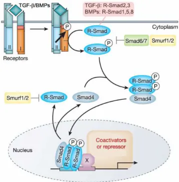
Figure 8. General mechanism of TGF-β superfamily signaling and Smad activation. At the cell surface, a TGF-β superfamily ligand binds a complex of transmembrane receptor serine/threonine kinases (types I and II) and induces transphosphorylation of the type I receptor by the type II receptor kinases. The consequently activated type I receptors phosphorylate selected Smads (Smad2 or -3 for TGF-β and Smad1, -5 or -8 for BMPs), and these receptor-activated Smads (R-Smads) then form a complex with a common Smad4. Activated Smad complexes translocate into the nucleus, where they regulate transcription of target genes, through physical interaction and functional cooperation with DNA-binding transcription factors (X) and CBP or p300 coactivators. Activation of R-Smads by type I receptor kinases is inhibited by Smad6 or Smad7. R-Smads and Smad4 shuttle between nucleus and cytoplasm. The E3 ubiquitin ligases Smurf1 and Smurf2 mediate ubiquitination and consequent degradation of R-Smads. Figure and legend adapted from Derynck R, Zhang YE. Nature 200378.
or activate EMT-related genes (Table 2). Of note, other intracellular signaling pathways besides the Smad signaling pathway respond to TGF-β superfamily ligands such as the MAPK, Erk and JNK pathways78.
Table 2. EMT-promoting Smad complexes and their target genes
Cofactor Smad partner Target genes Transcriptional
activity type Ref.
Snail1 Smad3 or -4 E-cadherin, occludin,
claudin-3, claudin-4 Repression
80
Zeb1 Smad2, -3 or -4 E-cadherin, claudin-3,
claudin-4 Repression
81
Zeb2 Smad1, -2, -3 or -5 E-cadherin, claudin-3,
claudin-4 Repression
82
Sp1 Smad3 Endoglin, collagen, vimentin Activation 74 Hmga2 Smad2 or -4 Snail1 Activation 75
The TGF-β superfamily signaling is inhibited by molecules such as the Sloan-Kettering Institute proto-oncogene (Ski), the Ski-related novel gene, non-Alu-containing (SnoN) and the transforming growth interacting factor (TGIF) which have been shown to prevent gene transcription through inhibition of R-Smads83-84. Also, two other Smad protein family members, Smad6 and Smad7, are called inhibitory Smads as they antagonize R-Smad phosphorylation by blocking their access to type I receptors and/or promote the degradation of the receptor complex85-86. Additionally, TGF-β superfamily signaling is inhibited by Smurf1 (Smad-ubiquitination-regulatory factor 1) and Smurf2 which interact with R-Smads and target them for degradation87.
3. miR-200 family
to two subfamilies, whose seed regions (nucleotides 2 to 7 of the mature active strand, which is the most important region for miRNA target recognition), differ by a single nucleotide (Figure 9).
Figure 9. miR-200 family. A. Stem-loop structure of hsa-mir-200c (precursor miRNA). B. The miR-200 family members. The human miR-200 family is located in two fragile chromosomal regions on 1p36.33 (200b, 200a and 429) and 12p13.31 (200c and 141), respectively. It consists of two clusters based on seed sequence similarity: miR-200bc/429 (red) and 200a/141 (blue), distinguished by a single nucleotide change (U to C). Figure and legend reproduced from Mutlu M SÖ, Raza U, Eyüpoglu E, Yurdusev E, Sahin Ö. Atlas Genet Cytogenet Oncol Haematol 201588.
3.1. Published miR-200 family target genes
During the progression of this project various miR-200 family target genes have been described in humans and mice (Table 3) in addition to those identified in this project89 (see Chapter 2).
Table 3. Published miR-200 target genes. Only genes experimentally validated to be directly targeted by miR-200 were considered. Species (Human (H), Mouse (M) or Rat (R)) in which gene was experimentally validated and miR-200 family members used for validation are shown.
Gene Species miR-200 family member Ref.
1 ACVR2B H 200c, 141 90
2 AP-2Α
(TCFAP2A) H 200c, 200b, 429
91
3 ARHGAP19 H 200c 92
4 BCL-2 H
H
200b, 200c, 429 429 93 94 5 BMI1 H H H M 200b 200c 141 200c 95-96 97-99 100 101
6 BRD3 H 141 102
7 BRD7 H 200c 103
8 CAGE H 200b 104
9 CD47 H 141 105
10 CDC25B H 141 106
11 CDH11 H
M
200c 141
107 108
12 CDK2 H 200b 109
13 CDK6 H 200a 110
14 CDKN1B
(p27/kip1) H 200b
111
15 CFL2 M
H
200c 200b
112 109
16 c-Maf M 200c 113
17 c-MYB H 200b, 200c, 429 114
18 C-MYC H 429 115
19 CREB1 H 200b 116
20 (β-CATENIN) CTNNB1
Table 3 (cont.). Published miR-200 target genes. Only genes experimentally validated to be directly targeted by miR-200 were considered. Species (Human (H), Mouse (M) or Rat (R)) in which gene was experimentally validated and miR-200 family members used for validation are shown.
21 CUL3 H 141 105
22 CXCL1 H 200a, 200b 122
23 CXCL12β H, M 141 123
24 CYCLIN E2 H 200a, 200b, 200c 124
25 DLC-1 H 141 125
26 Dlx5 M 141, 200a 126
27 DNMT3A H 200b, 200c 127
28 DNMT3B H 200b, 200c 127
29 E2f3 M 200c 128
30 eIF4E H 141 129
31 EPHA2 H 200a 130
32 EPHRIN-A1
(EFNA1) H
200c 131
33 ERRFI-1 H 200c 132
34 ETAR
(EDNRA) H 200c
133
35 ETS-1 M
H
200c 200b
134 135
36 (PTPN13) FAP-1 H 200c 136
37 FBLN5 H 200c 137
38 FHOD1 H 200c 132
39 FLK1 (KDR, VEGFR2) M M H H 200c 200b 200b 200c 134 138 139 140
40 (VEGFR1) FLT1
H H H 200b 200c 200c 139 141 142 41 FN1 H M H 200c 200c/141 200b 92 143 144
42 (ZFPM2) FOG2 H 200a, 200b, 200c 141, 429 145 43 FTH1
(Ferritin) H 200b
146
44 GATA2 H 200b 138
45 GATA4 H H 200b 200c
147 148
46 GNA13 H 200a
141
149
47 Grb2 M 200a 111,150
48 HDAC4 H 200a 151
49 HDGF H 141 152
50 IKKΒ
(Ikbkb) H 200c
Table 3 (cont.). Published miR-200 target genes. Only genes experimentally validated to be directly targeted by miR-200 were considered. Species (Human (H), Mouse (M) or Rat (R)) in which gene was experimentally validated and miR-200 family members used for validation are shown.
51 IL-8
(Cxcl15) H 200a, 200b
122
52 JAGGED1 (Jag1) H 200c, 141 153-155 53 KEAP1 H H M 141 200a 200c/141 156 157 143 54 KINDLIN-2
(FERMT2) H 200b
109
55 KLF4 H 200b 96
56 KLF9 H 200c 141
57 KRAS H 200c 158
58 LEPR H 200c 92
59 LPAR1 H 200c 133
60 Lrp1 M 200c 112
61 MAML2 H 200c 154
62 MAML3 H 200c 154
63 MAP4K4 H 141 159
64 MAPK14
(P38Α) H 200a, 141
160
65 MARCKS H 200c 161
66 MKK4 H 141 162
67 MSN H H 200c 200b
92 163
68 MUC16 H 200c 164
69 MUC4 H 200c 164
70 NCAM1 H 200c 165
71 Noggin
(Nog) M 200c
166
72 NOTCH1 H
H
200b, 200c 200b
167 168
73 NOXA H 200c 169
74 Nrp1 M 200a 108
75 NTF3 H 200c 170
76 (TRKB) NTRK2 H 200c 92
77 ONECUT2 H 429 171
78 OREBP H 200b 172
79 OXR1 H 200b 173
80 P53 H 200a 174
81 PAF H 200b 109
82 PIN1 H 200b 175
83
Pkd1 (polycystic kidney disease
1)
Table 3 (cont.). Published miR-200 target genes. Only genes experimentally validated to be directly targeted by miR-200 were considered. Species (Human (H), Mouse (M) or Rat (R)) in which gene was experimentally validated and miR-200 family members used for validation are shown.
84 PLCG1 H 200b,200c,429 177
85 PPARA H 141 178
86 PPM1F H 200c 179
87 PRKAR2B H 200b 180
88 PSMD1 H 200b 181
89 PSMD1 H 200b 181
90 PTEN
H H H M 141 200a 200c 200a 102 182 183 184
91 RAB18 H H 200b 429
185 186-187
92 RAB21 H 200b 186-187
93 RAB23 H 200b 186-187
94 RAB3B H 200b 186-187
95 RBBP4 H 429 188
96 RHOA H 200c 133
97 RND3 (RhoE) H H H 200b 200b 200c 189 111 190
98 ROCK2 H 200b, 200c 191
99 SEC23A H
M
200c 200c
107,192 112
100 Serca2b
(Atp2a2) M 200b
193
101 SHP
(NR0B2) H 141
194
102 SIRT1 H 200a 195
103 Slc25a3 M 141 196
104 Slug
(Snai2) M 200b
197 105 SMAD2 H M H 141, 200c 200b 200b 198 199 200
106 Snail
(Snai1) M 200b
199
107 SOX17 H
H
141, 200a 200a
155 201
108 SOX2 H
M
429 200c
202 128
109 SP1 H
H
200b, 200c 429
127 94
110 Srf R 200b 203
111 STAT5B H 200a 204
112 SUZ12 H
H
200b 200b, 200c
96,205 191
Table 3 (cont.). Published miR-200 target genes. Only genes experimentally validated to be directly targeted by miR-200 were considered. Species (Human (H), Mouse (M) or Rat (R)) in which gene was experimentally validated and miR-200 family members used for validation are shown.
114 TGF-Β2
H R M R 141 200a 200c/141 200a 207 208 143 118
115 TGFΒR1 H 141, 200a 198
116 THBS1 H 200a 209
117 TIAM1 H 141 210
118 TIMP2 H 200b 211
119 TIMP2 H 200c 137
120 TM4SF1 H 141 212
121 TUBB3 H
H
200c 200c
213 214
122 UBAP1 H 141 102
123 UBASH3B H 200a 215
124 UBQLN1 H 200b 181
125 UBQLN1 H 200b 181
126 VEGF-A H, M H, R H H H 200b 200b 200b 200c 200c 216 217 139 141 137 127 WAVE3
(WASF3) H 200b
218
128 Wnt1 M 200b 219
129 XIAP H 200b, 200c, 429 93 130 YAP1 H H 200a 141
220 221-222
131 YWHAG
(14-3-3γ) H 141
223
132 ZEB1 H
M 200a, 200b 200b, 200c 224 225 133 ZEB2 (SIP1) H M 200a, 200b 200b, 200c 224 225
134 ZMPSTE24 H 141 226
3.2. miR-200 family role in EMT and metastasis
Because of its role as a master EMT regulator, miR-200 has been extensively studied. Besides targeting EMT-inducers Zeb1 and Zeb2, miR-200 also targets other important EMT activators such as Ets1, Snail1 (Snail) and Snail2 (Slug) (Table 3). miR-200 has also been shown to regulate cell-cell adhesion207,224,228, cancer stem cell self-renewal and differentiation96,229-230, cell division and apoptosis136,147,177,189 and chemoresistance93,231 (Figure 10).
Figure 10. miR-200c targets several genes regulating numerous processes involved in cancer development and progression. Figure and legend reproduced from Mutlu M SÖ, Raza U, Eyüpoglu E, Yurdusev E, Sahin Ö. Atlas Genet Cytogenet Oncol Haematol 201588.
between miR-200b/c/429 and miR-200a/141 subfamily target genes, we chose for this project to focus on miR-200c as a representative member of the miR-200 family.
Even though miR-200 was shown to regulate metastasis formation, its effect is controversial as miR-200 expression enhances metastasis in some cancer models,112,202,225,233-234 but reduces it in others95,186,235. Different cancer cell models may have different factors that alter miR-200’s role in metastasis. Additionally, the microenvironment surrounding the primary tumor may also contribute to these differences. Extracellular cytokines at the invasive front, such as TGF-β, may activate miR-200 repressors such as Zeb1/2 that repress miR-200 expression at the transcriptional level. Finally, the microenvironment at secondary metastatic sites may also cooperate or counteract miR-200’s colonization-promoting effect.
Circulating miR-200 levels can also be used as a prognostic tool in oncology, as high levels of miR-200 in the bloodstream of cancer patients are associated with poor prognosis in several cancers, including ovarian, prostate, pancreatic and metastatic colorectal cancers236-239.
4. Thesis Scope
The main goals of the work within this thesis were to provide the first genome-wide map of miR-200c target genes and investigate their biological role. Although miR-200 family was already known to regulate EMT, a genome-wide list of its targets and a comprehensive map of miR-200-regulated pathways was yet to be obtained.
miR-200c putative targets. This genome-wide list of miR-200c target genes was then bioinformatically analyzed to provide a global list of miR-200-regulated pathways. Some of these putative target genes were selected for experimental validation. Results show that this high-throughput method for miRNA target discovery has a low false-positive rate. Additionally, biochemical assays were performed to study the role of some novel target genes in miR-200-dependent processes. Some of these target genes had novel and unexpected biological functions.
Overall, the framework developed in this thesis allowed us to generate the most complete genome-wide map of miR-200-regulated pathways to date and discover new genes involved in EMT. Moreover, this framework can be used to identify the genome-wide targets of other miRNAs.
5. References
1 Thomas M, Lieberman J, Lal A. Desperately seeking microRNA targets. Nat Struct Mol Biol 2010;
17: 1169-1174.
2 Winter J, Jung S, Keller S, Gregory RI, Diederichs S. Many roads to maturity: microRNA biogenesis pathways and their regulation. Nat Cell Biol 2009; 11: 228-234.
3 Ardekani AM, Naeini MM. The Role of MicroRNAs in Human Diseases. Avicenna J Med Biotechnol
2010; 2: 161-179.
4 Friedman RC, Farh KK, Burge CB, Bartel DP. Most mammalian mRNAs are conserved targets of microRNAs. Genome Res 2009; 19: 92-105.
5 Lee Y, Kim M, Han J, Yeom KH, Lee S, Baek SH et al. MicroRNA genes are transcribed by RNA polymerase II. EMBO J 2004; 23: 4051-4060.
6 Hata A, Lieberman J. Dysregulation of microRNA biogenesis and gene silencing in cancer. Sci Signal 2015; 8: re3.
7 Baskerville S, Bartel DP. Microarray profiling of microRNAs reveals frequent coexpression with neighboring miRNAs and host genes. RNA 2005; 11: 241-247.
8 Kim VN, Nam JW. Genomics of microRNA. Trends Genet 2006; 22: 165-173.
9 Yang JS, Lai EC. Alternative miRNA biogenesis pathways and the interpretation of core miRNA pathway mutants. Mol Cell 2011; 43: 892-903.
10 Hutvagner G, Zamore PD. A microRNA in a multiple-turnover RNAi enzyme complex. Science
2002; 297: 2056-2060.
11 Liu J, Carmell MA, Rivas FV, Marsden CG, Thomson JM, Song JJ et al. Argonaute2 is the catalytic engine of mammalian RNAi. Science 2004; 305: 1437-1441.
12 Jones-Rhoades MW, Bartel DP, Bartel B. MicroRNAS and their regulatory roles in plants. Annu Rev Plant Biol 2006; 57: 19-53.
13 Bartel DP. MicroRNAs: target recognition and regulatory functions. Cell 2009; 136: 215-233.
14 Hutvagner G, Simard MJ. Argonaute proteins: key players in RNA silencing. Nat Rev Mol Cell Biol
2008; 9: 22-32.
15 Zekri L, Huntzinger E, Heimstadt S, Izaurralde E. The silencing domain of GW182 interacts with PABPC1 to promote translational repression and degradation of microRNA targets and is required for target release. Mol Cell Biol 2009; 29: 6220-6231.
16 Jonas S, Izaurralde E. Towards a molecular understanding of microRNA-mediated gene silencing.
Nat Rev Genet 2015; 16: 421-433.
17 Lewis BP, Shih IH, Jones-Rhoades MW, Bartel DP, Burge CB. Prediction of mammalian microRNA targets. Cell 2003; 115: 787-798.
18 Nielsen CB, Shomron N, Sandberg R, Hornstein E, Kitzman J, Burge CB. Determinants of targeting by endogenous and exogenous microRNAs and siRNAs. RNA 2007; 13: 1894-1910.
19 Rehmsmeier M, Steffen P, Hochsmann M, Giegerich R. Fast and effective prediction of microRNA/target duplexes. RNA 2004; 10: 1507-1517.
21 Ahmadi H, Ahmadi A, Azimzadeh-Jamalkandi S, Shoorehdeli MA, Salehzadeh-Yazdi A, Bidkhori G
et al. HomoTarget: a new algorithm for prediction of microRNA targets in Homo sapiens.
Genomics 2013; 101: 94-100.
22 Zisoulis DG, Lovci MT, Wilbert ML, Hutt KR, Liang TY, Pasquinelli AE et al. Comprehensive discovery of endogenous Argonaute binding sites in Caenorhabditis elegans. Nat Struct Mol Biol
2010; 17: 173-179.
23 Chi SW, Zang JB, Mele A, Darnell RB. Argonaute HITS-CLIP decodes microRNA-mRNA interaction maps. Nature 2009; 460: 479-486.
24 Lal A, Navarro F, Maher CA, Maliszewski LE, Yan N, O'Day E et al. miR-24 Inhibits cell proliferation by targeting E2F2, MYC, and other cell-cycle genes via binding to "seedless" 3'UTR microRNA recognition elements. Mol Cell 2009; 35: 610-625.
25 Shin C, Nam JW, Farh KK, Chiang HR, Shkumatava A, Bartel DP. Expanding the microRNA targeting code: functional sites with centered pairing. Mol Cell 2010; 38: 789-802.
26 Johnson CD, Esquela-Kerscher A, Stefani G, Byrom M, Kelnar K, Ovcharenko D et al. The let-7 microRNA represses cell proliferation pathways in human cells. Cancer Res 2007; 67: 7713-7722.
27 Vella MC, Choi EY, Lin SY, Reinert K, Slack FJ. The C. elegans microRNA let-7 binds to imperfect let-7 complementary sites from the lin-41 3'UTR. Genes Dev 2004; 18: 132-137.
28 Tarang S, Weston MD. Macros in microRNA target identification: a comparative analysis of in silico, in vitro, and in vivo approaches to microRNA target identification. RNA Biol 2014; 11: 324-333.
29 Sandberg R, Neilson JR, Sarma A, Sharp PA, Burge CB. Proliferating cells express mRNAs with shortened 3' untranslated regions and fewer microRNA target sites. Science 2008; 320: 1643-1647.
30 Ji Z, Lee JY, Pan Z, Jiang B, Tian B. Progressive lengthening of 3' untranslated regions of mRNAs by alternative polyadenylation during mouse embryonic development. Proc Natl Acad Sci U S A
2009; 106: 7028-7033.
31 Mayr C, Bartel DP. Widespread shortening of 3'UTRs by alternative cleavage and polyadenylation activates oncogenes in cancer cells. Cell 2009; 138: 673-684.
32 Wang ET, Sandberg R, Luo S, Khrebtukova I, Zhang L, Mayr C et al. Alternative isoform regulation in human tissue transcriptomes. Nature 2008; 456: 470-476.
33 Nicolas FE. Experimental validation of microRNA targets using a luciferase reporter system.
Methods Mol Biol 2011; 732: 139-152.
34 Baek D, Villen J, Shin C, Camargo FD, Gygi SP, Bartel DP. The impact of microRNAs on protein output. Nature 2008; 455: 64-71.
35 Hu R, Wallace J, Dahlem TJ, Grunwald DJ, O'Connell RM. Targeting human microRNA genes using engineered Tal-effector nucleases (TALENs). PLoS One 2013; 8: e63074.
36 Krutzfeldt J, Rajewsky N, Braich R, Rajeev KG, Tuschl T, Manoharan M et al. Silencing of microRNAs in vivo with 'antagomirs'. Nature 2005; 438: 685-689.
37 Kloosterman WP, Wienholds E, de Bruijn E, Kauppinen S, Plasterk RH. In situ detection of miRNAs in animal embryos using LNA-modified oligonucleotide probes. Nat Methods 2006; 3: 27-29.
40 Khan AA, Betel D, Miller ML, Sander C, Leslie CS, Marks DS. Transfection of small RNAs globally perturbs gene regulation by endogenous microRNAs. Nat Biotechnol 2009; 27: 549-555.
41 Stenvang J, Petri A, Lindow M, Obad S, Kauppinen S. Inhibition of microRNA function by antimiR oligonucleotides. Silence 2012; 3: 1.
42 Beitzinger M, Peters L, Zhu JY, Kremmer E, Meister G. Identification of human microRNA targets from isolated argonaute protein complexes. RNA Biol 2007; 4: 76-84.
43 Easow G, Teleman AA, Cohen SM. Isolation of microRNA targets by miRNP immunopurification.
RNA 2007; 13: 1198-1204.
44 Landthaler M, Gaidatzis D, Rothballer A, Chen PY, Soll SJ, Dinic L et al. Molecular characterization of human Argonaute-containing ribonucleoprotein complexes and their bound target mRNAs. RNA 2008; 14: 2580-2596.
45 Hong X, Hammell M, Ambros V, Cohen SM. Immunopurification of Ago1 miRNPs selects for a distinct class of microRNA targets. Proc Natl Acad Sci U S A 2009; 106: 15085-15090.
46 Karginov FV, Conaco C, Xuan Z, Schmidt BH, Parker JS, Mandel G et al. A biochemical approach to identifying microRNA targets. Proc Natl Acad Sci U S A 2007; 104: 19291-19296.
47 Hendrickson DG, Hogan DJ, Herschlag D, Ferrell JE, Brown PO. Systematic identification of mRNAs recruited to argonaute 2 by specific microRNAs and corresponding changes in transcript abundance. PLoS One 2008; 3: e2126.
48 Su H, Trombly MI, Chen J, Wang X. Essential and overlapping functions for mammalian Argonautes in microRNA silencing. Genes Dev 2009; 23: 304-317.
49 Ipsaro JJ, Joshua-Tor L. From guide to target: molecular insights into eukaryotic RNA-interference machinery. Nat Struct Mol Biol 2015; 22: 20-28.
50 Maniataki E, Mourelatos Z. Human mitochondrial tRNAMet is exported to the cytoplasm and associates with the Argonaute 2 protein. RNA 2005; 11: 849-852.
51 Zhang X, Graves PR, Zeng Y. Stable Argonaute2 overexpression differentially regulates microRNA production. Biochim Biophys Acta 2009; 1789: 153-159.
52 Diederichs S, Haber DA. Dual role for argonautes in microRNA processing and posttranscriptional regulation of microRNA expression. Cell 2007; 131: 1097-1108.
53 Hafner M, Landthaler M, Burger L, Khorshid M, Hausser J, Berninger P et al. Transcriptome-wide identification of RNA-binding protein and microRNA target sites by PAR-CLIP. Cell 2010; 141:
129-141.
54 Sugimoto Y, Konig J, Hussain S, Zupan B, Curk T, Frye M et al. Analysis of CLIP and iCLIP methods for nucleotide-resolution studies of protein-RNA interactions. Genome Biol 2012; 13:
R67.
55 Lal A, Thomas MP, Altschuler G, Navarro F, O'Day E, Li XL et al. Capture of microRNA-bound mRNAs identifies the tumor suppressor miR-34a as a regulator of growth factor signaling. PLoS Genet 2011; 7: e1002363.
56 Orom UA, Nielsen FC, Lund AH. MicroRNA-10a binds the 5'UTR of ribosomal protein mRNAs and enhances their translation. Mol Cell 2008; 30: 460-471.
(miR-21), which down-regulates expression of family of dedicator of cytokinesis (DOCK) proteins.
J Biol Chem 2012; 287: 3976-3986.
59 Cloonan N, Wani S, Xu Q, Gu J, Lea K, Heater S et al. MicroRNAs and their isomiRs function cooperatively to target common biological pathways. Genome Biol 2011; 12: R126.
60 Tan SM, Kirchner R, Jin J, Hofmann O, McReynolds L, Hide W et al. Sequencing of captive target transcripts identifies the network of regulated genes and functions of primate-specific miR-522.
Cell Rep 2014; 8: 1225-1239.
61 Kozomara A, Hunt S, Ninova M, Griffiths-Jones S, Ronshaugen M. Target repression induced by endogenous microRNAs: large differences, small effects. PLoS One 2014; 9: e104286.
62 Subramanian M, Li XL, Hara T, Lal A. A biochemical approach to identify direct microRNA targets.
Methods Mol Biol 2015; 1206: 29-37.
63 Kalluri R, Weinberg RA. The basics of epithelial-mesenchymal transition. J Clin Invest 2009; 119:
1420-1428.
64 Duband JL, Monier F, Delannet M, Newgreen D. Epithelium-mesenchyme transition during neural crest development. Acta Anat (Basel) 1995; 154: 63-78.
65 Kang P, Svoboda KK. Epithelial-mesenchymal transformation during craniofacial development. J Dent Res 2005; 84: 678-690.
66 Yang J, Weinberg RA. Epithelial-mesenchymal transition: at the crossroads of development and tumor metastasis. Dev Cell 2008; 14: 818-829.
67 Thiery JP. Epithelial-mesenchymal transitions in tumour progression. Nat Rev Cancer 2002; 2:
442-454.
68 Thiery JP, Acloque H, Huang RY, Nieto MA. Epithelial-mesenchymal transitions in development and disease. Cell 2009; 139: 871-890.
69 Zeisberg M, Neilson EG. Biomarkers for epithelial-mesenchymal transitions. J Clin Invest 2009;
119: 1429-1437.
70 Weigelt B, Peterse JL, van 't Veer LJ. Breast cancer metastasis: markers and models. Nat Rev Cancer 2005; 5: 591-602.
71 Lee JM, Dedhar S, Kalluri R, Thompson EW. The epithelial-mesenchymal transition: new insights in signaling, development, and disease. J Cell Biol 2006; 172: 973-981.
72 Wells A, Yates C, Shepard CR. E-cadherin as an indicator of mesenchymal to epithelial reverting transitions during the metastatic seeding of disseminated carcinomas. Clin Exp Metastasis 2008;
25: 621-628.
73 Wan L, Pantel K, Kang Y. Tumor metastasis: moving new biological insights into the clinic. Nat Med 2013; 19: 1450-1464.
74 Wu Y, Zhang X, Salmon M, Lin X, Zehner ZE. TGFbeta1 regulation of vimentin gene expression during differentiation of the C2C12 skeletal myogenic cell line requires Smads, AP-1 and Sp1 family members. Biochim Biophys Acta 2007; 1773: 427-439.
75 Thuault S, Tan EJ, Peinado H, Cano A, Heldin CH, Moustakas A. HMGA2 and Smads co-regulate SNAIL1 expression during induction of epithelial-to-mesenchymal transition. J Biol Chem 2008;
283: 33437-33446.