Article/Artigo
INTRODUCTION
1. Institute of Biological Sciences, Federal University of Pará, Belém, PA, Brazil. 2. Laboratory of Molecular Biology, Center of Hematology and Hemotherapy of Pará, Belém, PA, Brazil. 3. Program of Viral Hepatitis, State Department of Public Health , Belém, PA, Brazil. 4. Section of Tropical Medicine, Federal University of Pará, Belém, PA, Brazil. 5. Campus of Marajó, Federal University of Pará, Breves, PA, Brazil.
Address to: Dr. Aldemir Branco de Oliveira Filho. FCN/UFPA. Avenida Anajás s/n, Conjunto Bandeirantes, Aeroporto 68800-000 Breves, PA, Brazil.
Phone: 55 91 3273 1129 ext: 213 e-mail: [email protected]
Received in 27/03/2010
Accepted in 13/10/2010
Distribution of hepatitis C virus genotypes among diferent exposure
categories in the State of Pará, Brazilian Amazon
Distribuição dos genótipos do vírus da hepatite C em diferentes categorias de exposição no
Estado do Pará, Amazônia Brasileira
Leila Sawada
1,2, Andréia Cristina Costa Pinheiro
1,2, Daiane Locks
1,2, Adriana do Socorro Coelho Pimenta
2,
Priscila Rocha Rezende
2, Deborah Maia Crespo
3, José Ângelo Barleta Crescente
4, José Alexandre Rodrigues
de Lemos
1,2and Aldemir Branco de Oliveira Filho
1,2,5ABSTACT
Introduction: Epidemiological studies concerning HCV genotypic distribution in the Brazilian Amazon are scarce. hus, this study determined the paterns of distribution of HCV genotypes among diferent exposure categories in the State of Pará, Brazilian Amazon. Methods: A cross-sectional study was conducted on 312 HCV-infected individuals belonging to diferent categories of exposure, who were atended at the HEMOPA, CENPREN and a private hemodialysis clinic in Belém. hey were tested for HCV antibodies using an immunoenzymatic test, RNA-HCV, using real-time PCR and HCV genotyping through phylogenetic analysis of the 5' UTR. he population groups were epidemiologically characterized according to data collected in a brief interview or medical consultation. Results: Genotype 1 predominated in all the diferent categories of HCV exposure. HCV genotypic distribution among blood donors comprised genotypes 1 (94%) and 3 (6%). All patients with chronic hematologic diseases had HCV genotype 1. he genotypic distribution in illicit-drug users comprised genotypes 1 (59.6%) and 3 (40.4%). In patients under hemodialysis, genotypes 1 (90.1%), 2 (3.3%), and 3 (6.6%) were detected. Finally, the frequency of genotypes 1 and 3 was signiicantly diferent between the groups: BD and DU, PUH and DU, PUH and PCHD and PCHD and DU. Conclusions: he genotypic frequency and distribution of HCV in diferent categories of exposure in the State of Pará showed a predominance of genotype 1, regardless of the possible risk of infection.
Keywords: HCV. Genotype. Risk group. Pará. Brazilian Amazon.
RESUMO
Introdução: Estudos epidemiológicos sobre a distribuição genotípica do HCV na Amazônia Brasileira são escassos. Baseado nisto, determinamos o padrão de distribuição genotípica do HCV em diferentes categorias de exposição no Estado do Pará, Amazônia Brasileira. Métodos:
Estudo transversal foi realizado com 312 indivíduos infectados pelo HCV, pertencentes a diferentes categorias de exposição atendidas pelo HEMOPA, CENPREN e uma clínica privada de hemodiálise em Belém. Eles foram testados quanto à presença de anticorpos anti-HCV por teste imunoenzimático, RNA-HCV utilizando PCR em tempo real e genotipados através de análise ilogenética da 5’ UTR. Os grupos de populações foram caracterizados epidemiologicamente de acordo com dados coletados em breve entrevista ou consulta de prontuários médicos.
Resultados: Em todas as diferentes categorias de exposição ao HCV, foram encontrados predomínio do genótipo 1. A distribuição genotípica do HCV em doadores de sangue (BD) foi constituída pelos genótipos 1 (94%) e 3 (6%). Todos os pacientes com doenças hematológicas crônicas (PCHD) possuíam genótipo 1. A distribuição genotípica em usuários de drogas ilícitas (DU) foi constituída pelos genótipos 1 (59,6%) e 3 (40,4%). Em pacientes em hemodiálise (PUH) foram detectados os genótipos 1 (90,1%), 2 (3,3%) e 3 (6,6%). Finalmente, a frequência entre os genótipos 1 e 3 foi signiicativamente diferente entre os grupos: BD e DU, PUH e DU, PUH e PCHD, e PCHD e DU. Conclusões: A frequência genotípica e distribuição de HCV em diferentes categorias de exposição no Estado do Pará mostraram predominância do genótipo 1, independentemente do possível risco de infecção.
Palavras-chaves: HCV. Genótipo. Grupo de risco. Pará. Amazônia Brasileira.
Hepatitis C virus (HCV) infection is a global health problem. HCV is one of the leading causes of acute and chronic hepatitis, cirrhosis of the liver and hepatocellular carcinoma1. he prevalence of
HCV infection varies from 0.1% to 0.2% in the West, to 3% in Mediterranean countries and well over 10% in some countries and regions of Africa, Asia and Europe, such as the Chinese provinces of Hubei (30.1%) and Mongolia (31.9%), and in Egypt (28%)1,2. Oicial data on the prevalence of
HCV in the Brazilian population are limited. It is estimated that the seroprevalence in blood donors is approximately 1.6%, characterizing Brazil as an area with low endemicity. he HCV prevalence in northern Brazil (Brazilian Amazon) is the highest among the Brazilian regions (2.2%) and in this region the highest rates are observed in the States of Acre (5.9%) and Pará (0.2 - 2%)3-5.
Phylogenetic analysis of nucleotide sequences resulted in the classiication of six genotypes and several subtypes. HCV genotypes and their subtypes coexist in various geographical locations, but show different prevalence levels6,7. The prevalence of
genotypes is also associated with the transmission route of the infection. For example, infections with subtypes 1a and 3a have been shown to be significantly associated with intravenous drug abuse8. In addition, HCV genotypes possess diferent
biological potentials. Certain HCV genotypes are more amenable to interferon treatment and are more frequently associated with severe forms of liver disease9,10. In Brazil, genotype 1 predominates,
followed by genotypes 3 and 211,12. However,
RESULTS METHODS
donors) is genotype 1, at 78%, 64% and 93%, respectively5,12,13. his
work reports on the paterns of distribution of HCV genotypes among patients in diferent categories of exposure in the State of Pará, Brazilian Amazon.
Individuals with HCV infection who were attended by the Blood Bank of Pará (HEMOPA)5, the Center for Prevention and
Treatment of Chemical Dependency (CENPREN) and a private hemodialysis clinic, from January 2004 to February 2008 in the State of Pará, Brazilian Amazon, were selected. he population groups were epidemiologically characterized according to data collected in a brief interview or medical consultation. All of the plasma samples were tested for HCV antibodies using an immunoenzyme test (Murex anti-HCV version 4.0, Murex Biotech SA, Kyalami, South Africa). A conirmatory test for viral infection was performed on the seroreactive samples using real-time PCR (ABI Prism 7,000, Applied Biosystems). Viral RNA was extracted using the commercial extraction kit QIAmp Viral RNA Mini Kit (Qiagen). Molecular diagnosis was based on detection of the 67 base pairs of 5’ UTR with the commercial kit TaqMan EZ RT-PCR Core Reagents (Applied Biosystems)5. Following molecular diagnosis of viral infection, all
positive samples were selected for ampliication of the 5’ UTR using nested-PCR5. he product of this second ampliication was run on a
2% agarose gel bufered with TBE; the gel was stained with ethidium bromide and observed under ultraviolet light. he ampliied fragment was sequenced in both directions using the dideoxynucleotide chain terminator method with an ABI Prism 377 and the commercial kit Big Dye Cycle Sequencing Standard, both from Applied Biosystems.
All nucleotide sequences obtained were edited and aligned using BioEdit software (http://www.mbio.ncsu.edu/BioEdit/bioedit. htm). his alignment was entered in the DnaSP sotware version 5.1014 to identify likely identical nucleotide sequences, and in the
Modelgenerator sotware version 0.8515 to select the best model to
apply to phylogenetic analyses, according to the corrected Akaike information criterion. hese parameters were used in the PHYML program version 2.4.416 to infer trees, according to the
maximum-likelihood method. To test the robustness of the tree topologies, 1,000 bootstrap replicates were performed. he inal phylogenetic tree was obtained by majority-rule consensus and then edited using the graphical resources contained in the FigTree sotware version 1.3.1 (http://tree.bio.ed.ac.uk/software/figtree). Nucleotide sequences obtained from the National Center of Biotechnology Information were added to the alignment and used to construct the phylogenetic tree to identify HCV genotypes (Genotype 1: U45476, AJ132997, M62321. Genotype 2: D49757, D49754, AB030907, D10077, D49745, AB031663, D00944, AF169003, D50409, D49755, AY746460. Genotype 3: D17763, D28917, D37840, D49374, D49747, D49753. Genotype 4: D45193. Genotype 5: D50466. Genotype 6: D88476, D88473, D88475). Univariate analyses, comparing the distribution of HCV genotypes among the diferent exposure categories, were performed using BioEstat sotware version 5.0. he Chi square test was used and the results were considered to be statistically signiicant when p < 0.05. Nucleotide sequence data obtained in this work are available in the DDBJ/EMBL/GenBank databases under the accession numbers FJ696418 to FJ696533, and HM042983 to HM043178.
Ethical considerations
his study was approved by the Research Ethics Commitee of the Tropical Medicine Section of the Federal University of Pará, Belém, Pará, Brazil (Acession number: 041/2004-CEP/NMT).
he diferent HCV exposure categories consisted of 151 blood donors who were selected from 298,259 donors attended at the HEMOPA, 47 of the 94 illicit drug users under the care of the CENPREN, 53 of the 98 patients with chronic hematological disease who were receiving treatment at the HEMOPA and 61 of the 271 patients under hemodialysis in a private hemodialysis clinic, located in the City of Belém. A total of 312 nucleotide sequences of HCV 5’ UTR were isolated; however, 293 identical nucleotide sequences were identiied and only one representative was maintained in the alignment. hus, the inal alignment consisted of 19 nucleotide sequences: 5 isolates in blood donors, 5 isolates in illicit drug users, 3 isolates in patients with chronic hematological disease and 6 isolates in patients under hemodialysis. Based on the Modelgenerator sotware, the most appropriate evolutionary model for this data matrix was the Tamura-Nei model, adjusted by the parameters proportion of invariable sites (0.451)
and rate of gamma distribution (0.327). The base frequencies
(A = 0.22459, C = 0.27331, G = 0.27534, T = 0.22677), transition/ transversion ratio for purines (3.923) and transition/transversion ratio for pyrimidines (4.885) were estimated by the PHYML sotware during the phylogenetic analysis. Of the diferent HCV exposure categories, genotype 1 predominated (Figure 1 and Table 1). A likely predominance of subtype 1b was also detected in all the risk groups. Due to the low values of grouping, it is necessary to analyze another region of the virus genome to conirm this inding.
he group of infected donor blood was comprised mostly of males (66%), with a signiicantly higher frequency of infection in individuals aged 30 to 49 years-old (30-39 years-old: 27%; 40-49 years-old: 39%). he genotypic distribution was formed by genotypes 1 (approximately 94%) and 3 (approximately 6%) (Figure 1 and Table 1). he group of infected multitransfused patients had one of the following chronic hematological diseases: hemophilia A (52%), hemophilia B (12.3%), sickle-cell anemia (20.4%), deiciency of coagulation factors (8.2%) and others (7.1%). he average age of infected patients was 36 years-old (23-56 years-old), and the average number of blood transfusions or blood products received was around 45 (min-max: 6-352). All these patients presented HCV genotype 1 (Figure 1 and Table 1). he group of infected drug users consumed preferably non-injected drugs (82.1%) including marijuana, crack cocaine, and cocaine. Most drug users were males (62.3%), with an average age of around 28 old (18-52 years-old). Among the drug users, the genotypic distribution was formed by genotypes 1 (59.6%) and 3 (40.4%) (Figure 1 and Table 1). In patients under hemodialysis, genotypes 1 (90.2%), 2 (3.3%), and 3 (6.5%) were detected. Most patients under hemodialysis were males (66.6%), with an average age around 47 years-old (28-65 years-old). he mean period that they had received dialysis therapy was 1.5 years (min-max: 0.25-5.5 years) (Figure 1 and Table 1). Finally, the HCV genotypic frequency (1 and 3) was signiicantly diferent between the groups: blood donors and illicit drug users (χ2 = 18.58, p-value < 0.001), hemodialysis patients
and illicit drug users (χ2 =13.83; p-value < 0.001), hemodialysis
patients and multitransfused patients (χ2 = 4.87; p-value = 0.02),
and multitransfused patients and illicit drug users (χ2 = 25.57, p < 0.001).
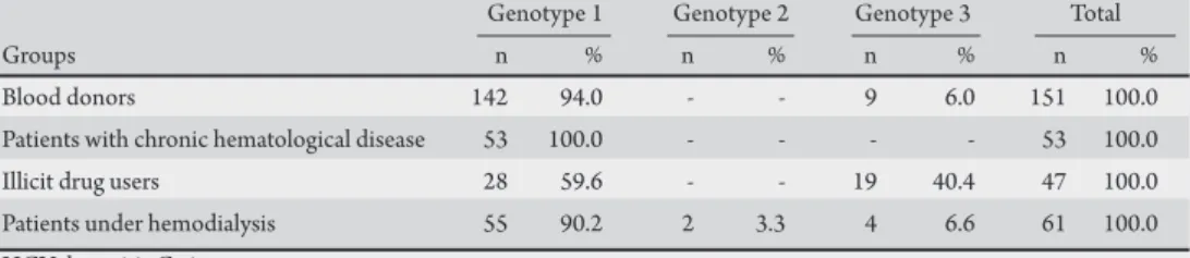
TABLE 1 -Distribution of HCV genotypes among groups of patients in diferent exposure categories in the Brazilian Amazon.
Genotype 1 Genotype 2 Genotype 3 Total
Groups n % n % n % n %
Blood donors 142 94.0 - - 9 6.0 151 100.0 Patients with chronic hematological disease 53 100.0 - - - - 53 100.0 Illicit drug users 28 59.6 - - 19 40.4 47 100.0 Patients under hemodialysis 55 90.2 2 3.3 4 6.6 61 100.0 HCV: hepatitis C virus.
FINANCIAL SUPPORT
REFERENCES
he authors declare that there are no conlicts of interest.
CONFLICT OF INTEREST DISCUSSION
Some genotypes of HCV (1a, 1b, 2a, 2b, and 3a) are widely distributed all over the world. Others have a more restricted distribution, such as genotype 4 in the Middle East and Africa, genotype 5a in South Africa and genotype 6 in Southeast Asia7.
Certain factors, such as migration and travel paterns, may alter the current genotypic map. In Sardinia, Coppola et al17 identiied
genotype 4, which was rare in continental Italy, at a level of 13%, and suggested that this high prevalence was associated with a connection between Northern Africa and the Southern Mediterranean basin with the nearby Italian island. In Brazil, genotype 1 predominates, followed by genotypes 3 and 211,12. The distribution of HCV
genotypes in diferent exposure categories in Brazil is similar, with a higher frequency of genotypes 1 and 3, followed by genotype 2 and sporadic cases of genotype 418. his study showed a predominance
of genotype 1, followed by genotypes 3 and 2, while genotypes 4 and 5 were not detected. his result probably relects the high frequency of genotypes 1 and 3 in the Brazilian Amazon5,11-13,19.
Furthermore, the risk of HCV infection between the groups was observed in the distribution and frequency of the viral genotypes. he high frequency of genotype 1 in blood donors inluenced the complete dominance of genotype 1 in multitransfused patients, which was probably transferred by blood/blood product transfusion. In the Netherlands, the transfusion of blood/blood products was responsible for infection in 17.5% of genotype 1 patients. Among patients with genotype 1b, transfusion of blood/blood products was the main route of transmission20.
Some studies have reported that changes may occur in the HCV subtype patern. In some countries of Europe, genotype 1a and 3a are increasing, while 2a, 2c and 1b are decreasing, especially in young patients. These results probably stem from the high prevalence of genotype 1a and 3a among intravenous drug users, the reservoir from which the general population is then afected21. Other studies have
reported that a growing number of young drug addicts are infected with subtype 3a. hese results indicate that changes in social behavior afect the epidemiological trends of infectious diseases to a signiicant extent22,23. In the present study, these epidemiological characteristics can
be seen in the group of users of illicit drugs, relected in the high frequency of genotype 3 (3a/3b) and the low average age of those infected. In the group of patients under hemodialysis, the longer exposure time (the higher average age) to possible risk factors may have been responsible for this group showing the highest genotypic diversity of HCV. his study also provided evidence that the 5’ UTR is conserved and limited in its ability to discriminate subtypes within genotypes 1, 2, 3, 4, and 624,25. Some of the genotype-speciic motifs that were
initially identiied in the 5' UTR are no longer found to be conserved. For example, the G residue at position 243 of the 5' UTR, originally considered to be representative of subtype 1b, is now known to occur in some proportion of subtype 1a viruses. Several subtypes that oten share the same 5’ UTR sequence have been described26-28.
Despite this, the 5' UTR contains suicient variation to resolve HCV classiications at the level of viral genotype. hus, this study illustrated the need for sequencing of other regions of the HCV genome to improve the resolution of viral subtypes.
Based on the results of this study, it can be concluded that most hepatitis C infections in the Brazilian Amazon consist of genotype 1, regardless of the possible risk factors for infection. his implies
that patients infected with HCV in the Brazilian Amazon should preferentially be treated with a speciic clinical protocol for genotype 1. Moreover, this study shows the need for sequencing of other regions of the HCV genome, to achieve improved resolution of the viral subtypes circulating in the Brazilian Amazon. In the future, more extensive surveys should be conducted to assess geographical diferences in the distribution of HCV genotypes among diferent exposure categories in other Brazilian regions.
FAPESPA, PPSUS, CNPq, and PN-DST/AIDS.
1. Alter MJ. Epidemiology of hepatitis C virus infection. World J Gastroenterol 2007;13:2436-2441.
2. Shepard CW, Finelli L, Alter MJ. Global epidemiology of hepatitis C virus infection. Lancet Infect Dis 2005; 5:558-567.
3. Grupo de Estudo da Sociedade Brasileira de Hepatologia. Epidemiologia da infecção pelo vírus da hepatite C no Brasil. Gastroenterol End Digest 1999; 18 (suppl.1):S3-S8.
4. Da Fonseca JCF, Brasil LM. Hepatitis C virus infection in the Amazon Brazilian Region. Rev Soc Bras Med Trop 2004; 37 (supl II):1-8.
5. Oliveira-Filho AB, Pimenta ASC, Rojas MFM, Chagas MCM, Crespo DM, Crescente JAB, et al. Prevalence and genotyping of hepatitis C virus in blood donors in the state of Pará, Northern Brazil. Mem Inst Oswaldo Cruz 2010; 105:103-106.
6. Simmonds P, Bukh J, Combet C, Deléage G, Enomoto N, Feinstone S, et al. Consensus proposals for a uniied system of nomenclature of hepatitis C genotypes. Hepatology 2005; 42:962-973.
7. Kuiken C, Hraber P, hurmond J, Yusim K. he hepatitis C sequence database in Los Alamos. Nucleic Acids Res 2008; 36:512-526.
8. van Asten L, Verhaest I, Lamzira S, Hernandez-Aguado I, Zangerle R, Boufassa F, et al. Spread of hepatitis C virus among European injection drug users infected with HIV: a phylogenetic analysis. J Infect Dis 2004; 189:292-302.
9. Hadziyannis SJ, Koshina JS. Differences in epidemiology, liver disease and treatment response among HCV genotypes. Hepatol Res 2004; 29: 129-135.
10. Zeuzem S. Heterogeneous virologic response rates to interferon-based therapy in patients with chronic hepatitis C: who responds less well. Ann Intern Med 2004; 140:370-381.
11. Busek S, Oliveira G. Molecular epidemiology of the hepatitis C virus in Brazil. Genet Mol Res 2003; 2:117-123.
12. Campiotto S, Pinho JR, Carrilho FJ, Da Silva LC, Souto FJ, Spinelli V, et al. Geographic distribution of hepatitis C virus genotypes in Brazil. Braz J Med Biol Res 2005; 38:41-49.
13. Paraná R, Paiva T, Leite MR, Oliveira FN, Kali N, Lobato C, et al. Infection with hepatitis C virus among health care workers in the Brazilian Western Amazon Region (Rio Branco, state of Acre). Am J Trop Med Hyg 2007; 76:165-169. 14. Librado P, Rozas J. DnaSP v5: A sotware for comprehensive analysis of DNA
polymorphism data. Bioinformatics 2009; 25:1451-1452.
15. Keane TM, Creevey CJ, Pentony MM, Naughton TJ, Mclnerney JO. Assessment of methods for amino acid matrix and their use on empirical data shows that ad hoc assumptions for choice of matrix are not justified. BMC Evol Biol 2006; 6:29.
17. Coppola RC, Masia G, Pradat P, Trepò C, Carboni G, Argiolas F, et al. Impact of hepatitis C virus infection on healthy subjects on an Italian island. J Viral Hepat 2000; 7:130-137.
18. Oliveira ML, Bastos FI, Sabino RR, Paetzold U, Schreier E, Pauli G, et al. Distribution of HCV genotypes among diferent exposure categories in Brazil. Braz J Med Biol Res 1999; 32:279-282.
19. Torres KL, Malheiro A , Tateno A , de-Lima TA , Viana-Maia LP, Diniz-Pimentel JP, et al. Hepatitis C virus in blood donors, Brazil. Emerg Infect Dis 2009; 15: 676-678.
20. De Vries MJ, Te Rijdt B, Van Nieuwkerk CMJ. Transmission of hepatitis C genotypes in the Netherlands amongst recently genotyped patients. Neth J Med 2008; 66:40-41.
21. Ross RS, Viazov S, Renzing-Köhler K, Roggendorf M. Changes in the epidemiology of hepatitis C infection in Germany: shit in the predominance of hepatitis C subtypes. J Med Virol 2000; 60:122-125.
22. Kovalev SI, Maliushenko OI, Glinskikh NP. Genetic variations of hepatitis C virus circulating in the Ural region. Vopr Virusol 2003;48:11-14.
23. Cantaloube JF, Gallian P, Atoui H, Biagini P, De Micco P, de Lamballerie X. Genotype Distribution and Molecular Epidemiology of Hepatitis C Virus in Blood Donors from Southeast France. J Clin Microbiol 2005, 43:3624-3629. 24. Hraber PT, Fischer W, Bruno WJ, Leitner T, Kuiken C. Comparative analysis
of hepatitis C virus phylogenies from coding and non-coding regions: the 5' untranslated region (UTR) fails to classify subtypes. Virol J 2006; 3:103. 25. Murphy DG, Willems B, Deschênes M, Hilzenrat N, Mousseau R, Sabbah S.
Use of Sequence Analysis of the NS5B Region for Routine Genotyping of Hepatitis C Virus with Reference to C/E1 and 5’ Untranslated Region Sequences. J Clin Microbiol 2005;45:1102-1112.
26. Cantaloube JF, Laperche S, Gallian P, Bouchardeau F, de Lamballerie X, de Micco P. Analysis of the 5’ noncoding region versus the NS5b region in genotyping hepatitis C virus isolates from blood donors in France. J Clin Microbiol 2006; 44:2051-2056.
27. Tamalet C, Colson P, Tissot-Dupont H, Henry M, Tourres C, Tivoli N, et al. Genomic and phylogenetic analysis of hepatitis C virus isolates: a survey of 535 strains circulating in southern France. J Med Virol 2003; 71:391-398. 28. Chen Z, Weck KE. Hepatitis C virus genotyping: interrogation of the 5’