Cop
yright
© ABE&M t
odos os dir
eit
os r
eser
vados
.
Prevalence of thyroid diseases
in patients with acromegaly –
Experience of a Brazilian center
Prevalência das doenças tireoidianas em pacientes com acromegalia – Experiência de um centro brasileiro
Helena Bandeira de Melo Paiva Uchoa1, Giovanna Aparecida Balarini Lima1,2, Lívia Lugarinho Corrêa1, Ana Paula Sieiro Vidal1, Suzana Aquino Cavallieri3, Mário Vaisman1, Alexandru Buescu1, Mônica Roberto Gadelha1
ABSTRACT
Objectives: Acromegaly is frequently associated with thyroid diseases. In this study, we evaluated the frequency of thyroid disorders in a series of acromegalic patients. Subjects and methods:
We evaluated 106 acromegalic patients using thyroid ultrasonography (US) and measurements of GH, IGF-I, free T4, TSH and anti-thyroperoxidase antibody levels. IGF-I was expressed in mass units and age-related standard deviation scores (SD-scores). Fine-needle aspiration biopsy (FNAB) was performed on thyroid nodules with a diameter greater than one centimeter or with suspicious cha-racteristics. Results: Thyroid disorders were found in 75 patients. Eleven patients had diffuse goiter, 42 patients had nodular goiter, and 22 patients had unspeciic morphological abnormalities. Four pa-tients (3.8%) had thyroid carcinoma. Considering the papa-tients with diffuse or nodular goiter, thyroid volume was greater in patients with active acromegaly, and was positively correlated with GH, IGF-I, and IGF-I SD-score. Conclusions: Our study conirmed that benign thyroid diseases are frequent in acromegalic patients. The prevalence of thyroid cancer was higher than in the overall population. We suggest that thyroid US should be routinely performed in patients with acromegaly. Arq Bras Endocrinol Metab. 2013;57(9):685-90
Keywords
Acromegaly; thyroid; goiter; cancer
RESUMO
Objetivos: Acromegalia está frequentemente associada a doenças tireoidianas. Neste estudo, ava-liamos a presença de tireoidopatias em uma série de pacientes acromegálicos. Sujeitos e méto-dos: Foram avaliados 106 pacientes por ultrassonograia (US) e dosagens de GH, IGF-1, T4 livre, TSH e anticorpo antitireoperoxidase. O IGF-I foi expresso em unidades de massa e desvio-padrão (DP-IGF-I). Punção aspirativa por agulha ina (PAAF) foi realizada quando os nódulos eram maiores que um centímetro ou tinham características suspeitas. Resultados: Alterações tireoidianas foram encontradas em 75 pacientes. Onze apresentavam bócio difuso, 42, bócio nodular e 22, alterações morfológicas inespecíicas. Houve quatro casos (3,8%) de câncer diferenciado de tireoide. Consi-derando os pacientes com bócio difuso ou nodular, o volume tireoidiano foi maior naqueles com acromegalia em atividade e correlacionou-se positivamente com os níveis de GH, IGF-1 e DP-IGF-1.
Conclusões: Nosso estudo conirmou que as doenças tireoidianas benignas são frequentes nos pa-cientes acromegálicos. A prevalência de câncer diferenciado de tireoide foi maior que na população geral. Sugerimos que US de tireoide seja realizado rotineiramente nos pacientes com acromegalia.
Arq Bras Endocrinol Metab. 2013;57(9):685-90
Descritores
Acromegalia; tireoide; bócio; câncer
1 Division of Endocrinology, Hospital Universitário Clementino Fraga Filho, Universidade Federal do Rio de Janeiro (HUCFF/UFRJ), Rio de Janeiro, RJ, Brazil
2 Division of Endocrinology, Hospital Universitário Antônio Pedro, Universidade Federal Fluminense (UFF), Niterói, RJ, Brazil 3 Labs D’Or Laboratório e Imagem, Rio de Janeiro, RJ, Brazil
Correspondence to:
Helena Bandeira de Melo Paiva Uchoa Rua Professor Rodolpho Paulo Rocco, 255, 9º andar, 9 E23
21941-913 – Rio de Janeiro, RJ, Brazil [email protected]
Received on Mar/25/2013 Accepted on Aug/29/2013
INTRODUCTION
A
cromegaly is an uncommon disease that is usual ly caused by a growth hormone (GH)secreting pituitary adenoma (1), and is associated with a 1.7foldCop
yright
© ABE&M t
odos os dir
eit
os r
eser
vados
.
It is wellestablished that acromegaly is associa ted with an increased prevalence of goiter (1219). Thyroid follicular cells express IGFI receptors (21), and the continuous exposure of thyroid cells to the chronic effect of high IGFI levels may be involved in goiter development. Several authors found a positive relationship between thyroid volume and high serum levels of IGFI (1220). Several studies have found an increased prevalence of nodular goiter and thyroid car cinoma (14,16,1820,22,23) in acromegalic patients. A Brazilian study of 34 consecutive patients with acro megaly recently demonstrated that 67% of the patients had thyroid nodules and 11% had differentiated thyroid carcinoma (24). In this study, we evaluated the fre quency of thyroid disorders in a series of acromegalic patients and correlated the occurrence of thyroid disor ders with disease activity.
METHODS
Patients
The study group consisted of 106 patients with acro megaly recruited from the outpatient endocrinology clinic of the Hospital Universitário Clementino Fraga Filho (HUCFF) of the Universidade Federal do Rio de Janeiro (UFRJ) over a 12month period. Reasons for ineligibility were pregnancy, thyroid nodular diseases diagnosed before acromegaly, and patients previously submitted to total thyroidectomy. All subjects entered the study after written informed consent was signed, according to a protocol approved by the Ethics Com mittee of the HUCFF.
Laboratory diagnosis of acromegaly
Acromegaly diagnosis was based on the following crite ria: 1) a lack of suppression of GH to below 1 ng/mL after the oral administration of 75 g glucose; and 2) high levels of serum IGFI.
Clinical parameters
After documentation of age, sex, estimated acromegaly duration (determined from the time of the onset of signs and symptoms to the time of inclusion in the stu dy), and an inquiry concerning the signs and symptoms of thyroid disorders, all patients underwent clinical exa mination of the thyroid gland by the same physician (H.B.M.P.U.).
Hormone assays
Serum GH, IGFI, free T4 (fT4), TSH and anti thyroperoxidase antibody (antiTPO Ab) levels were determined by chemiluminescent immunometric assays (Diagnostic Products CorporationDPC, Los Angeles, CA) with the IMMULITE 2000 analyzer. IGFI level was expressed in mass units and agerelated standard deviation scores (SDscores). The IGFI SDscore was calculated according to Elmlinger and cols. (25). The reference values were 0.891.76 ng/dL for fT4, 0.35 5.50 for TSH and < 35 UI/mL for antiTPO Ab.
All serum samples were collected early in the mor ning, after an eighthour fasting period.
Thyroid ultrasound
Thyroid ultrasonography was performed by the same practitioner (S.A.C.) using a HDI 5000, ATL (Advan ced Technology Laboratories, Bothell, WA), Phillips, 2003, with a 712 MHz transducer. Thyroid volume was calculated by the elliptical shape volume formula (π/6 x length x width x thickness). Total volume was determined by the sum of each lobe and isthmus. Goi ter was deined as thyroid volume exceeding 12.6 cm3
for women, and 17.1 cm3 for men (26).
Morphology was classiied in four categories: nor mal gland, unspeciic morphological abnormalities (colloid cyst or heterogeneous texture) and diffuse and nodular goiter (uni or multinodular).
Fine-needle aspiration biopsy (FNAB)
FNAB was performed on all thyroid nodules with a diameter greater than one centimeter, or on nodules that displayed two or more suspicious characteristics in the ultrasound. All FNABs were performed by the same practitioner (S.A.C.), and were guided by ultrasono graphy. The cytopathological analysis was performed by the same pathologist (A.P.A.V.), and the results were classiied according to the Bethesda system (27).
When the patients were submitted to thyroidec tomy, all specimens were reviewed by the same patholo gist (A.P.A.V.). Thyroid cancer was staged using TNM classiication (tumor, lymph node, and distant metasta sis) (28).
Statistical analysis
Cop
yright
© ABE&M t
odos os dir
eit
os r
eser
vados
.
median (minimummaximum). Comparisons between categorical variables were performed using Fisher’s exact test. Comparisons between numerical variables were performed using the MannWhitney test. Corre lations were determined by calculating Spearman’s rank correlation coeficient. P values < 0.05 were considered statistically signiicant.
RESULTS
Study population
The main characteristics of the study population are described in table 1.
Table 1. Clinical characteristics of the 106 acromegalic patients
Patient characteristics
Sex Female: 62 (58.5%)
Age 46.5 (25 – 81) years
GH 3.5 (0.15 – 68) ng/mL
IGF-I 381.5 (56 – 1,600) ng/mL
IGF-I SD-score 3.5 (-4.5 – 8.5)
Acromegaly duration 10.5 (1 – 25) years
Tumor size Macroadenoma*: 91 (85.8%)
Acromegaly control** Yes: 32 (30.2%)
Data are shown as medians (minimum-maximum). * Adenoma > 10 mm. ** Basal GH < 2.5 ng/mL and normal IGF-I.
Thyroid abnormalities
Functional and morphological thyroid evaluations were performed in all 106 patients. Nineteen patients pre sented secondary hypothyroidism, and nine patients presented primary hypothyroidism (antiTPO Ab was positive in eight of them). Of the patients with primary hypothyroidism, two had subclinical hypothyroidism and were not being treated. Two patients had history of hyperthyroidism.
Thyroid morphology abnormalities at ultrasound were observed in 75 patients (71.0%). Eleven patients (10.4%) had diffuse goiter, 42 (40.0%) had nodular goiter, and 22 patients (20.6%) had unspeciic mor phology abnormalities (colloid cyst or heterogeneous texture). The two patients with TSH levels above the reference value presented subclinical hypothyroidism and unspeciic morphology abnormalities.
Fineneedle aspiration biopsy was performed in 22 patients, and the other 20 patients did not meet the cri teria for FNAB. The Bethesda system of classiication showed one unsatisfactory result (Bethesda I); 10 benign
(Bethesda II); three follicular lesions of undetermined signiicance (Bethesda III); four follicular neoplasms (Be thesda IV); one suspicious for malignancy (Bethesda V); and three malignant lesions (Bethesda VI). Among the patients with follicular lesions, three patients underwent thyroidectomy: two had nodular hyperplasia, one had papillary carcinoma, and ive were not submitted to sur gery (two refused it, and three are in a waiting list).
Considering the ive patients that were not opera ted on, three of them presented Bethesda III lesions, and two presented Bethesda IV lesions. Morphological thyroid evaluation is described in table 2. Four patients (3.8%) had thyroid carcinoma (two multifocal papilla ry carcinomas, one papillary variant of follicular carci noma, and one papillary microcarcinoma). Two of the thyroid carcinoma patients were females, with a median age of 51 years, and two of the patients had active acro megaly at the time of the thyroid carcinoma diagnosis. The clinical characteristics of the patients with thyroid carcinoma are described in table 3.
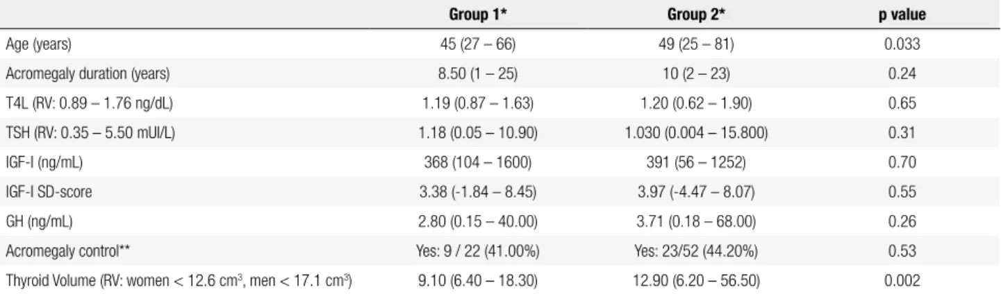
The group of patients with morphological abnorma lities (group 2, n = 75) showed a greater number of women (52 vs. 10, p = 0.001), older patients (49 vs. 45
yearsold, p = 0.033), and greater thyroid volume (12.9
vs. 9.1 cm3, p = 0.002) compared with the group of pa
tients with a normal thyroid gland at ultrasound (group 1, n = 31). No difference was found considering the following parameters: disease control, estimated acro megaly duration, IGFI, IGFI SDscore, GH, fT4 or TSH (Table 4).
Table 2. Morphological thyroid evaluation of 106 acromegalic patients
Thyroid morphology in
ultrasonography n (%) n (Female) n (Male)
Normal 31/106 (29.0%) 11 20
Diffuse goiter 11/106 (10.4%) 11 0
Nodular goiter 42/106 (40.0%) 24 18
Uninodular goiter 8/106 (7.5%) 4 4
Multinodular goiter 34/106 (32.5%) 20 14
Unspeciic morphology abnormalities
22/106 (20.6%) 12 10
Fine-needle aspiration biopsy
Benign 10/22 (45.5%) 6 4
Follicular lesions 8/22 (36.4%) 2 6
Papillary carcinoma 3/22 (13.6%) 2 1
Unsatisfactory 1/22 (4.6%) 0 1
Histological examination
Benign nodular hyperplasia 2/6 (33.3%) 0 2
Cop
yright
© ABE&M t
odos os dir
eit
os r
eser
vados
.
Table 3. Characteristics of the patients with thyroid carcinoma
Patient Sex Age (years)
Duration of acromegaly
(years)
Acromegaly
control FNAB Histopathology
#1 Male 49 13 Yes Papillary carcinoma Multifocal papillary carcinoma
#2 Female 56 1/3 No Papillary carcinoma Multifocal papillary carcinoma
#3 Female 63 2/9 Yes Papillary carcinoma Papillary carcinoma (follicular variant)
#4 Male 36 13 No Follicular tumor Papillary microcarcinoma
FNAB: ine-needle aspiration biopsy.
Table 4. Comparison of acromegalic patients without (n = 31, group 1) and with (n = 75, group 2) thyroid diseases (nodular goiter, diffuse goiter, and unspeciic morphology abnormalities)
Group 1* Group 2* p value
Age (years) 45 (27 – 66) 49 (25 – 81) 0.033
Acromegaly duration (years) 8.50 (1 – 25) 10 (2 – 23) 0.24
T4L (RV: 0.89 – 1.76 ng/dL) 1.19 (0.87 – 1.63) 1.20 (0.62 – 1.90) 0.65
TSH (RV: 0.35 – 5.50 mUI/L) 1.18 (0.05 – 10.90) 1.030 (0.004 – 15.800) 0.31
IGF-I (ng/mL) 368 (104 – 1600) 391 (56 – 1252) 0.70
IGF-I SD-score 3.38 (-1.84 – 8.45) 3.97 (-4.47 – 8.07) 0.55
GH (ng/mL) 2.80 (0.15 – 40.00) 3.71 (0.18 – 68.00) 0.26
Acromegaly control** Yes: 9 / 22 (41.00%) Yes: 23/52 (44.20%) 0.53
Thyroid Volume (RV: women < 12.6 cm3, men < 17.1 cm3) 9.10 (6.40 – 18.30) 12.90 (6.20 – 56.50) 0.002
* Data are shown as medians (minimum-maximum). ** Basal GH < 2.5 ng/mL and normal IGF-I. RV: reference value.
Considering only the patients with morphological abnormalities, thyroid volume was weakly correlated with IGFI levels (r = 0.255, p = 0.032) and IGFI SD score (r = 0.250, p = 0.036). There was no correlation between thyroid volume and age, estimated acromegaly duration, fT4 or TSH. Thyroid volume in this group of patients was greater in the patients with active acrome galy (13.7 vs. 8.9 cm3, p = 0.033). In the patients with
diffuse or nodular goiter, thyroid volume was greater in patients with active acromegaly (15.30 vs. 8.50 cm3, p
= 0.028), and was weakly correlated with GH levels (p = 0.032; r = 0.304), IGFI (p = 0.013; r = 0.347), and the IGFI SDscore (p = 0.017; r = 0.335). There was no correlation between thyroid volume and the dura tion of acromegaly. TSH levels were lower in patients with nodular disease (0.73 mU/L vs. 1.18 mU/L, p =
0.007). Five of the patients with lower TSH levels and nodular diseases (n = 6) had a pituitary macroadenoma, and all the six patients had secondary hypothyroidism with normal fT4 levels.
In the group of patients with morphological abnor malities, the patients with nodular or diffuse goiter (n = 53) had greater thyroid volume (14.7 vs. 12.9 cm3, p <
0.001) and longer estimated acromegaly duration than
patients with unspeciic morphology abnormalities (6
vs. 3 years, p = 0.019).
DISCUSSION
Cop
yright
© ABE&M t
odos os dir
eit
os r
eser
vados
.
se patients had a longer history of acromegaly. Cheung and Boyages (12) and Miyakawa and cols. (20) descri bed an important positive correlation between IGFI levels and thyroid volume, while Kasagi and cols. (13) found decreased TSH levels in acromegalic patients with nodular goiter.
In this study, the frequency of thyroid cancer in acromegalic patients was 3.8%, which may be underes timated because three patients had not yet undergo ne surgery. This frequency is approximately 300 times higher than the one reported in the overall Brazilian population (30), and in the majority of studies that have evaluated thyroid cancer in acromegalic patients (5,8,10,15,3133). Balkany and Cushing (17) and Marchisotti and cols. (34) found an approximately 3% prevalence of thyroid cancer in acromegalic patients, which is similar to our indings. Tita and cols. (14) des cribed seven cases of thyroid cancer in 125 acromega lic patients (5.6%). The use of routine ultrasonography might explain the higher frequency of thyroid cancer in acromegalic patients described in recent studies. However, dos Santos and cols. (35) demonstrated a significantly increased prevalence of thyroid cancer in acromegalic patients compared with a control group. The study by dos Santos and cols. included 124 acro megalic patients from northeast Brazil and 263 age and sexmatched control subjects. Nine acromegalic patients presented thyroid carcinoma (7.2%), and only two (0.7%) in the control group, which conirmed the higher prevalence of thyroid carcinoma in acromegaly. Rogozinski and cols. found four cases of differentia ted thyroid carcinoma among 34 acromegalic patients (11%) (24).
In this study, the patients with diffuse or nodular goiter that presented active acromegaly had greater thyroid volume than the patients with adequately trea ted acromegaly. This inding suggests that sustained ex posure to high GH and IGFI levels has a role in goiter development in the acromegalic population.
In conclusion, this study conirms the high fre quency of benign thyroid diseases in acromegalic pa tients. The prevalence of thyroid cancer was greater than expected for the overall population, which stron gly supports the indication of routine thyroid ultraso nography in patients with acromegaly.
Disclosure: no potential conlict of interest relevant to this article was reported.
REFERENCES
1. Melmed S. Acromegaly. N Engl J Med. 1990;322:966-77. 2. Dekkers OM, Biermasz NR, Pereira AM, Romijn JA, Vandenbroucke
JP. Mortality in acromegaly: a metaanalysis. J Clin Endocrinol Metab. 2008;93:61-7.
3. Wright AD, Hill DM, Lowy C, Fraser TR. Mortality in acromegaly. Q J Med. 1970;39:1-16.
4. Alexander L, Appleton D, Hall R, Ross WM, Wilkinson R. Epidemiology of acromegaly in the Newcastle region. Clin Endocrinol. 1980;12:71-9.
5. Nabarro JDN. Acromegaly. Clin Endocrinol. 1987;26:481-512. 6. Bengtsson B, Eden S, Ernest I, Oden A, Sjogren B. Epidemiology and
long-term survival in acromegaly. A study of 166 cases diagnosed between 1955 and 1984. Acta Med Scand. 1988;223:327-35. 7. Rajasoorya C, Holdaway IM, Wrightson P, Scott DJ, Ibbertson HK.
Determinants of clinical outcome and survival in acromegaly. Clin Endocrinol. 1994;41:95-102.
8. Orme SM, Mcnally RJQ, Cartwright RA, Belchetz PE. Mortality and cancer incidence in acromegaly: a retrospective cohort study. J Clin Endocrinol Metab. 1998;83:2730-4.
9. Barzilari J, Heatley GJ, Cushing GW. Benign and Malignant tumors in patients with acromegaly. Arch Intern Med. 1991;151(8):1629-32. 10. Popovic V, Damjanovic S, Micic D, Nesovic M, Djurovic M,
Petakov M; and the Pituitary Study Group. Increased incidence of neoplasia in patients with pituitary adenomas. Clin Endocrinol. 1998;49:441-5.
11. Jenkins PJ, Besser M. Clinical perspective: acromegaly and cancer: a problem. J Clin Endocrinol Metab. 2001;86(7):2935-41. 12. Cheung NW, Boyages SC. The thyroid gland in acromegaly: an
ultrasonographic study. Clin Endocrinol. 1997;46:545-9.
13. Kasagi K, Shimatsu A, Miyamoto S, Misaki T, Sakahara H, Konishi J. Goiter associated with acromegaly: sonographic indings of the thyroid gland. Thyroid. 1999;9:792-6.
14. Tita P, Ambrosio MR, Scollo C, Carta A, Gangemi P, Bondanelli M, et al. High prevalence of differentiated thyroid carcinoma in acromegaly. Clin Endocrinol. 2005;63:161-7.
15. Gasperi M, Martino E, Manetti L, Arosio M, Porretti S, Faglia G, et al. Prevalence of thyroid diseases in patients with acromegaly: results of an Italian multicenter study. J Endocrinol Invest. 2002;25:240-5.
16. Herrmann BL, Baumann H, Janssen OE, Görges R, Schmid KW, Mann K. Impact of disease activity on thyroid disease in patients with acromegaly: basal evaluation and follow up. Exp Clin Endocrinol Diabetes. 2004;112:225-30.
17. Balkany C, Cushing GW. An association between acromegaly and thyroid carcinoma. Thyroid. 1995;5:47-50.
18. Cannavò S, Squadrito S, Finocchiaro MD, Curtò L, Almoto B, Vieni A, et al. Goiter and impairment of thyroid function in acromegalic patients: basal evaluation and follow-up. Horm Metab Res. 2000;32(5):190-5.
19. Kurimoto M, Fukuda I, Hizuka N, Takano K. The prevalence of benign and malignant tumors in patients with acromegaly in a single institute. Endocr J. 2008;55(1):67-71.
20. Miyakawa M, Saji M, Tsushima M, Wakai K, Shizume K. Thyroid volume and serum thyroglobulin levels in patients with acromegaly: correlation with plasma insulin-like growth factor I levels. J Clin Endocrinol Metab. 1988;67(5):973-8.
21. Derwahl M, Broecker M, Kraiem Z. Clinical review 101: thyrotropin may not be the dominant growth factor in benign and malignant thyroid tumors. J Clin Endocrinol Metab. 1999;84:829-34. 22. Ruchala M, Skiba A, Gurgul E, Uruski P, Wasko R, Sowinski J. The
Cop
yright
© ABE&M t
odos os dir
eit
os r
eser
vados
.
aspiration biopsy in patients with acromegaly due to an increased risk of thyroid cancer. Neuro Endocrinol Lett. 2009;30(3):382-6. 23. Cheung NW, Boyages SC. Increased incidence of neoplasia in
females with acromegaly. Clin Endocrinol. 1997;47(3):323-7. 24. Rogozinski A, Furioso A, Glikman P, Junco M, Laudi R, Reyes A,
et al. Thyroid nodules in acromegaly. Arq Bras Endocrinol Metab. 2012;56(5):300-4.
25. Elmlinger MW, Kühnel W, Weber MM, Ranke MB. Reference ranges for two automated chemiluminescent assays for serum insulin-like growth factor I (IGF-I) and IGF-binding protein 3 (IGFBP3). Clin Chem Lab Med. 2004;42:654-64.
26. Prando D. Anatomia ultrassonográica normal da tireoide. In: Brandão LG. Tireóide e paratireóide: estudo de casos. São Paulo: Brandão LG; 2002. p. 311-6.
27. Cibas ES, Ali SZ. The Bethesda system for reporting thyroid cytopathology. Thyroid. 2009;19(11):1159-65.
28. Greene FL, Page D, Fleming ID, Fritz AG, Balch CM, Haller DG, et al. (eds). AJCC Cancer Staging Manual, 6th ed. Springer-Verlag; 2002. p. 77-87.
29. Wüster C, Steger G, Schmelzle A, Gottswinter J, Minne HW, Ziegler R. Increased incidence of euthyroid and hyperthyroid
goiters independently of thyrotropin in patients with acromegaly. Horm Metab Res. 1991;23(3):131-4.
30. Coeli MC, Brito AS, Barbosa FS, Ribeiro MG, Sieiro APAV, Vaisman M. Incidência e mortalidade por câncer de tireóide no Brasil. Arq Bras Endocrinol Metab. 2005;49(4):503-9.
31. Kauppinen-Mäkelin R, Sane T, Välimäki MJ, Markkanen H, Niskanen L, Ebeling T, et al. Increased cancer incidence in acromegaly – A nationwide survey. Clin Endocrinol (Oxf). 2009;72:277-9.
32. Baris D, Gridley G, Ron E, Weiderpass E, Mellemkjaer L, Ekbom A, et al. Acromegaly and cancer risk: a cohort study in Sweden and Denmark. Cancer Causes Control. 2002;13:395-400.
33. Barzilari J, Heatley GJ, Cushing GW. Benign and malignant tumors in patients with acromegaly. Arch Intern Med. 1991;151(8):1629-32.
34. Marchisotti FG, Umeda LM, Zach PL, Saldanha MD, First OS, Liberman B. [Acromegaly and thyroid disease: prevalence of thyroid cancer]. Arq Bras Endocrinol Metabol. 2005;49(5):843-9. 35. dos Santos MC, Nascimento GC, Nascimento AG, Carvalho VC,