Early diagnosis of atherosclerosis with panoramic radiographs:
a review
O diagnóstico precoce de aterosclerose através de radiografias panorâmicas:
uma revisão
Daiane Landim Borba1, Ulisses Vilela Hipólito1, Yamba Carla Lara Pereira1
*
Abstract
Carotid artery disease has been linked with cerebral vascular accident, also known as stroke, cerebral hemorrhage, or cerebral ischemia. It is caused by narrowing or obstruction of arteries in the neck (the carotid arteries) that are responsible for transporting blood from the aorta to the brain. Panoramic radiographs are used in dentistry to show both dental arches as a supplement to the clinical dental examination. he objective of this study is to highlight the importance of panoramic radiographs for diagnosis of arterial disease, by means of a bibliographic review. he PubMed database was searched using the keywords “atherosclerosis” and “panoramic”, with the ilters “last 5 years” and “humans”. Twenty articles were identiied, six of which were chosen for this study because they were open access. he review concluded that panoramic radiographs enable early diagnosis of carotid artery calciication, resulting in earlier interventions, and ofer an accessible cost.
Keywords: panoramic radiographs; atherosclerosis; early diagnosis.
Resumo
Doenças das artérias carótidas estão associadas a casos de acidente vascular cerebral, também chamados de derrame ou isquemia cerebral. Ocorrem devido ao estreitamento ou obstrução das artérias do pescoço, ou artérias carótidas, responsáveis por levar o sangue da aorta para o cérebro. A radiograia panorâmica é um exame de ambos os arcos dentários que ajuda no exame clínico odontológico. Destacar a importância da radiograia panorâmica no diagnóstico de doença arterial é o objetivo deste trabalho, que foi realizado através de pesquisa bibliográica na PubMed, com as palavras-chave “atherosclerosis” e “panoramic”, e os iltros: últimos 5 anos e humanos. Vinte artigos foram elencados, dos quais seis formaram a base para este estudo, graças ao acesso livre ao seu conteúdo. Conclui-se que a radiograia panorâmica permite o diagnóstico precoce de calciicação da artéria carótida, levando a uma intervenção precoce a um custo acessível.
Palavras-chave: radiograia panorâmica; aterosclerose; diagnóstico precoce.
1Faculdade de Ciências do Tocantins – FACIT, Curso de Odontologia, Araguaína, TO, Brazil.
Financial support: None.
Conlicts of interest: No conlicts of interest declared concerning the publication of this article. Submitted: October 02, 2016. Accepted: December 16, 2016.
INTRODUCTION
Cerebrovascular accidents (strokes) are the third
greatest cause of death in industrialized countries.
Atherosclerosis is a pathology that is related to strokes
and consists of formation calcium-rich fatty plaques
on the walls of arteries and their ramiications, which
can be diffuse and/or localized and contribute to
narrowing and hardening of the arteries, in combination
with accumulation of fat in the artery walls, which
is known as atheroma.
1They are generally seen radiographically in
individuals over the age of 50 and affect both sexes.
2Carotid artery disease can cause cerebrovascular
and encephalic accidents (also known as strokes or
cerebral ischemia), since they are directly associated
with narrowing or blockage of the arteries in the neck
(carotid arteries) that carry blood from the aorta to
the brain.
3In many cases, the disease is related to traditional
risk factors, such as high blood pressure, diabetes,
high cholesterol, smoking, and obesity, in which
the symptoms only appear when blood vessels are
almost completely blocked.
4Atherosclerosis has
chronic inlammatory characteristics and can cause
death and disability, making it a serious public
health problem because of the high cost of efforts
to rehabilitate patients.
5Of the different methods
to diagnose atherosclerotic disease, angiography is
considered the “gold standard”.
6However, since 1981,
certain radiopaque images seen in panoramic dental
X-rays have been described as a sign of the presence
of calciied carotid atheroma plaques.
7Panoramic
radiography provides images of the middle third of
the face, obtained using an extra-oral technique in
which the X-ray machine rotates around the patient,
acquiring a virtual image.
8This technique enables
examination of both dental arches and the neighboring
structures in a single X-ray. Their practicality and
comprehensiveness means they are considered the
diagnostic examination of choice, in combination
with the clinical examination.
9In asymptomatic individuals who are at risk of
stroke, atherosclerosis of the carotid artery (ACA) can
be identiied in a panoramic radiograph as a diffuse
bilateral radiopaque image extending from the region
of the ramus and angle of the mandible to the base of
the neck.
5This examination therefore has an important
role to play in early diagnosis of ACA.
10When ACA
is diagnosed, atherosclerosis treatment can be initiated
by the appropriate professional, with the objective
of repairing or mitigating acute or chronic ischemic
lesions, thereby preventing serious manifestations
and preserving the patient’s quality of life.
11In 2006, Friedlander assessed 94 people (mean
age of 65.6 years) for the presence of atheromatous
plaques using panoramic radiographs and in 50% of
them atheromatous plaques were found in carotid
arteries.
12This subset had occult metabolic syndrome,
which comprises a combination of abdominal obesity,
elevated triglycerides, reduced HDL levels, high blood
pressure, insulin resistance, and atheroma plaques.
12The common carotid artery ascends through the
mid-cervical area, where it bifurcates giving rise to the
external and internal carotid arteries.
13The location of
this bifurcation varies slightly and, on rare occasions,
may occur so far below the normal level that it is no
longer visible on a panoramic radiograph.
14Therefore,
a risk of stroke may go undetected in such patients
if this type of imaging examination is employed.
5However, if the region between cervical vertebrae C3
and C4 is observed carefully, and structures located
in the same region are differentiated, it is possible to
detect signs of atherosclerosis in the carotid arteries
using panoramic radiographs, thereby anticipating
treatment and reducing patient morbidity and mortality.
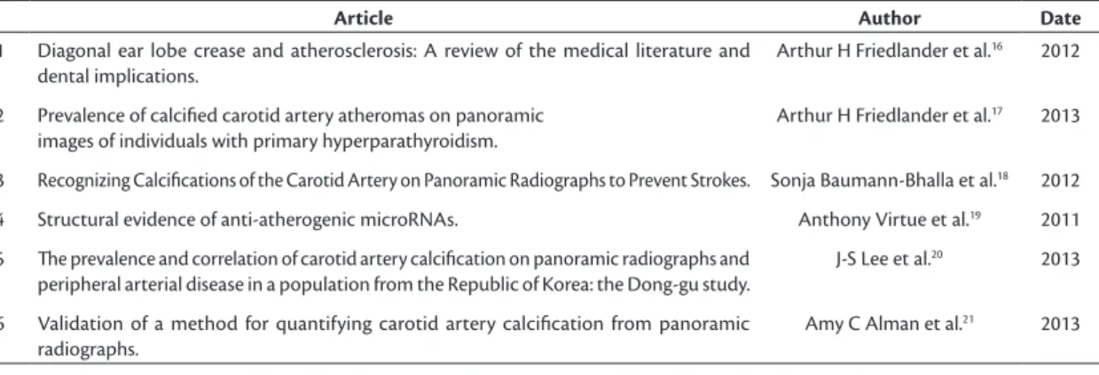
15Table 1. Descriptive table of items found in the database, with authors and year of publication.
Article Author Date
1 Diagonal ear lobe crease and atherosclerosis: A review of the medical literature and
dental implications.
Arthur H Friedlander et al.16 2012
2 Prevalence of calciied carotid artery atheromas on panoramic
images of individuals with primary hyperparathyroidism.
Arthur H Friedlander et al.17 2013
3 Recognizing Calciications of the Carotid Artery on Panoramic Radiographs to Prevent Strokes. Sonja Baumann-Bhalla et al.18 2012
4 Structural evidence of anti-atherogenic microRNAs. Anthony Virtue et al.19 2011
5 he prevalence and correlation of carotid artery calciication on panoramic radiographs and
peripheral arterial disease in a population from the Republic of Korea: the Dong-gu study.
J-S Lee et al.20 2013
6 Validation of a method for quantifying carotid artery calciication from panoramic
radiographs.
Detection of atheroma plaques in the carotid
artery by examination with panoramic radiographs
is valuable for early diagnosis and for minimizing
patient risk, contributing to early referral of patients
for treatment.
MATERIAL AND METHOD
This is a review of bibliography identiied by
searching the PubMed Web site with the keywords:
“atherosclerosis” and “panoramic”. The ilters used
were “last 5 years” and “humans”.
RESULT
The search results identiied 20 articles, 6 of which
were reviewed for this study, chosen because they
were open access. Four publications were from 2013
and two were from 2012 (Table 1 and
Figure 1
).
DISCUSSION
Atherosclerosis is one of the leading causes of
mortality in Brazil.
22It is a chronic inlammatory
disease of multifactorial origin, in which there is
accumulation of fat, cholesterol, and other substances
on the artery walls, restricting blood flow and
causing many different health problems.
23Factors
that contribute to the emergence of arterial disease
include: high cholesterol, diabetes, obesity, smoking,
family history of heart disease, physical inactivity,
chronic kidney disease, and others.
24One of the manifestations of atherosclerosis is
formation of atheroma, which are characterized by
large accumulations of lipids, ibrous tissues, calcium
deposits, blood, and blood products, among others.
25Plaque formation occurs after endothelial damage
followed by tissue repair.
26The risk factors can
damage the vascular endothelium, causing endothelial
dysfunction and mediating entry of monocytes, which
include modiied proteins such as oxidative LDL, for
example, causing foam cells.
27Inlammatory mediators
are released, extending the process and forming the
plaques.
28Cerebrovascular accidents (strokes), also
known as encephalic vascular accidents,
29are one of
the leading causes of death and disability all over the
world.
30They are characterized by blood leakage in
brain tissues.
31When blood circulation is interrupted,
functional and structural changes also occur in the
region involved, establishing a complex “ischemic
cascade”, the inal result of which is neuronal death.
32In these cases, the diagnosis of the disease is based
on the patient’s clinical status associated with a
neurological examination.
33The irst X-ray was performed on November 8,
1895, by Rontgen, who used the left hand of his wife,
Anna Bertha Rontgen, placed between a frame holding
photographic ilm and a cathode ray tube that emitted
radiation for 15 minutes. After the ilm was developed,
the shape of his wife’s hand was revealed, showing
the bones inside less dense soft tissue.
34Radiographic
examinations are of fundamental importance in dentistry
because they supplement the clinical examination
and are essential for diagnosis and for planning and
monitoring dental treatments.
35Dentistry relies on
two different types of X-ray techniques: intra-oral
and extra-oral. In intra-oral methods, the X-ray ilm
or sensor is placed inside the patient’s mouth. This
category in turn can be subdivided into periapical,
occlusal, and interproximal, which aim to provide
detailed views of dental elements and adjacent bone
tissue.
36Extra-oral techniques consist of taking X-rays
with the radiographic ilm or sensor exposed outside
of the patient’s mouth.
37The most widely used of
the extra-oral techniques is panoramic radiography,
which offers visualization of both dental arches and
their surrounding structures in a single X-ray.
38There
are both analog and digital methods.
39Panoramic radiography is of fundamental
importance in early and incidental diagnosis of
carotid atheroma.
40It is the dental surgeon’s duty to
recognize atherosclerosis when inspecting panoramic
radiography images and to instruct patients to seek
conirmation with other examination methods and
to refer them to the relevant health professional for
Figure 1. Images compatible with carotid atherosclerotic lesion
appropriate treatment.
41This is in order to ensure
patients’ quality of life and well-being.
42In a 2012 article, Friedlander et al. discussed the
need to identify atherosclerosis and its risk factors
before the complications set in. They stated that
atherosclerosis is the leading cause of deaths in Spain
and drew an analogy of the prevalence of the disease
to extra-vascular signs related to earlobe creases.
16They emphasize that such creases are incipient signs of
circulatory problems in the head and neck area and that
their etiology is associated with atherosclerotic disease.
They recommended that, although further research
is advisable, dentists should conduct examinations
of their patients’ ears, checking for diagonal earlobe
creases and, in conjunction with clinical history,
vital signs, and panoramic radiographs, decide on
the need for a medical assessment of the patient.
16In 2013, the same group of researchers presented
reports associating primary hyperparathyroidism
(PHPT) to carotid complications. They stated that
calciied carotid artery atheroma are often recorded
in panoramic images of patients with PHPT and so
health professionals should be vigilant.
17Baumann-Bhalla et al. consider that it is important
to examine more closely the panoramic radiographs
that are taken every day in Switzerland, especially in
relation to arterial calciication, and to direct affected
patients to a specialist in order to conirm or rule
out this diagnosis. They stressed that on panoramic
radiographs, it not only teeth and jaws that should
be analyzed, but also the areas at the sides of the
image, especially in patients over the age of 50 and
in patients with risk factors, thereby making early
recognition of calciications more likely and preventing
cerebrovascular events.
18Virtue et al.
19have studied regulation of
pro-inlammatory genes inluencing inhibition of
production of miRNAs with inlammatory potential.
Their study suggests a treatment avenue, based on an
understanding of the protective mechanism provided
by miRNAs, especially through suppression of the
atherogenic effects of certain genes, and also shows
that individual patient characteristics are risk factors
for triggering atherosclerotic processes.
Data published in 2013 illustrate the prevalence
of carotid artery calciication (CAC), detected with
panoramic radiographs and associated with peripheral
arterial disease (PAD). They analyzed the difference
in PAD prevalence between patients with and patients
without CAC detectable on panoramic radiographs.
The study sample comprised 4078 subjects aged
50 or older (1410 males and 2668 females) who had
undergone medical and dental examinations in the city
of Gwangju, South Korea. Panoramic radiographs and
presence of carotid artery calciication were analyzed.
Presence of PAD was determined by measuring the
ankle-brachial index (ABI). An ABI of 0.9 in any leg
was considered evidence of PAD. The prevalence of
CAC in panoramic radiographs was 6.2 and the PAD
prevalence was 2.6, in middle-aged or older patients.
It is known that it is important to detect CAC and
peripheral artery disease to prevent fatal events such
as ischemic stroke and myocardial infarction.
20Digital panoramic radiographs were used to evaluate
the area of carotid artery calciication using tools
available in ImageJ, while inpatient and outpatient
discharge records were reviewed to identify patients
who had undergone Doppler ultrasound examination
of the carotid arteries. Area under the curve analysis
showed that quantiication of carotid artery calciication
correlated well with degree of stenosis. They concluded
that quantiication of carotid artery calciication using
digital panoramic images can identify patients who
need further evaluation, such as with conventional
medical tests.
21CONCLUSION
It is concluded that panoramic radiography, a
routine examination used by dentists, offers the
opportunity for early diagnosis of carotid artery
calciication, enabling interventions while disorders
are still incipient, ensuring preservation of patients’
quality of life at an accessible cost, accomplished
using an inexpensive imaging exam that is widely
available for dentists, neurologists, and angiologists.
REFERENCES
1. Friedlander AH, El Saden SM, Hazboun RC, Chang TI, Wong WK, Garrett NR. Detection of carotid artery calcification on the panoramic images of post-menopausal females is significantly associated with severe abdominal aortic calcification: a risk indicator of future adverse vascular events. Dentomaxillofac Radiol. 2015;44(7):20150094. PMid:25945511. http://dx.doi.org/10.1259/ dmfr.20150094.
2. Ramesh A, Pabla T. Panoramic radiographs: a screening tool for calcified carotid atheromatous plaque. J Mass Dent Soc. 2007;56(2):20-1. PMid:17691505.
3. Zétola A, Ferreira FM, Larson R, Shibli JA. Recombinant human bone morphogenetic protein-2 (rhBMP-2) in the treatment of mandibular sequelae after tumor resection. Oral Maxillofac Surg. 2011;15(3):169-74. PMid:20571845. http://dx.doi.org/10.1007/ s10006-010-0236-7.
5. Abreu TQ, Ferreira EB, Brito SB Fo, Sales KP, Lopes FF, Oliveira AE. Prevalence of carotid artery calcifications detected on panoramic radiographs and confirmed by Doppler ultrasonography: Their relationship with systemic conditions. Indian J Dent Res. 2015;26(4):345-50. PMid:26481878. http://dx.doi.org/10.4103/0970-9290.167644. 6. Romano-Sousa CM, Krejci L, Medeiros FM, et al. Diagnostic agreement between panoramic radiographs and color Doppler images of carotid atheroma. J Appl Oral Sci. 2009;17(1):45-8. PMid:19148405. http://dx.doi.org/10.1590/S1678-77572009000100009.
7. Khambete N, Kumar R, Risbud M, Joshi A. Reliability of digital panoramic radiographs in detecting calcified carotid artery atheromatous plaques: a clinical study. Indian J Dent Res. 2014;25(1):36-40. PMid:24748296. http://dx.doi.org/10.4103/0970-9290.131052.
8. Friedman MH, Weisberg J. The craniocervical connection: a retrospective analysis of 300 whiplash patients with cervical and temporomandibular disorders. Cranio. 2000;18(3):163-7. PMid:11202833. http://dx.doi.org/10.1080/08869634.2000.1174 6128.
9. Ramesh A, Soroushian S, Ganguly R. Coincidence of calcified carotid atheromatous plaque, osteoporosis, and periodontal bone loss in dental panoramic radiographs. Imaging Sci Dent. 2013;43(4):235-43. PMid:24380062. http://dx.doi.org/10.5624/ isd.2013.43.4.235.
10. Martinez-Cruz S, Manson-Hing LR. Comparison of focal trough dimensions and form by resolution measurements in panoramic radiography. J Am Dent Assoc. 1987;114(5):639-42. PMid:3474266. http://dx.doi.org/10.14219/jada.archive.1987.0139.
11. Hoke M, Schmidt B, Schillinger T, et al. Evidence of carotid atherosclerosis in orthopantomograms and the risk for future cardiovascular events. Vasa. 2010;39(4):298-304. PMid:21104618. http://dx.doi.org/10.1024/0301-1526/a000053.
12. Friedlander AH, Golub MS. The significance of carotid artery atheromas on panoramic radiographs in the diagnosis of occult metabolic syndrome. Oral Surg Oral Med Oral Pathol Oral Radiol Endod. 2006;101(1):95-101. PMid:16360613. http://dx.doi. org/10.1016/j.tripleo.2005.04.027.
13. Sobotta, J. Sobotta atlas de anatomia humana. 23 ed. Rio de
Janeiro: Guanabara Koogan; 2012. 3 v.
14. Khalil H. A basic review on the inferior alveolar nerve block techniques. Anesth Essays Res. 2014;8(1):3-8. PMid:25886095. http://dx.doi.org/10.4103/0259-1162.128891.
15. Mathew S, Lund RJ, Strebeck F, Tustison KS, Geurs T, Hruska KA. Reversal of the adynamic bone disorder and decreased vascular calcification in chronic kidney disease by sevelamer carbonate therapy. J Am Soc Nephrol. 2007;18(1):122-30. PMid:17182886. http://dx.doi.org/10.1681/ASN.2006050490.
16. Friedlander AH, López-López J, Velasco-Ortega E. Diagonal ear lobe crease and atherosclerosis: a review of the medical literature and dental implications. Med Oral Patol Oral Cir Bucal. 2012;17(1):e153-9. PMid:21743392. http://dx.doi.org/10.4317/medoral.17390.
17. Friedlander AH, Aghazadehsanai N, Chang TI, Harada N, Garrett NR. Prevalence of calcified carotid artery atheromas on panoramic images of individuals with primary hyperparathyroidism. Dentomaxillofac Radiol. 2013;42(8):20130118. PMid:23775925. http://dx.doi.org/10.1259/dmfr.20130118.
18. Baumann-Bhalla S, Meier RM, Burow A, et al. Recognizing calcifications of the carotid artery on panoramic radiographs to prevent strokes. Schweiz Monatsschr Zahnmed. 2012;122(11):1016-29. PMid:23184365.
19. Virtue A, Mai J, Yin Y, et al. Structural evidence of anti-atherogenic microRNAs. Front Biosci. 2011;16(1):3133-45. PMid:21622224. http://dx.doi.org/10.2741/3901.
20. Lee JS, Kim OS, Chung HJ, et al. The prevalence and correlation of carotid artery calcification on panoramic radiographs and peripheral arterial disease in a population from the Republic of Korea: the Dong-gu study. Dentomaxillofac Radiol. 2013;42(3):29725099. PMid:22752323. http://dx.doi.org/10.1259/dmfr/29725099.
21. Alman AC, Johnson LR, Calverley DC, Grunwald GK, Lezotte DC, Hokanson JE. Validation of a method for quantifying carotid artery calcification from panoramic radiographs. Oral Surg Oral Med Oral Pathol Oral Radiol. 2013;116(4):518-24. PMid:24035118. http://dx.doi.org/10.1016/j.oooo.2013.06.026.
22. Rabelo LM, Viana RM, Schimith MA, et al. Risk factors for atherosclerosis in students of a private university in São Paulo-Brazil. Arq Bras Cardiol. 1999;72(5):569-80. PMid:10668227. http:// dx.doi.org/10.1590/S0066-782X1999000500004.
23. Ashley KE, Geraci SA. Ischemic heart disease in women. South Med J. 2013;106(7):427-33. PMid:23820324. http://dx.doi.org/10.1097/ SMJ.0b013e31829b9eab.
24. Buttar HS, Li T, Ravi N. Prevention of cardiovascular diseases: Role of exercise, dietary interventions, obesity and smoking cessation. Exp Clin Cardiol. 2005;10(4):229-49. PMid:19641674.
25. Singh RB, Mengi SA, Xu YJ, Arneja AS, Dhalla NS. Pathogenesis of atherosclerosis: A multifactorial process. Exp Clin Cardiol. 2002;7(1):40-53. PMid:19644578.
26. van Hinsbergh VW. Endothelium--role in regulation of coagulation and inflammation. Semin Immunopathol. 2012;34(1):93-106. PMid:21845431. http://dx.doi.org/10.1007/s00281-011-0285-5.
27. Singh N, Dhalla AK, Seneviratne C, Singal PK. Oxidative stress and heart failure. Mol Cell Biochem. 1995;147(1-2):77-81. PMid:7494558. http://dx.doi.org/10.1007/BF00944786.
28. Galkina E, Ley K. Immune and inflammatory mechanisms of atherosclerosis. Annu Rev Immunol. 2009;27(1):165-97. PMid:19302038. http://dx.doi.org/10.1146/annurev.immunol.021908.132620. 29. Herrmann N, Mamdani M, Lanctôt KL. Atypical antipsychotics and
risk of cerebrovascular accidents. Am J Psychiatry. 2004;161(6):1113-5. PMid:15169702. http://dx.doi.org/10.1176/appi.ajp.161.6.1113.
30. Percudani M, Barbui C, Fortino I, Tansella M, Petrovich L. Second-generation antipsychotics and risk of cerebrovascular accidents in the elderly. J Clin Psychopharmacol. 2005;25(5):468-70. PMid:16160623. http://dx.doi.org/10.1097/01.jcp.0000178414.14685.c4.
31. Chodobski A, Zink BJ, Szmydynger-Chodobska J. Blood-brain barrier pathophysiology in traumatic brain injury. Transl Stroke Res. 2011;2(4):492-516. PMid:22299022. http://dx.doi.org/10.1007/ s12975-011-0125-x.
32. Lee JM, Grabb MC, Zipfel GJ, Choi DW. Brain tissue responses to ischemia. J Clin Invest. 2000;106(6):723-31. PMid:10995780. http:// dx.doi.org/10.1172/JCI11003.
33. Tillema JM, Pirko I. Neuroradiological evaluation of demyelinating disease. Ther Adv Neurol Disorder. 2013;6(4):249-68. PMid:23858328. http://dx.doi.org/10.1177/1756285613478870.
34. Kundel HL, Nodine CF. A visual concept shapes image perception. Radiology. 1983;146(2):363-8. PMid:6849084. http://dx.doi. org/10.1148/radiology.146.2.6849084.
35. Gross H, Nilsson M, Hellén-Halme K. Detectability of normal anatomy in digital panoramic radiographs. Swed Dent J. 2014;38(4):179-85. PMid:25771652.
36. Jones KB, Jordan R. White lesions in the oral cavity: clinical presentation, diagnosis, and treatment. Semin Cutan Med Surg. 2015;34(4):161-70. PMid:26650693. http://dx.doi.org/10.12788/j. sder.2015.0180.
into the middle cranial fossa. J Can Dent Assoc. 2002;68(11):676-80. PMid:12513935.
38. Shah N, Bansal N, Logani A. Recent advances in imaging technologies in dentistry. World J Radiol. 2014;6(10):794-807. PMid:25349663. http://dx.doi.org/10.4329/wjr.v6.i10.794.
39. Kiefer H, Lambrecht JT, Roth J. Dose exposure from analog and digital full mouth radiography and panoramic radiography. Schweiz Monatsschr Zahnmed. 2004;114(7):687-93. PMid:15360104.
40. Patil SR. Prevalence of carotid artery calcification in postmenopausal women and its correlation with atherogenic risk factors. J Nat Sci Biol Med. 2015;6(Suppl 1):S1-6. PMid:26604593. http://dx.doi. org/10.4103/0976-9668.166048.
41. Friedlander AH. Panoramic radiography: the differential diagnosis of carotid artery atheromas. Spec Care Dentist. 1995;15(6):223-7. PMid:9002923. http://dx.doi.org/10.1111/j.1754-4505.1995. tb00522.x.
42. Friedlander AH, Friedlander IK. Identification of stroke prone patients by panoramic radiography. Aust Dent J. 1998;43(1):51-4.
PMid:9583227. http://dx.doi.org/10.1111/j.1834-7819.1998. tb00153.x.
*
Correspondence
Yamba Carla Lara Pereira Rua Águas Claras, 59/703 CEP 77824-230 - Araguaína (TO), Brazil Tel.: +55 (63) 99996-7311 E-mail: [email protected]
Author information
DLB - Undergraduate student, Faculdade de Ciências do Tocantins (FACIT). UVH - Nurse; MSc and PhD in Sciences; post-doctoral studies in Pediatric Nursing; Professor at Faculdade de Ciências do Tocantins (FACIT) and Universidade Federal do Tocantins. YCLP - DDS; PhD in Oral-Dental Biology; MSc in Sciences; Specialist in Endodontics and Dental Radiology (focus on Public Health and Oral Rehabilitation); MBA candidate in School Management at PECEGE/ESALQ/USP.
Author contributions
Conception and design: UVH, YCLP Analysis and interpretation: DLB Data collection: DLB Writing the article: DLB, UVH, YCLP Critical revision of the article: DLB, UVH, YCLP Final approval of the article*: DLB, UVH, YCLP Statistical analysis: N/A. Overall responsibility: DLB, UVH, YCLP