Original article
reconstruction: factors inluencing occurrences of complications
and the inal outcome
Retalho miocutâneo de peitoral maior em reconstruções de cabeça e pescoço: fatores que
inluenciam a ocorrência de complicações e o resultado inal
Fábio Roberto Pinto
I, Carina Rosa Malena
II, Christiana Maria Ribeiro Salles Vanni
III, Fábio de Aquino Capelli
IV,
Leandro Luongo de Matos
V, Jossi Ledo Kanda
VIDepartment of Head and Neck Surgery, Hospital de Ensino Padre Anchieta, Faculdade de Medicina do ABC (FMABC), São Bernardo do Campo, São Paulo, Brazil
IMD, PhD. Attending physician, Discipline of Head and Neck Surgery, Faculdade de Medicina do ABC (FMABC), São Bernardo do Campo, São Paulo, Brazil. IIMD. Resident, Discipline of Plastic Surgery, Faculdade de Medicina do ABC (FMABC), São Bernardo do Campo, São Paulo, Brazil.
IIIMD. Attending physician, Discipline of Head and Neck Surgery, Faculdade de Medicina do ABC (FMABC), São Bernardo do Campo, São Paulo, Brazil. IVMD. Attending physician, Discipline of Head and Neck Surgery, Faculdade de Medicina do ABC (FMABC), São Bernardo do Campo, São Paulo, Brazil. VMD, MSc. Resident, Discipline of Head and Neck Surgery, Faculdade de Medicina do ABC (FMABC), São Bernardo do Campo, São Paulo, Brazil.
ABSTRACT
CONTEXT AND OBJECTIVE: Pedicled laps play an important role in cancer treatment centers, particularly in developing and emerging countries. The aim of this study was to identify factors that may cause complications and inluence the inal result from reconstructions using pectoralis major myocutaneous laps (PMMFs) for head and neck defect repair following cancer resection.
DESIGN AND SETTING: Cross-sectional study at the Hospital de Ensino Padre Anchieta of Faculdade de Medicina do ABC (FMABC).
METHODS: Data on 58 patients who underwent head and neck defect reconstruction using PMMFs were reviewed. The inal result from the reconstruction (success or failure) and the complications observed were evaluated in relation to the patients’ ages, area reconstructed, disease stage, previous oncological treatment and need for blood transfusion.
RESULTS: There were no total lap losses. The reconstruction success rate was 93.1%. Flap-related complications occurred in 43.1% of the cases, and half of them were considered major. Most of the complications were successfully treated. Defects originating in the hypopharynx were correlated with the development of major complications (P = 0.02) and with reconstruction failure (P < 0.001). Previous oncological treatment negatively inluenced the reconstruction success (P = 0.04).
CONCLUSIONS: Since the risk factors for developing major complications and reconstruction failure are known, it is important to heed the technical details and provide careful clinical support for patients in a more critical condition, so that better results from using PMMFs can be obtained.
RESUMO
CONTEXTO E OBJETIVO: Retalhos pediculados têm grande importância nos centros de tratamento do câncer, principalmente em países emergentes e em desenvolvimento. O objetivo deste trabalho foi identiicar fatores que determinam complicações e inluenciam no resultado inal das reconstruções com retalho miocutâneo de peitoral maior (RMPM) no reparo de defeitos cirúrgicos por ressecções de cânceres de cabeça e pescoço.
TIPO DE ESTUDO E LOCAL: Estudo transversal no Hospital de Ensino Padre Anchieta da Faculdade de Medicina do ABC.
MÉTODOS: Dados de 58 pacientes submetidos a reconstruções de defeitos de cabeça e pescoço com RMPM foram revisados. O resultado inal da reconstrução (sucesso ou falha) e as complicações observadas foram avaliados de acordo com a idade do paciente, área reconstruída, estágio da doença, tratamento oncológico prévio e necessidade de transfusão sanguínea.
RESULTADOS: Não houve perda total de retalho. A taxa de sucesso da reconstrução foi de 93,1%. Complicações relacionadas ao retalho ocorreram em 43,1% dos casos e 50% delas foram considerados como complicações maiores. A maioria das complicações foi tratada com sucesso. Os defeitos originados da hipofaringe relacionaram-se com o desenvolvimento de complicações maiores (P = 0,02) e falhas na reconstrução (P < 0,001). Tratamento oncológico prévio inluenciou negativamente no sucesso da reconstrução (P = 0,04).
CONCLUSÕES: Como os fatores de risco para o desenvolvimento de complicações maiores e de falha na reconstrução são conhecidos, é importante atentar para detalhes técnicos e empregar suporte clínico cuidadoso aos pacientes mais críticos para que sejam obtidos melhores resultados com o uso do RMPM.
KEY WORDS:
Surgical laps.
Reconstructive surgical procedures. Head and neck neoplasms. Postoperative complications. Treatment outcome.
PALAVRAS-CHAVE: Retalhos cirúrgicos. Procedimentos cirúrgicos reconstrutivos.
INTRODUCTION
Reconstruction of complex head and neck defects resulting from cancer resection remains a challenge for head and neck and plastic sur-geons. Free laps, which are considered to be the “gold standard” for this kind of reparative procedure, are not available in many centers that treat head and neck tumors, because of the high costs and the highly spe-cialized technology involved in microsurgical techniques.1 Furthermore, the extended period of anesthesia required for constructing the micro-surgical laps2 means that this kind of reconstructive tool is sometimes not the best choice for critically ill patients.
For these reasons, pedicled laps continue to play an important role in many institutions worldwide,3-6 with special regard to cancer treat-ment centers in developing and emerging countries. Among these laps, pectoralis major myocutaneous laps (PMMFs) are undoubtedly the most reliable and versatile type of lap and are still considered to be the “workhorse” in head and neck reconstruction. hirty years after the use of PMMFs for head and neck defect repair was irst described by Ariyan,7 PMMFs are still being used on a large scale in many centers, and they have been the subject of many published papers.8-12
he literature shows that the PMMF complication rate is between 17% and 63%.10,12-16 Despite some authors’ assertion that, in skilled hands, free laps result in fewer complications than do PMMFs,15,17 there is a consensus that total lap necrosis is a rare complication when PMMFs are used, even when an inexperienced surgeon harvests the lap.10,18-21 In addition, most of the remaining complications associated with PMMFs, including mild skin lap necrosis, are treated by means of a conservative approach with satisfactory resolution.14,16,21,22 Hence, in the institutions where PMMFs are widely used, it is important to inves-tigate the results and the complications associated with their use.
OBJECTIVE
he objective of this paper was to analyze our experience with PMMFs for head and neck reconstruction and try to determine risk factors for developing complications and reconstruction outcome pre-dictors when this lap is employed.
METHODS
Patients and data collection
We conducted a cross-sectional study on all cases of head and neck cancer that were surgically treated at our institution (Department of Head and Neck Surgery, Faculdade de Medicina do ABC) between 2000 and 2009 and underwent reconstruction of postablative defects using PMMFs. he medical iles were reviewed and the tumors were re-staged in accordance with the 2000 TNM criteria (Union Internation-ale Contre le Cancer, UICC), as stages I to IV. Patients were eligible for the study if they had presented malignant tumors of the head and neck and underwent immediate or delayed reconstruction using PMMFs. he protocol described here was approved by our Department’s Ethics
Committee and was therefore implemented in accordance with the ethi-cal standards laid down in the 1964 Declaration of Helsinki.
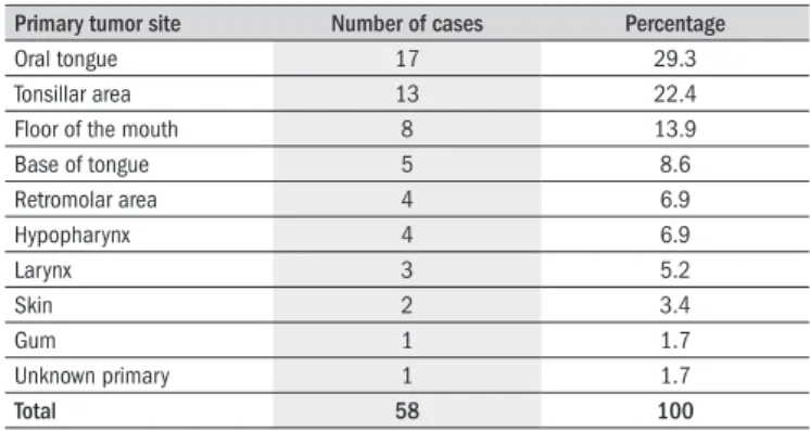
We identiied 61 cases for analysis. hree cases had to be excluded because these patients died within the irst six postoperative days due to clinical complications and thus, investigation of lap-related compli-cations and reconstruction outcomes was impossible. Hence, 58 cases were studied. Among these, 55 (94.8%) were men and the mean age of the group was 54.1 ± 8.5 years. Almost all the tumors were squamous cell carcinomas (57/58; 98.2%), mainly located in the oral cavity and oropharynx, and were at an advanced stage of disease (stages III and IV). Other locations included the hypopharynx, skin and unknown primary tumor (Tables 1 and 2). One patient was afected by a liposarcoma lo-cated in the supraclavicular fossa. he areas that were reconstructed were divided into four categories to enable analysis: tongue/loor of mouth; retromolar area/oropharynx; hypopharynx; and skin.
We also categorized patients according to whether they had received any previous oncological treatment (surgery, radiation or chemothera-py) or not. he need for blood transfusion was recorded as well. All pa-tients undergoing oral cavity and pharynx reconstruction were followed up by a speech therapist for speech and swallowing rehabilitation.
he surgical technique used to harvest the PMMF was as described in the literature,7,10,18,23 and is depicted in Figure 1 (A-C). In most cas-es, the vascular pedicle was dissected under direct viewing, after mak-ing an incision in the chest skin from the upper border of the cutane-ous paddle of the lap to the midclavicular line. Likewise, in most of the cases (n = 57; 98.2%), the supraclavicular route was used to transfer the lap to the defect. Figure 1D shows an intraoral transferred lap three months after the surgery.
he inal result from the reconstruction (reconstruction outcome) and the presence and severity of complications were analyzed. he re-construction outcome was divided into two categories: success, when the goal of the reconstructive procedure was achieved; and failure, when the
Primary tumor site Number of cases Percentage
Oral tongue 17 29.3
Tonsillar area 13 22.4 Floor of the mouth 8 13.9
Base of tongue 5 8.6
Retromolar area 4 6.9
Hypopharynx 4 6.9
Larynx 3 5.2
Skin 2 3.4
Gum 1 1.7
Unknown primary 1 1.7
Total 58 100
Table 1. Distribution of the primary tumor sites (n = 58)
Disease stage Number of cases Percentage
IV 37 63.8
III 15 25.9
I + II 6 10.3
II 5 8.6
I 1 1.7
Table 2. Disease stage, in accordance with the UICC (Union Internacionale
Data analysis
In each case, the parameters “inal result from the reconstruction” and “presence or absence of major and minor complications” were com-pared in relation to the following variables: age, disease stage, area re-constructed, previous oncological treatment and need for blood trans-fusion. he age variable was deined in relation to the mean age, i.e. < 54 years or ≥ 54 years. he disease stage was separated into stages I and II (considered together because of the low numbers of cases classi-ied in these stages), stage III and stage IV. Previous oncological treat-ment and the need for blood transfusion had two possible responses: “yes” or “no.”
Associations between the inal result from the reconstruction and the variables of age, previous oncological treatment and need for blood transfusion were evaluated by means of Fisher’s exact test. his statistical test was also used to evaluate the relationships between the presence of major and minor complications and the same variables as above. Associ-ations presented by the inal result from the reconstruction and the pres-ence of major and minor complications in relation to disease stage and area reconstructed were evaluated by means of the chi-square test. Find-ings were taken to be statistically signiicant when P ≤ 0.05. he SPSS (Statistical Package for the Social Sciences) statistical software, version 13.0 for Windows (SPSS Inc; Illinois, USA), was used for all the statis-tical analyses.
RESULTS
he length of hospital stay ranged from 2 to 32 days (mean: 10.3 ±
6.9 days; median: 8 days). he mean time taken to perform the opera-tion of harvesting the lap and transferring it toward the defect was ap-proximately 50 minutes. he distribution of the cases regarding the area reconstructed, previous oncological treatment and need for blood trans-fusion is shown in Table 3.
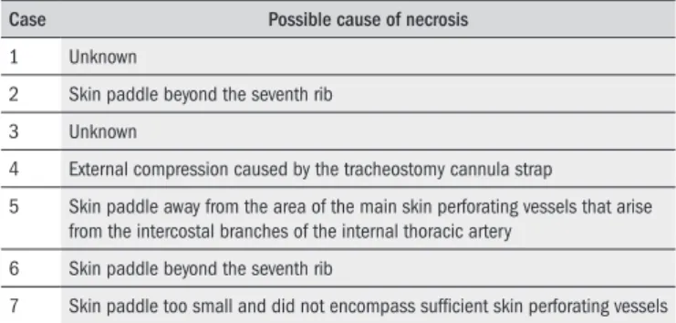
he reconstruction was a success in 54 cases (93.1%) and a failure in the remaining four cases. here were seven cases of partial lap ne-crosis (12%), although most of them were limited to less than 25% of the cutaneous component of the lap. In all of these cases, the inal out-come was successful. here were no cases of total lap loss. Among the seven laps that developed partial loss, we were able to identify a pos-sible technical basis that could explain the necrosis observed, in ive of them (Table 4).
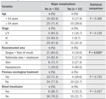
Some kind of complication occurred in 38 cases (65.5%), and lap-related complications were observed in 25 cases (43.1%). Major com-plications were observed in 14 patients (24.1%) and minor complica-tions were seen in 12 cases (20.7%). In one case, independent major and minor complications were recorded. All lap-related complications and other complications are listed in Table 5. Most patients (n = 41; 70.7%) received adjuvant radiotherapy and no further lap-related complications were recorded in this group. A statistically signiicant association was ob-served between the presence of major complications and the hypophar-ynx as the area reconstructed (P = 0.020; chi-square test). Among the ive patients who underwent hypopharynx reconstruction using PMMFs, four (80%) developed major complications. No statistical relationship
Figure 1. Surgical steps involved in pectoralis major myocutaneous laps
harvesting and delayed appearance of an intraoral lap: A. Skin paddle incision; B. Flap rotation through supraclavicular route; C. Flap inserted into oral cavity to repair the defect; D. Reconstructed oral cavity and oropharynx three months after the surgery.
reparative objectives were not reached or the patient died because of sur-gical complications. For example, for intraoral and pharyngeal defects, the objective of the reconstruction procedure was to enable intelligible speech and restore swallowing ability, in order to enable exclusive oral intake without the aid of a feeding tube.
he complications were classiied as lap-related complications if they were directly associated with the lap, the repaired area or the donor site.
Other complications were those that were not directly related to the re-parative procedure, including surgical and clinical complications. he lap-related complications were categorized as suggested by Chepeha et al.,15 as major complications or minor complications. Major complications were those that required reoperation in a surgical theater, or resulted in failure to attain the reconstruction goal. Minor complications were those that were treated successfully by means of conservative manage-ment, i.e. without reoperation in the surgical theater, and from which the result was successful reconstruction. Conservative management in-cluded packing, small drainage, debridement and medication.
Area reconstructed Number of cases Percentage
Tongue + loor of mouth 25 43.1 Retromolar area + oropharynx 17 29.3
Skin 11 19.0
Hypopharynx 5 8.6
Previous oncological treatment Number of cases Percentage
Yes 14 24.1
No 44 75.9
Blood transfusion Number of cases Percentage
Yes 12 20.7
No 46 79.3
Table 3. Case distribution in relation to the area reconstructed, previous
treatment and blood transfusion (n = 58)
Case Possible cause of necrosis
1 Unknown
2 Skin paddle beyond the seventh rib 3 Unknown
4 External compression caused by the tracheostomy cannula strap
5 Skin paddle away from the area of the main skin perforating vessels that arise from the intercostal branches of the internal thoracic artery
6 Skin paddle beyond the seventh rib
7 Skin paddle too small and did not encompass suficient skin perforating vessels
Table 4. Possible causes of development of partial necrosis, in each case
affected
*In this case the area reconstructed was the skin.
Major complications Number
of cases %
Minor complications
Number
of cases %
Other complications
Number
of cases %
No 44 75.9 No 46 79.3 No 36 62.1
Yes 14 24.1 Yes 12 20.7 Yes 22 37.9
Orocutaneous or pharyngocutaneous istula 3 5.2 Orocutaneous or
pharyngocutaneous istula 6 10.3 Neck skin dehiscence 9 15.7 Partial lap loss + intraoral lap dehiscence
and/or donor site dehiscence 3 5.2 Neck skin dehiscence 2 3.5 Lymphatic istula 2 3.5 Partial lap loss 2 3.5 Partial lap loss 2 3.5 Neck skin dehiscence and hyperkalemia 1 1.7 Death 2 3.5 Donor site hematoma 1 1.7 Neck skin dehiscence and ischemic stroke 1 1.7 Donor site abscess 1 1.7 Reconstructed area infection 1 1.7 Neck abscess 1 1.7
Titanium plate exposure 1 1.7 Neck seroma 1 1.7
Intraoral lap dehiscence 1 1.7 Orocutaneous istula* and mandibular
osteomyelitis 1 1.7
Venous congestion of the lap 1 1.7
Cardiac arrhythmia and pneumonia 1 1.7 Prerenal acute renal failure 1 1.7 Hypertensive crisis 1 1.7 Pneumothorax 1 1.7 Lymphatic istula + neck skin dehiscence
+ pneumonia 1 1.7
Pleural empyema 1 1.7
Table 5. Flap-related complications (major and minor complications) and other complications (n = 58)
was observed between this parameter and the other variables of interest (Table 6). In the analysis on minor complications, no statistical associa-tion was observed between this parameter and the variables of interest.
A statistically signiicant association between the inal result from the reconstruction and the area reconstructed was observed. Again, when the hypopharynx was the area reconstructed, the chance of re-parative failure was higher (P < 0.001; chi-square test). Previous onco-logical treatment also negatively inluenced the reconstruction outcome (P = 0.04; Fisher’s exact test). No statistical relationship was observed between the inal result from the reconstruction and the other variables of interest (Table 7).
DISCUSSION
Despite the recent advances in microsurgical techniques, local and regional laps are still acceptable options for reconstruction of complex head and neck defects. Among these laps, PMMFs are the most reliable and versatile type and may be considered to be the irst reparative choice for patients disigured by cancer or presenting severe medical morbidi-ties. In these situations, it is advisable to opt for a lap type that, in ad-dition to reliability, requires shorter operative time. Since many head and neck cancer patients are diagnosed at an advanced stage and, con-sequently, present signiicant weight loss,24 PMMFs continue to be used on a large scale in many centers.3,10-12
Upon analyzing our cases, what stood out was that we had used PMMFs for a wide spectrum of defects, encompassing almost all mu-cosal sites and diferent locations on the face and neck skin. hus, this diversity of applications attests to the versatility of PMMFs, as already pointed out by other researchers.18,19,25,26 A high success rate for recon-struction purposes was observed using PMMFs (54/58; 93.1%), and this rate compares favorably with several papers in the literature.9,13,15,27
From evaluation of the inal result from the reconstruction, the vari-ables that statistically correlated with reconstruction failure were the hy-popharynx as the site of the defect and other oncological treatment prior
had received some kind of treatment prior to the repair procedure.9,12 Scar tissue from previous surgery and microcirculation compromised by radiotherapy are factors that may be involved in this condition. More-over, when the hypopharynx is reconstructed, enhanced nutritional sup-port must be provided. Both for hypopharyngeal defects and for situa-tions in which patients have undergone previous treatment, care must
be taken during cancer resection and PMMF harvesting in order to pre-serve the vascular integrity of other locoregional laps, such as deltopec-toral laps, which may be useful in the event of PMMF failure.
We did not observe any cases of total lap loss and our rate of partial loss was acceptable, with good evolution in all of these cases. hese re-sults are also comparable to those in the literature.3,9,10,12,27,30 From anal-ysis on the seven cases in which partial lap loss occurred, we were able to identify the possible cause of this complication in ive of them. As pointed out by Rikimaru et al.,31 positioning the skin island just medi-ally to the nipple, over the fourth, ifth and sixth intercostal spaces, is essential for encompassing the skin perforator vessels that arise from the intercostal branches of the internal thoracic artery. hese cutaneous ves-sels are supplied by the pectoralis branch of the thoracoacromial artery, through open choke vessels, when the main blood low through the in-ternal thoracic artery is interrupted during PMMF elevation. Hence, a totally axial myocutaneous lap may be created respecting this anatomi-cal condition. Below the seventh rib, the vascular supply for the skin comes from the cutaneous branches of the superior epigastric artery,31 and therefore, when portions of skin beyond this limit are included in the lap, this creates an axial lap with a distal random portion, thereby increasing the risk of partial loss. In two cases, the skin island extended below the seventh rib and, in one case, the skin paddle was located away from the position of the main skin perforator vessels. One lap had a small skin paddle that probably did not encompass a suicient number of skin perforator vessels, thus resulting in unstable blood circulation.32 In the other case, external compression caused by the tracheostomy can-nula strap led to lap vascular impairment and necrosis. Hence, we con-clude that attention should be paid to avoiding technical mistakes and, particularly, to placing the PMMF skin island over the cutaneous perfo-rator vessel area, in order to ensure minimum risk of lap necrosis.
Another pitfall, described by Cunha-Gomes et al.,33 relates to the lateral pectoralis nerve division. hese authors observed that this nerve may lie parallel or oblique to the PMMF vascular pedicle. When running obliquely to the pedicle, the lateral thoracic nerve becomes taut after the lap is rotated through 180º and presses against the vascular pedicle, thus leading to PMMF vascular impairment. hese authors observed this phe-nomenon in 30% of their cases and recommended that this nerve should be dissected and divided when the above situation is observed.
Regarding the complications recorded in the present study, the lap-related complication rate (43.1%) was comparable to other series.3,9,10,13,16,19 An acceptable number of major complications were re-corded, and most of these cases evolved successfully after proper treat-ment. here were only four instances, representing 28.6% of the major complications recorded, in which the objectives of the reparative proce-dure were not achieved. he presence of major complications was posi-tively correlated with the hypopharynx as the reconstruction site. Again, the clinical characteristics of the hypopharyngeal cancer patients, along with the particular features of hypopharynx reconstruction discussed above, explain these results.
Most of our patients received adjuvant radiotherapy and no delayed complications were recorded. hese data prove that PMMFs tolerate ra-diotherapy well. Moreover, a recent study has suggested that
modiica-Variables Final result Statistical
comparison
Success (n = 54) Failure (n = 4)
Age n (%) n (%)
< 54 years 23 (100.0) 0 (0.0) P = 0.144 ≥ 54 years 31 (88.6) 4 (11.4)
Stage n (%) n (%)
I/II 6 (100.0) 0 (0.0) P = 0.295 III 15 (100.0) 0 (0.0)
IV 33 (89.2) 4 (10.8)
Reconstructed area n (%) n (%)
Tongue + loor of mouth 25 (100.0) 0 (0.0) P < 0.0001*
Retromolar area + oropharynx 17 (100.0) 0 (0.0) Skin 10 (90.9) 1 (9.1) Hypopharynx 2 (40.0) 3 (60.0)
Previous oncological treatment n (%) n (%)
Yes 11 (78.6) 3 (21.4) P = 0.040†
No 43 (97.7) 1 (2.3)
Blood transfusion n (%) n (%)
Yes 11 (91.7) 1 (8.3) P = 1.000 No 43 (93.5) 3 (6.5)
*Statistical relationship between the inal result from reconstruction and the area reconstructed area, using chi-square test. †Statistical relationship between the inal result from reconstruction and the history of previous
oncological treatment, using Fisher’s exact test.
Table 6. Comparisons between the inal result from reconstruction and the
variables of interest (age, area reconstructed, previous treatment and need for blood transfusion). The values that showed statistical signiicance are depicted in bold (n = 58)
Variables Major complications comparisonStatistical
No (n = 32) Yes (n = 10)
Age n (%) n (%)
< 54 years 19 (82.6) 4 (17.4) P = 0.369 ≥ 54 years 25 (71.4) 10 (28.6)
Stage n (%) n (%)
I/II 5 (83.3) 1 (16.7) P = 0.130 III 14 (93.3) 1 (6.7)
IV 25 (67.6) 12 (32.4)
Reconstructed area n (%) n (%)
Tongue + loor of mouth 21 (84.0) 4 (16.0) P = 0.020*
Retromolar area + oropharynx 14 (82.4) 3 (17.6) Skin 8 (72.7) 3 (27.3) Hypopharynx 1 (20.0) 4 (80.0)
Previous oncological treatment n (%) n (%)
Yes 10 (71.4) 4 (28.6) P = 0.725 No 34 (77.3) 10 (22.7)
Blood transfusion n (%) n (%)
Yes 8 (66.7) 4 (33.3) P = 0.457 No 36 (78.3) 10 (21.7)
*Statistical relationship between the presence of major complications and the stage, using chi-square test.
Table 7. Comparisons between the presence of major complications
tion of the intraoral environment through the efects of ionizing radia-tion may predispose towards some kind of metaplasia of the cutaneous portion of PMMFs, in relation to the adjacent mucosa. his phenom-enon may represent an advantageous adaptation for patients.34
CONCLUSIONS
he results from the present study emphasize that PMMFs are a liable, versatile and feasible type of pedicled lap for head and neck re-construction. Since advanced disease in the head and neck is the rule, it can be expected that large numbers of clinically jeopardized patients will continue to be treated using this method. Although high numbers of lap-related complications were observed with the use of PMMFs in the present study, the reparative goals were achieved in the cases of most of the patients. Since the risk factors for developing major complica-tions and outcome failure may be anticipated, we are convinced that if the technical pitfalls listed throughout this paper are given due heed and judicious clinical and nutritional support is provided for patients in a more critical condition, better results from PMMFs can be obtained.
REFERENCES
1. Petruzzelli GJ, Brockenbrough JM, Vandevender D, Creech SD. The inluence of reconstructive modality on cost of care in head and neck oncologic surgery. Arch Otolaryngol Head Neck Surg. 2002;128(12):1377-80.
2. Smeele LE, Goldstein D, Tsai V, et al. Morbidity and cost differences between free lap re-construction and pedicled lap rere-construction in oral and oropharyngeal cancer: Matched control study. J Otolaryngol. 2006;35(2):102-7.
3. Vartanian JG, Carvalho AL, Carvalho SM, et al. Pectoralis major and other myofascial/myo-cutaneous laps in head and neck cancer reconstruction: experience with 437 cases at a single institution. Head Neck. 2004;26(12):1018-23.
4. Andrews BT, McCulloch TM, Funk GF, Graham SM, Hoffman HT. Deltopectoral lap revisited in the microvascular era: a single institution 10-year experience. Ann Otol Rhinol Laryngol. 2006;115(1):35-40.
5. Uğurlu K, Ozçelik D, Hüthüt I, et al. Extended vertical trapezius myocutaneous lap in head and neck reconstruction as a salvage procedure. Plast Reconstr Surg. 2004;114(2): 339-50.
6. Pinto FR, de Magalhães RP, Capelli F de A, Brandão LG, Kanda JL. Pedicled temporo-parietal galeal lap for reconstruction of intraoral defects. Ann Otol Rhinol Laryngol. 2008;117(8):581-6.
7. Ariyan S. The pectoralis major myocutaneous lap. A versatile lap for reconstruction in the head and neck. Plast Reconstr Surg. 1979;63(1):73-81.
8. Salvatori P, Motto E, Paradisi S, et al. Oromandibular reconstruction using titanium plate and pectoralis major myocutaneous lap. Acta Otorhinolaryngol Ital. 2007;27(5):227-32. 9. El-Marakby HH. The reliability of pectoralis major myocutaneous lap in head and neck
reconstruction. J Egypt Natl Canc Inst. 2006;18(1):41-50.
10. Milenović A, Virag M, Uglesić V, Aljinović-Ratković N. The pectoralis major lap in head and neck reconstruction: irst 500 patients. J Craniomaxillofac Surg. 2006;34(6):340-3. 11. Croce A, Moretti A, D’Agostino L, Neri G. Continuing validity of pectoralis major muscle lap
25 years after its irst application. Acta Otorhinolaryngol Ital. 2003;23(4):297-304. 12. Liu R, Gullane P, Brown D, Irish J. Pectoralis major myocutaneous pedicled lap in head
and neck reconstruction: retrospective review of indications and results in 244 consecutive cases at the Toronto General Hospital. J Otolaryngol. 2001;30(1):34-40.
13. Shah JP, Haribhakti V, Loree TR, Sutaria P. Complications of the pectoralis major myocuta-neous lap in head and neck reconstruction. Am J Surg. 1990;160(4):352-5.
14. Castelli ML, Pecorari G, Succo G, et al. Pectoralis major myocutaneous lap: analysis of complications in dificult patients. Eur Arch Otorhinolaryngol. 2001;258(10):542-5. 15. Chepeha DB, Annich G, Pynnonen MA, et al. Pectoralis major myocutaneous lap vs
revas-cularized free tissue transfer: complications, gastrostomy tube dependence, and hospitali-zation. Arch Otolaryngol Head Neck Surg. 2004;130(2):181-6.
16. Wadwongtham W, Isipradit P, Supanakorn S. The pectoralis major myocutaneous lap:
ap-plications and comap-plications in head and neck reconstruction. J Med Assoc Thai. 2004;87 Suppl 2:S95-9.
17. Tsue TT, Desyatnikova SS, Deleyiannis FW, et al. Comparison of cost and function in recons-truction of the posterior oral cavity and oropharynx. Free vs pedicled soft tissue transfer. Arch Otolaryngol Head Neck Surg. 1997;123(7):731-7.
18. Ariyan S. Further experiences with the pectoralis major myocutaneous lap for the imme-diate repair of defects from excisions of head and neck cancers. Plast Reconstr Surg. 1979;64(5):605-12.
19. Baek SM, Lawson W, Biller HF. An analysis of 133 pectoralis major myocutaneous laps. Plast Reconstr Surg. 1982;69(3):460-9.
20. Nagral S, Sankhe M, Patel CV. Experience with the pectoralis major myocutaneous lap for head and neck reconstruction in a general surgical unit. J Postgrad Med. 1992;38(3):119-23. 21. Koh KS, Eom JS, Kirk I, Kim SY, Nam S. Pectoralis major musculocutaneous lap in
oropharyn-geal reconstruction: revisited. Plast Reconstr Surg. 2006;118(5):1145-9; discussion 1150. 22. Saussez S, Cuno A, Urbain F, Chantrain G, Lequeux T. Reconstruction of circumferential oro- and hypopharyngeal defects with U-shaped pectoralis major myocutaneous lap. Otolaryn-gol Head Neck Surg. 2006;134(5):823-9.
23. Freeman JL, Walker EP, Wilson JS, Shaw HJ. The vascular anatomy of the pectoralis major myocutaneous lap. Br J Plast Surg. 1981;34(1):3-10.
24. Jager-Wittenaar H, Dijkstra PU, Vissink A, et al. Critical weight loss in head and neck can-cer--prevalence and risk factors at diagnosis: an explorative study. Support Care Cancer. 2007;15(9):1045-50.
25. Chew CT, Stanley R, Peck R, Chew SC. Pectoralis major myocutaneous lap reconstruc-tion in head and neck surgery--experience with 60 cases. Ann Acad Med Singapore. 1991;20(5):570-80.
26. Vendrell Marqués JB, Zapater Latorre E, Ferrandis Perepérez E, Estellés Ferriol E, Brotons Durbán S. Colgajo pediculado de pectoral mayor: nuestra experiencia en 76 casos con-secutivos [Pedicled pectoralis major musculocutaneous laps]. Acta Otorrinolaringol Esp. 2002;53(1):39-45.
27. Pfuetzenreiter Júnior EG, Andrade CRA, Lehn CN, Dedivitis RA. O retalho músculo-cutâneo peitoral maior na reconstrução do câncer de cabeça e pescoço [The pectoralis major musculocutaneous lap in head and neck cancer reconstruction]. Arq Bras Ciênc Saúde. 2008;33(3):151-4.
28. Triboulet JP, Mariette C, Chevalier D, Amrouni H. Surgical management of carcinoma of the hypopharynx and cervical esophagus: analysis of 209 cases. Arch Surg. 2001;136(10): 1164-70.
29. Clark JR, Gilbert R, Irish J, et al. Morbidity after lap reconstruction of hypopharyngeal de-fects. Laryngoscope. 2006;116(2):173-81.
30. Magrin J, Kowalski LP, Sabóia M, Sabóia RP. Major glossectomy: end results of 106 cases. Eur J Cancer B Oral Oncol. 1996;32B(6):407-12.
31. Rikimaru H, Kiyokawa K, Inoue Y, Tai Y. Three-dimensional anatomical vascular distribution in the pectoralis major myocutaneous lap. Plast Reconstr Surg. 2005;115(5):1342-52; discussion 1353-4.
32. Rikimaru H, Kiyokawa K, Watanabe K, et al. New method of preparing a pectoralis major myocutaneous lap with a skin paddle that includes the third intercostal perforating branch of the internal thoracic artery. Plast Reconstr Surg. 2009;123(4):1220-8.
33. Cunha-Gomes D, Choudhari C, Kavarana NM. Vascular compromise of the pectora-lis major musculocutaneous lap in head and neck reconstruction. Ann Plast Surg. 2003;51(5):450-4.
34. Pinto FR, Kanda JL, Diniz MT, Santos CP, Zveibil DK. Delayed histological changes in cuta-neous portion of pectoralis major laps employed in upper aero-digestive tract reconstruc-tion. Eur Arch Otorhinolaryngol. 2009;266(4):553-8.
Sources of funding: None
Conlict of interest: None
Date of irst submission: March 23, 2010
Last received: June 20, 2010
Accepted: September 21, 2010
Address for correspondence: