Original article
vulvovaginitis and skin color
Relação entre agentes infecciosos de vulvovaginites e cor da pele
Rosekeila Simoes Nomelini
I, Ana Paula Borges Carrijo
II, Sheila Jorge Adad
III,
Altacílio Aparecido Nunes
IV, Eddie Fernando Candido Murta
VInstituto de Pesquisa em Oncologia (IPON), Discipline of Gynecology and Obstetrics, Universidade Federal do Triângulo Mineiro (UFTM), Uberaba, Minas Gerais, Brazil
IMD. Professor in the Instituto de Pesquisa em Oncologia (IPON), Discipline of Gynecology and Obstetrics, Universidade Federal do Triângulo Mineiro (UFTM), Uberaba, Minas Gerais, Brazil. IIMedical student, Universidade Federal do Triângulo Mineiro (UFTM), Uberaba, Minas Gerais, Brazil.
IIIPhD. Professor in the Discipline of Special Pathology, Universidade Federal do Triângulo Mineiro (UFTM), Uberaba, Minas Gerais, Brazil.
IVMD. Professor in the Department of Social Medicine, Faculdade de Medicina de Ribeirão Preto, Universidade de São Paulo (FMRP/USP), Ribeirão Preto, Brazil.
ABSTRACT
CONTEXT AND Objective: Many factors inluence occurrences of vulvovaginitis. The aims here were to assess skin color and age-related differences in the vaginal lora and occurrences of vulvovaginitis.
DESIGN AND SETTING: Cross-sectional study; tertiary referral hospital (Universidade Federal do Triângulo Mineiro, Uberaba).
METHODS: Healthy women who underwent routine outpatient gynecological assessments were assessed for vulvovaginitis and vaginal lora and then divided into whites (n = 13,881) and nonwhites (n = 5,295). Statistical analysis was performed using the X2 test, logistic regression and odds ratios.
RESULTS: The vaginal microlora was skin-color dependent, with greater occurrence of clue cells, Trichomonas vaginalis and coccobacilli in nonwhite women (P < 0.0001). Döderlein bacilli and cytolytic lora were more prevalent in white women (P < 0.0001 and P < 0.05, respectively). The vaginal
microlora was age-dependent within the skin color groups. Among the nonwhite women, clue cells were more prevalent in women aged 21 to 50 years; Trichomonas in women up to 40 years and coccobacili in women between 21 and 40 years (P < 0.05). During the proliferative and secretory
phases, the nonwhite women were more likely to present clue cells, Trichomonas, Candida and coccobacilli (OR, proliferative phase: 1.31, 1.79, 1.6 and 1.25 respectively; secretory phase: 1.31, 2.88, 1.74 and 1.21 respectively), while less likely to present Döderlein lora (OR, proliferative phase:
0.76; secretory phase: 0.66), compared with white women, irrespective of age.
CONCLUSIONS: There are differences in vulvovaginitis occurrence relating to skin color, which may be associated with variations in vaginal lora.
RESUMO
CONTEXTO E OBJETIVO: Muitos fatores inluenciam a ocorrência de vulvovaginites. Os objetivos foram avaliar diferenças relacionadas à cor da pele e idade na lora vaginal e vulvovaginites.
TIPO DE ESTUDO E LOCAL: Estudo transversal; hospital de referência terciário (Universidade Federal do Triângulo Mineiro, Uberaba).
MÉTODOS: Mulheres saudáveis em atendimento de rotina para exames ginecológicos foram divididas em brancas (n = 13.881) e não brancas (n = 5.295) e avaliadas quanto a vulvovaginites e lora vaginal. Para análise estatística, foram utilizados teste X2, regressão logística e odds ratio. RESULTADOS: Microlora vaginal foi dependente da cor da pele, com maior ocorrência de “clue cells”, Trichomonas vaginalis e bacilos cocoides em não brancas (P < 0,0001); bacilos de Döderleine lora citolítica foram mais prevalentes em brancas (P < 0,0001 e P < 0,05, respectivamente). Flora
vaginal foi dependente da idade nos grupos de cor da pele. Entre não brancas, “clue cells”, Trichomonas e bacilos cocoides foram mais prevalentes nas idades: 21 a 50 anos, até 40 anos, e 21 a 40 anos respectivamente (P < 0,05). Durante as fases proliferativa e secretória, mulheres não
brancas tiveram maior probabilidade de apresentar “clue cells”, Trichomonas, Candida e cocoides (odds ratio, OR - fase proliferativa: 1,31; 1,79; 1,6 e 1,25 respectivamente; fase secretória: 1,31; 2,88; 1,74 e 1,21 respectivamente), e menor chance de apresentarem lora Döderlein (OR - fase
proliferativa: 0,76; fase secretória: 0,66) comparadas com brancas, independentemente da idade.
CONCLUSÕES: Há diferenças na ocorrência de vulvovaginites relacionadas com a cor da pele, podendo haver associação com variações na lora vaginal.
KEY WORDS:
Continental population groups. Vulvovaginitis.
Vaginosis, bacterial. Candidiasis.
Lactobacillus.
PALAVRAS-CHAVES:
Grupos de populações continentais.
Vulvovaginite. Vaginose bacteriana.
INTRODUCTION
Many factors may inluence occurrences of vulvovaginitis, including age, vaginal pH, hysterectomy, menstrual cycle phase, HPV (human pap-illomavirus) infection and skin color. Murta et al. demonstrated, via Pa-panicolaou staining, that occurrences of infectious agents for vaginitis cor-relate with age; and further, occurrences of Candida sp. in women over 60 years of age are inluenced by hysterectomy.1 he microlora are
interde-pendent, with a reported association between Gardnerella vaginalis and HPV infection.2 Moreover, the presence of vaginal lora is afected by the
phase of the menstrual cycle, as in the case of cytolytic lora, and by Can-dida albicans abundance,3 which in turn impacts local pH. During the
proliferative phase of the menstrual cycle and in post-menopausal wom-en, vaginal pH is correlated with endocervical pH.4
Racial diferences in vaginal lora ecology have been established, and numerous studies have reported diferences in occurrences of bac-terial vaginosis among racial groups, and speciically, higher prevalence among black women than among white women.5-7 However, the
major-ity of these studies evaluated bacterial vaginosis (and other types of vul-vovaginitis) solely in pregnant women, which in turn correlated with a greater number of preterm deliveries among black women than among white women.8 Bacterial vaginosis, Trichomonas vaginalis, Chlamydia trachomatis, Mycoplasma hominis and group B streptococcus coloniza-tion were also found to be more frequent in black women, and the pres-ence of one or more of these common microbial conditions was associ-ated with an increased risk of preterm birth.9
Bacterial vaginosis and candidiasis are infections relating to altera-tions in vaginal pH. Several studies have demonstrated that black wom-en have fewer lactobacilli, which maintain vaginal pH via anaerobic me-tabolism of vaginal glycogen to acidic products.10 his is possibly due
to overabundance of hydrogen peroxide-producing bacteria, which sup-presses the growth of anaerobic bacteria such as lactobacilli.11,12
Howev-er, the exact cause of this racial diference remains unknown.
OBJECTIVE
he aim of the present study was to assess potential diferences in diagnosing vaginal infectious agents, speciically clue cells and vaginal lora, among non-pregnant white and nonwhite women.
METHODS
Participants
A cross-sectional study was conducted at the Cytopathology Ser-vice of the Universidade Federal do Triângulo Mineiro (UFTM), Ubera-ba, Minas Gerais, Brazil, and the Instituto de Pesquisa em Oncologia (IPON), Discipline of Gynecology and Obstetrics (a tertiary referral hospital with free public attendance). he data were collected from the medical records by a single medical student who was participating in a scientiic initiation program (Fundação de Amparo à Pesquisa do Estado de Minas Gerais; FAPEMIG).
he presence of clue cells, Trichomonas vaginalis, Candida albicans, coccoid bacilli, Döderlein bacilli and cytolytic lora was determined via vaginal, cervical and endocervical cytological tests on healthy women who underwent outpatient gynecological assessments at the Discipline of Gynecology and Obstetrics at UFTM between January 1996 and December 2006.
he methodology for collecting cervicovaginal cytological samples is standard in our gynecology service, and the Papanicolaou method was used by the pathologist for the evaluations. During routine pelvic exam-ination on 19,176 non-pregnant women, the uterine cervix was exposed using a sterile and non-lubricated speculum and endocervical samples were obtained by means of a brush, and cervical and vaginal samples by means of a spatula. None of these women were undergoing hormone therapy, or contraception douching or taking vaginal medication. Hys-terectomized women were excluded. he cytological smears were ixed in ethyl alcohol and stained using the Papanicolaou method. he sam-ples were evaluated by trained and experienced cytopathologists for the presence of Trichomonas vaginalis, Candida sp., clue cells,Döderlein ba-cilli and vaginal lora using the established diagnosis criteria.
After the cytological analysis, the participant pool was divided into two groups according to skin color: (1) white women (described in the records as “white”); and (2) nonwhite women (described in the records as “black” or other mixed skin colors). Information concerning age, pregnancies, parity, smoking and phase of the menstrual cycle was ob-tained from the participants at the time of sample collection. he health professional who collected the samples described the subjects as white, black or other mixed colors.
For the menstrual cycle analysis, women with cycles of up to 30 days were included (proliferative phase, days 1 to 14; secretory phase, days 15 to 30). Out of the 19,176 patients, 14,442 (75.31%) were includ-ed. Among these, there were 6,863 patients (47.52%) in the prolifera-tive phase, of whom 4,987 (72.67%) were white and 1,876 (27.33%) were nonwhite; and 7,579 patients (52.48%) in the secretory phase, of whom 5,619 (74.14%) were white and 1,960 (25.86%) were nonwhite. Each woman was included only once in the series, and it was standard-ized that only the irst Pap smear from each patient would be recorded in the database.
his study was approved by the Research Ethics Committee of UFTM.
Cytological criteria
vesicular or crescent-shaped nucleus, lightly stained with hematoxylin, were also occasionally observed. Cytolysis was deined as a pale stain-ing; vesicular nuclei with little or no cytoplasm of the intermediate cells predominated in such smears. Coccobacilli were characterized as dif-fusely scattered bacilli and cocci organisms that occasionally occurred in clumps or microcolonies. Lactobacilli were characterized by the pres-ence of elongated bacillary structures.13-16
Statistical analysis
he results were assessed for statistical signiicance using the chi-squared test with the Yates correction, multivariate logistic regression and odds ratios, and a 95% conidence interval. he statistical signii-cance level was set at P ≤ 0.05.
RESULTS
A total of 19,176 participants were evaluated, and they were divid-ed into two groups basdivid-ed on skin color: white women (n = 13,881) and nonwhite women (n = 5,295). he mean age ± standard deviation (SD) was 33.0 ± 9.91 years among the white women, with a range from 13 to 82 years, and 31.1 ± 9.94 years among the nonwhite women, with a range from 12 to 98 years. he smear test was performed on older wom-en because they had symptoms. Table 1 shows the characteristics of each group of patients.
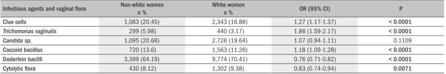
he adjusted odds ratio for age, taking into considering the expo-sure factor of nonwhite skin color is summarized in Table 2. Positive diagnoses of clue cells, Trichomonas vaginalis or coccobacilli were more frequent among the nonwhite women (P < 0.0001) than among the white women; while Döderlein and cytolytic lora were more frequent among the white women (P < 0.0001 and P < 0.05, respectively) than among the nonwhite women. he frequency of Candida sp.was not skin color-dependent.
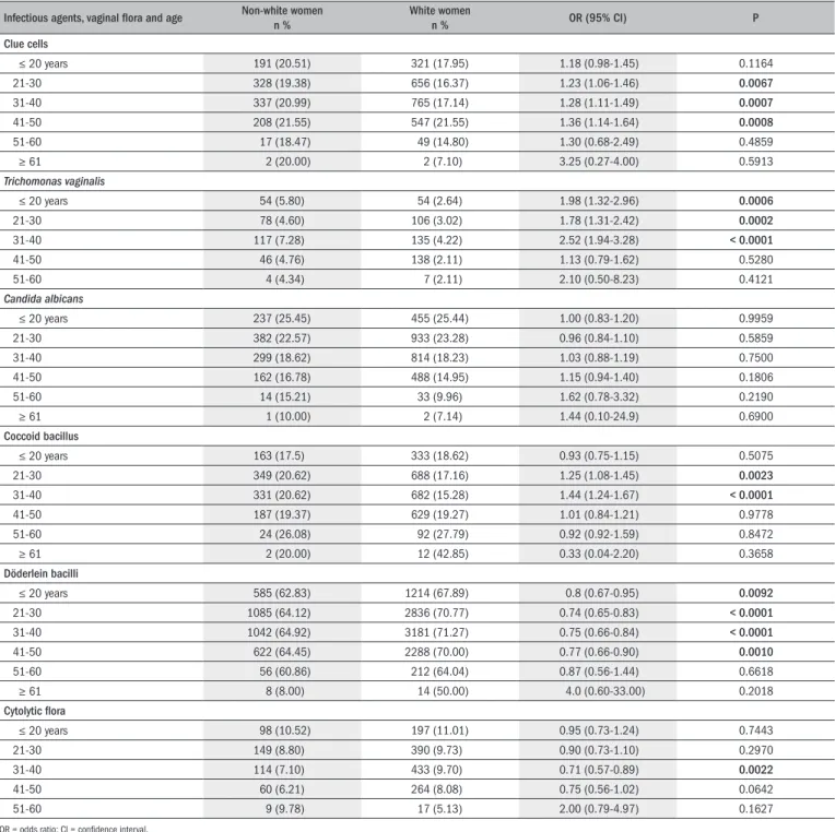
Yates correlation analysis revealed that skin color and age were sig-niicantly correlated in a vaginal lora-dependent manner. here were signiicant associations for occurrences of clue cells in nonwhite
wom-en betwewom-en 21 and 50 years of age, Trichomonas vaginalis in nonwhite women up to 40 years of age and coccobacilli in nonwhite women be-tween 21 and 40 years of age (P < 0.05). Döderlein and cytolytic lora were more prevalent in white women up to 50 years of age and between 31 and 40 years of age, respectively (P < 0.05). he occurrences of Can-dida sp. were not dependent on skin color or age (Table 3).
During the proliferative phase of the menstrual cycle, the nonwhite women were 1.31, 1.79, 1.6 and 1.25 times more likely to have a posi-tive diagnosis of clue cells, Trichomonas vaginalis,Candida sp. and coc-cobacilli, respectively. However, they were 24% and 22% less likely to be diagnosed with Döderlein lora and cytolytic lora, respectively, com-pared with the white women, regardless of age (Table 4).
During the secretory phase of the menstrual cycle, the nonwhite women were 1.31, 2.88, 1.74 and 1.21 times more likely to have a posi-tive diagnosis of clue cells, Trichomonas vaginalis,Candida sp. and coc-cobacilli, respectively. However, they were 34% less likely to be diag-nosed with Döderlein lora, compared with the white women, regardless of age. No statistically signiicant diferences were observed among the cytolytic lora during the secretory phase (Table 5).
DISCUSSION
he current evidence suggests that occurrences of bacterial vaginosis may be related to race. his includes recent reports of higher frequency of bacterial vaginosis in black women;17-19 relationships between
bacte-rial vaginosis, premature delivery20 and pelvic inlammatory disease;21
and higher risk of preterm birth among black women.9 Moreover,
ge-netic factors can inluence the susceptibility to infection.22 Macones et
al. demonstrated that an interaction between genetic susceptibilities (tu-mor necrosis factor-2 carriers) and environmental factors (bacterial vagi-nosis) was associated with a higher risk of spontaneous preterm birth.23
Ryckman et al. demonstrated that susceptibility to bacterial vaginosis was afected by patterns of genetic variation in stress-related genes, and that smoking played a major role.24 Giraldo et al. demonstrated that the
incidence of vulvovaginitis difered according to race among Brazilian women, and MBL2 codon 54 gene polymorphism was associated with both recurrent vulvovaginal candidiasis and bacterial vaginosis.25
Furthermore, other factors may be involved. A recent study showed a higher number of stressful life events (such as socioeconomic status and psychosocial stress) was signiicantly associated with higher bac-terial vaginosis prevalence, among both African-American and white American women.26 Signiicant diferences in cytokine concentrations
between women with and without bacterial vaginosis have been
dem-Epidemiological data Non-white women n = 5,295
White women n = 13,881 Age, years (mean ± SD) 31.1 ± 9.94 33.0 ± 9.91
Pregnancies (mean ± SD) 2.40 ± 2.34 2.27 ± 2.19
Parity (mean ± SD) 2.17 ± 1.90 2.06 ± 1.69
Smokers 1,519 (28.69%) 3,678 (26.49%)
SD = standard deviation.
Table 1. Characteristics of each group of patients
Infectious agents and vaginal lora Non-white women n %
White women
n % OR (95% CI) P
Clue cells 1,083 (20.45) 2,343 (16.88) 1.27 (1.17-1.37) < 0.0001
Trichomonas vaginalis 299 (5.98) 440 (3.17) 1.86 (1.59-2.17) < 0.0001 Candida sp. 1,095 (20.68) 2,726 (19.64) 1.07 (0.94-1.11) 0.1109
Coccoid bacillus 720 (13.6) 1,563 (11.26) 1.18 (1.09-1.28) < 0.0001
Doderlein bacilli 3,399 (64.19) 9,774 (70.41) 0.76 (0.71-0.82) < 0.0001
Cytolytic lora 430 (8.12) 1,302 (9.38) 0.83 (0.74-0.94) 0.0071
Table 3. Infectious agents for vaginitis and vaginal microbiota, correlated with skin color and age
Infectious agents, vaginal lora and age Non-white womenn % White womenn % OR (95% CI) P
Clue cells
≤ 20 years 191 (20.51) 321 (17.95) 1.18 (0.98-1.45) 0.1164 21-30 328 (19.38) 656 (16.37) 1.23 (1.06-1.46) 0.0067
31-40 337 (20.99) 765 (17.14) 1.28 (1.11-1.49) 0.0007
41-50 208 (21.55) 547 (21.55) 1.36 (1.14-1.64) 0.0008
51-60 17 (18.47) 49 (14.80) 1.30 (0.68-2.49) 0.4859
≥ 61 2 (20.00) 2 (7.10) 3.25 (0.27-4.00) 0.5913
Trichomonas vaginalis
≤ 20 years 54 (5.80) 54 (2.64) 1.98 (1.32-2.96) 0.0006
21-30 78 (4.60) 106 (3.02) 1.78 (1.31-2.42) 0.0002
31-40 117 (7.28) 135 (4.22) 2.52 (1.94-3.28) < 0.0001
41-50 46 (4.76) 138 (2.11) 1.13 (0.79-1.62) 0.5280
51-60 4 (4.34) 7 (2.11) 2.10 (0.50-8.23) 0.4121
Candida albicans
≤ 20 years 237 (25.45) 455 (25.44) 1.00 (0.83-1.20) 0.9959 21-30 382 (22.57) 933 (23.28) 0.96 (0.84-1.10) 0.5859 31-40 299 (18.62) 814 (18.23) 1.03 (0.88-1.19) 0.7500 41-50 162 (16.78) 488 (14.95) 1.15 (0.94-1.40) 0.1806 51-60 14 (15.21) 33 (9.96) 1.62 (0.78-3.32) 0.2190
≥ 61 1 (10.00) 2 (7.14) 1.44 (0.10-24.9) 0.6900
Coccoid bacillus
≤ 20 years 163 (17.5) 333 (18.62) 0.93 (0.75-1.15) 0.5075 21-30 349 (20.62) 688 (17.16) 1.25 (1.08-1.45) 0.0023
31-40 331 (20.62) 682 (15.28) 1.44 (1.24-1.67) < 0.0001
41-50 187 (19.37) 629 (19.27) 1.01 (0.84-1.21) 0.9778 51-60 24 (26.08) 92 (27.79) 0.92 (0.92-1.59) 0.8472
≥ 61 2 (20.00) 12 (42.85) 0.33 (0.04-2.20) 0.3658
Döderlein bacilli
≤ 20 years 585 (62.83) 1214 (67.89) 0.8 (0.67-0.95) 0.0092
21-30 1085 (64.12) 2836 (70.77) 0.74 (0.65-0.83) < 0.0001
31-40 1042 (64.92) 3181 (71.27) 0.75 (0.66-0.84) < 0.0001
41-50 622 (64.45) 2288 (70.00) 0.77 (0.66-0.90) 0.0010
51-60 56 (60.86) 212 (64.04) 0.87 (0.56-1.44) 0.6618
≥ 61 8 (8.00) 14 (50.00) 4.0 (0.60-33.00) 0.2018
Cytolytic lora
≤ 20 years 98 (10.52) 197 (11.01) 0.95 (0.73-1.24) 0.7443 21-30 149 (8.80) 390 (9.73) 0.90 (0.73-1.10) 0.2970 31-40 114 (7.10) 433 (9.70) 0.71 (0.57-0.89) 0.0022
41-50 60 (6.21) 264 (8.08) 0.75 (0.56-1.02) 0.0642
51-60 9 (9.78) 17 (5.13) 2.00 (0.79-4.97) 0.1627
OR = odds ratio; CI = conidence interval.
Infectious agents and
vaginal lora OR CI 95% P
Clue cells 1.31 1.14-1.50 < 0.0001
Trichomonas vaginalis 1.79 1.37-2.32 < 0.0001 Candida sp. 1.16 1.37-2.32 < 0.0001
Coccoid bacillus 1.25 1.09-1.44 < 0.0001
Döderlein bacilli 0.76 0.67-0.85 < 0.0001
Cytolytic lora 0.78 0.67-0.85 < 0.0001
Table 4. Adjusted odds ratio (OR) for age: an association between non-white women and vulvovaginitis/vaginal microbiota during the proliferative phase of the menstrual cycle
CI = conidence interval.
Infectious agents and
vaginal lora OR 95% CI P
Clue cells 1.31 1.14-1.49 < 0.0001
Trichomonas vaginalis 2.88 2.34-4.05 < 0.0001
Candida sp. 1.74 1.62-2.06 < 0.006
Coccoid bacilli 1.25 1.09-1.44 < 0.0001
Döderlein bacilli 0.66 0.59-0.74 < 0.0001
Cytolytic lora 0.92 0.77-1.09 0.34
Table 5. Adjusted odds ratio (OR) for age: association between nonwhite women and vulvovaginitis/vaginal microbiota during the secretory phase of the menstrual cycle
onstrated, and ethnic diferences in cytokine concentrations have also been observed in women with normal lora, thus showing that white and black women with normal lora have diferent cytokine levels, but respond to bacteriosis vaginal in a similar manner.27
Elevated vaginal pH, which is prevalent among black women, is also associated with preterm delivery,28,29 although racial diferences
re-lating to vaginal pH have yet to be deinitively identiied. Furthermore, black pregnant women are more susceptible to bacterial vaginosis than are white women,5-7 thus suggesting a possible link between bacterial
vaginosis and race. he relationship between genetic susceptibility and bacterial vaginosis has not been well deined. here are some studies showing this relationship, but others have not seen any correlation.23,30
In the present study, we investigated this hypothesis further and de-termined that occurrences of clue cells, Trichomonas vaginalis and coc-cobacilli were greater in nonwhite women (P < 0.0001) than in white women (non-pregnant). he results reported here are in agreement with previous reports on the prevalence of Mycoplasma hominis, Trichomonas vaginalis, bacterial vaginosis, group B streptococci, Neisseria gonorrhoeae
and Chlamydia trachomatis among African Americans.31 However, it is
important to note that additional factors, such as sexual behavior, hor-monal status, vaginal devices and abnormal vaginal bleeding may also inluence the vaginal microlora constituents.
Alterations to the vaginal microbiota that indirectly afect ovar-ian function, such as due to bacterial vaginosis and other vaginal in-fections, are typically associated with changes in pH.32 Vaginal pH
luctuations have been observed in premenopausal women during the menstrual cycle33 and in postmenopausal women after estrogen
administration,34 thus suggesting that vaginal pH is related to ovarian
hormones. Among premenopausal women, ovarian hormones facili-tate vaginal colonization with Döderlein lactobacilli, which convert vaginal glycogen to lactic acid, thereby maintaining vaginal acidity. After the menopause and the accompanying decrease in circulating es-trogens, the vaginal pH increases and glycogen and lactobacilli gradu-ally decrease.33 hus, vaginal pH is lower in premenopausal than in
postmenopausal women,4 and age is strongly associated with an
in-crease in vaginal pH.35 hese reports suggest that vaginal lora and pH
can inluence the incidence of vulvovaginitis, and the data reported here suggest that vulvovaginitis is directly associated with ethnicity, since clue cells, Trichomonas and coccobacilli were more frequent in nonwhite women. Furthermore, we observed that Döderlein and cy-tolytic lora were more frequent in white women than in nonwhite women (P < 0.0001 and P < 0.05, respectively), although occurrences of Candida sp. were independent of skin color. Hysterectomy can also inluence occurrences of vulvovaginitis in relation to changes in the vaginal lora.1,36
Among healthy adolescent women who do not have bacterial vagi-nosis or any other identiiable genital tract infection, the vaginal pH is more alkaline in black women than in white women.37 he equilibrium
of a healthy vaginal ecosystem is maintained by means of lactic acid pro-duction. Lactobacilli exert their growth inhibitory efect on other bac-teria via mechanisms involving the production of lactic acid, hydrogen peroxide and bacteriocin.38 On the other hand, Fiscella and Klebanof
demonstrated there was no signiicant diferences in vaginal pH levels between black and white women after controlling for confounding fac-tors, particularly vaginal lora.39
From analysis on the relationships between skin color and age, we found that there were signiicant correlations between the presence of clue cells, Trichomonas vaginalis and coccobacilli in nonwhite women aged 21-50 years, those aged up to 40 years and women aged 21-40 years, respectively (P < 0.05). Döderlein and cytolytic lora were associ-ated with white women aged up to 50 years and of 31-40 years of age, respectively (P < 0.05). Our patients were of premenopausal age, but it is likely that there is a gradual change in vaginal pH in perimenopaus-al women, regardless of skin color. Cauci et perimenopaus-al. demonstrated that the prevalence of bacterial vaginosis was signiicantly lower overall in post-menopausal women than in pre and peripost-menopausal women.40
How-ever, many other factors may be involved in this context and may inlu-ence occurrinlu-ences of vulvovaginitis, such as socioeconomic status.26
Garcia-Closas et al. demonstrated that the vaginal pH during men-struation (days 1-5) and the proliferative phase of the menstrual cycle tended to be higher than the pH during the secretory phase.35 In our
study, the nonwhite women were more likely to test positive for Can-dida during the secretory phase of the menstrual cycle than in the pro-liferative phase (1.74 versus 1.6), which may have been due to the high vaginal pH during the proliferative phase. Furthermore, the nonwhite women were 23% and 34% less likely to test positive for Döderlein lora during the proliferative and secretory phases of the menstrual cycle than were the white women. Moreover, during both menstrual cycle phas-es, the nonwhite women were more likely to have clue cells, Trichomo-nas vaginalis and coccobacilli. hese data strengthen the hypothesis that nonwhite women have fewer lactobacilli, which in turn results in the growth of anaerobic bacteria.11,12 In addition, black women remain at
a high risk of bacterial vaginosis and intermediate lora (OR 2.2, 95% CI 1.5-3.1), and are more likely to have speciic bacterial vaginosis re-lating to vaginal microlora, gonococcal or chlamydial cervicitis (OR 2.2, 95% CI 1.2-3.8), after adjustment for known bacterial vaginosis risk factors.41 From this, it may be possible to ascertain whether acid or
neutral pH might contribute towards prophylactic treatment for vagi-nal infections.
Skin color is diicult for examiners to deine, and this may lead to bias. Another limiting factor in this study was that no speciic meth-ods for detecting bacterial vaginosis were used, since the study was ret-rospective. Further research must be conducted in order to investigate the association of vaginal infections with skin color, the relationship be-tween skin color and vaginal lora and the possible contributions of oth-er factors, such as genetic altoth-erations, biological variation and socioeco-nomic status. hus, therapies could be used to correct the vaginal pH in nonwhite women as prophylaxis for vaginal infections.
CONCLUSION
REFERENCES
1. Murta EF, Silva AO, Silva EA, Adad SJ. Frequency of infectious agents for vaginitis in non- and hysterectomized women. Arch Gynecol Obstet. 2005;273(3):152-6.
2. Murta EF, Souza MA, Araújo Júnior E, Adad SJ. Incidence of Gardnerella vaginalis, Candida sp and human papilloma virus in cytological smears. Sao Paulo Med J. 2000;118(4):105-8. 3. Nomelini RS, Pansani PL, Murta EF. Frequency of cervical intraepithelial neoplasia and
infec-tious agents for vaginitis in menstrual cycle phase. Eur J Gynaecol Oncol. 2007;28(5):389-93.
4. Murta EF, Filho AC, Barcelos AC. Relation between vaginal and endocervical pH in pre- and post-menopausal women. Arch Gynecol Obstet. 2005;272(3):211-3.
5. Hillier SL, Nugent RP, Eschenbach DA, et al. Association between bacterial vaginosis and preterm delivery of a low-birth-weight infant. The Vaginal Infections and Prematurity Study Group. N Engl J Med. 1995;333(26):1737-42.
6. Goldenberg RL, Iams JD, Mercer BM, et al. The preterm prediction study: the value of new vs standard risk factors in predicting early and all spontaneous preterm births. NICHD MFMU Network. Am J Public Health. 1998;88(2):233-8.
7. Hay PE, Lamont RF, Taylor-Robinson D, et al. Abnormal bacterial colonisation of the genital tract and subsequent preterm delivery and late miscarriage. BMJ. 1994;308(6924):295-8. 8. Royce RA, Jackson TP, Thorp JM Jr, et al. Race/ethnicity, vaginal lora patterns, and pH during
pregnancy. Sex Transm Dis. 1999;26(2):96-102.
9. French JI, McGregor JA, Parker R. Readily treatable reproductive tract infections and pre-term birth among black women. Am J Obstet Gynecol. 2006;194(6):1717-26; discussion 1726-7.
10. Boskey ER, Telsch KM, Whaley KJ, Moench TR, Cone RA. Acid production by vaginal lo-ra in vitro is consistent with the lo-rate and extent of vaginal acidiication. Infect Immun. 1999;67(10):5170-5.
11. Antonio MA, Hawes SE, Hillier SL. The identiication of vaginal Lactobacillus species and the demographic and microbiologic characteristics of women colonized by these species. J Infect Dis. 1999;180(6):1950-6.
12. Hawes SE, Hillier SL, Benedetti J, et al. Hydrogen peroxide-producing lactobacilli and acqui-sition of vaginal infections. J Infect Dis. 1996;174(5):1058-63.
13. Adad SJ, de Lima RV, Sawan ZT, et al. Frequency of Trichomonas vaginalis, Candida sp and Gardnerella vaginalis in cervical-vaginal smears in four different decades. Sao Paulo Med J. 2001;119(6):200-5.
14. Gupta PK. Microbiology, inlammation, and viral infections. In: Bibbo M, editor. Comprehen-sive cytopathology. Philadelphia: WB Saunders; 1997. p. 125-60.
15. Solomon D. The Bethesda system for cervicovaginal cytopathology. In: Bibbo M, editor. Com-prehensive cytopathology. Philadelphia: WB Saunders; 1997. p. 93-100.
16. Wied GL, Bibbo M. Hormonal cytology. In: Bibbo M, editor. Comprehensive cytopathology. Philadelphia: WB Saunders; 1997. p. 101-24.
17. Tanaka VA, Fagundes LJ, Catapan A, et al. Peril epidemiológico de mulheres com vagi-nose bacteriana, atendidas em um ambulatório de doenças sexualmente transmissíveis, em São Paulo, SP [Epidemiological proile of women with bacterial vaginosis treated at a clinic for sexually transmitted diseases in the city of Sao Paulo, SP]. An Bras Dermatol. 2007;82(1):41-6.
18. Peipert JF, Lapane KL, Allsworth JE, et al. Bacterial vaginosis, race, and sexually transmitted infections: does race modify the association? Sex Transm Dis. 2008;35(4):363-7. 19. Cherpes TL, Hillier SL, Meyn LA, Busch JL, Krohn MA. A delicate balance: risk factors for
acquisition of bacterial vaginosis include sexual activity, absence of hydrogen peroxide-producing lactobacilli, black race, and positive herpes simplex virus type 2 serology. Sex Transm Dis. 2008;35(1):78-83.
20. Leitich H, Bodner-Adler B, Brunbauer M, et al. Bacterial vaginosis as a risk factor for preterm delivery: a meta-analysis. Am J Obstet Gynecol. 2003;189(1):139-47.
21. Hillier SL, Kiviat NB, Hawes SE, et al. Role of bacterial vaginosis-associated microorganisms in endometritis. Am J Obstet Gynecol. 1996;175(2):435-41.
22. Romero R, Chaiworapongsa T, Kuivaniemi H, Tromp G. Bacterial vaginosis, the inlammatory response and the risk of preterm birth: a role for genetic epidemiology in the prevention of preterm birth. Am J Obstet Gynecol. 2004;190(6):1509-19.
23. Macones GA, Parry S, Elkousy M, et al. A polymorphism in the promoter region of TNF and bacterial vaginosis: preliminary evidence of gene-environment interaction in the etiology of spontaneous preterm birth. Am J Obstet Gynecol. 2004;190(6):1504-8; discussion 3A. 24. Ryckman KK, Simhan HN, Krohn MA, Williams SM. Predicting risk of bacterial vaginosis: the
role of race, smoking and corticotropin-releasing hormone-related genes. Mol Hum Reprod. 2009;15(2):131-7.
25. Giraldo PC, Babula O, Gonçalves AK, et al. Mannose-binding lectin gene polymorphism, vulvovaginal candidiasis, and bacterial vaginosis. Obstet Gynecol. 2007;109(5):1123-8. 26. Paul K, Boutain D, Manhart L, Hitti J. Racial disparity in bacterial vaginosis: the role of
socioeconomic status, psychosocial stress, and neighborhood characteristics, and possible implications for preterm birth. Soc Sci Med. 2008;67(5):824-33.
27. Ryckman KK, Williams SM, Krohn MA, Simhan HN. Racial differences in cervical cytokine concentrations between pregnant women with and without bacterial vaginosis. J Reprod Immunol. 2008;78(2):166-71.
28. auth JC, Macpherson C, Carey JC, et al. Early pregnancy threshold vaginal pH and Gram stain scores predictive of subsequent preterm birth in asymptomatic women. Am J Obstet Gynecol. 2003;188(3):831-5.
29. Jazayeri A, Arnold H, Jazayeri MK, Spellacy WN. A prospective study of vaginal pH as a pre-dictor of preterm delivery. J Matern Fetal Neonatal Med. 2002;11(1):30-3.
30. De Seta F, Maso G, Piccoli M, et al. The role of mannose-binding lectin gene polymorphisms in women with recurrent bacterial vaginosis. Am J Obstet Gynecol. 2007;197(6):613.e1-3. 31. Newton ER, Piper JM, Shain RN, Perdue ST, Peairs W. Predictors of the vaginal microlora. Am
J Obstet Gynecol. 2001;184(5):845-53; discussion 853-5.
32. Lang WR. Vaginal acidity and pH; a review. Obstet Gynecol Surv. 1955;10(4):546-60. 33. Redondo-Lopez V, Cook RL, Sobel JD. Emerging role of lactobacilli in the control and
main-tenance of the vaginal bacterial microlora. Rev Infect Dis. 1990;12(5):856-72. 34. Molander U, Milsom I, Ekelund P, Mellström D, Eriksson O. Effect of oral oestriol on
va-ginal lora and cytology and urogenital symptoms in the post-menopause. Maturitas. 1990;12(2):113-20.
35. García-Closas M, Herrero R, Bratti C, et al. Epidemiologic determinants of vaginal pH. Am J Obstet Gynecol. 1999;180(5):1060-6.
36. Nai GA, Mello ALP, Ferreira AD, Barbosa RL. Freqüência de Gardnerella vaginalis em esfrega-ços vaginais de pacientes histerectomizadas [Frequency of Gardnerella Vaginalis in vaginal smears of hysterectomized women]. Rev Assoc Med Bras (1992). 2007;53(2):162-5. 37. Stevens-Simon C, Jamison J, McGregor JA, Douglas JM. Racial variation in vaginal pH among
healthy sexually active adolescents. Sex Transm Dis. 1994;21(3):168-72.
38. Aroutcheva A, Gariti D, Simon M, et al. Defense factors of vaginal lactobacilli. Am J Obstet Gynecol. 2001;185(2):375-9.
39. Fiscella K, Klebanoff MA. Are racial differences in vaginal pH explained by vaginal lora? Am J Obstet Gynecol. 2004;191(3):747-50.
40. Cauci S, Driussi S, De Santo D, et al. Prevalence of bacterial vaginosis and vaginal lora changes in peri- and postmenopausal women. J Clin Microbiol. 2002;40(6):2147-52. 41. Ness RB, Hillier S, Richter HE, et al. Can known risk factors explain racial differences in the
occurrence of bacterial vaginosis? J Natl Med Assoc. 2003;95(3):201-12.
Acknowledgements: The authors wish to acknowledge the funding received from Funda-ção de Amparo à Pesquisa do Estado de Minas Gerais (FAPEMIG)
Sources of funding: Fundação de Amparo à Pesquisa do Estado de Minas Gerais (FAPE-MIG, Grant no. 2293/07)
Conlict of interest: None
Date of irst submission: February 9, 2010 Last received: October 13, 2010 Accepted: October 13, 2010
Address for correspondence:
Rosekeila Simoes Nomelini Av. Getúlio Guaritá, s/no Abadia — Uberaba (MG) — Brasil CEP 38025-440