Original article
disease among premature infants
Um sistema de escore por tomograia computadorizada para avaliação da doença pulmonar
em lactentes prematuros
Márcia Cristina Bastos Boechat
I, Rosane Reis de Mello
II, Kátia Silveira da Silva
III, Pedro Daltro
I, Edson Marchiori
IV, Eloane
Guimarães Ramos
V, Maria Virgínia Peixoto Dutra
IIIInstituto Fernandes Figueira (IFF), Fundação Oswaldo Cruz (Fiocruz), Flamengo, Rio de Janeiro, Brazil
IMD. Pediatric radiologist, Instituto Fernandes Figueira (IFF), Fundação Oswaldo Cruz (Fiocruz), Flamengo, Rio de Janeiro, Brazil. IIMD. Neonatologist, Instituto Fernandes Figueira (IFF), Fundação Oswaldo Cruz (Fiocruz), Flamengo, Rio de Janeiro, Brazil. IIIMD. Epidemiologist, Instituto Fernandes Figueira (IFF), Fundação Oswaldo Cruz (Fiocruz), Flamengo, Rio de Janeiro, Brazil. IVMD. Radiologist, Universidade Federal Fluminense (UFF), Niterói, Rio de Janeiro, Brazil.
VBSc. Engineer and statistician, Instituto Fernandes Figueira (IFF), Fundação Oswaldo Cruz (Fiocruz), Rio de Janeiro, Brazil.
ABSTRACT
CONTEXT AND Objective: High-resolution computed tomography (HRCT) is considered to be the best method for detailed pulmonary evaluation. The aim here was to describe a scoring system based on abnormalities identiied on HRCT among premature infants, and measure the predictive validity
of the score in relation to respiratory morbidity during the irst year of life.
DESIGN AND SETTING: Prospective cohort study in Instituto Fernandes Figueira, Fundação Oswaldo Cruz.
Methods: Scoring system based on HRCT abnormalities among premature newborns. The affected lung area was quantiied according to the number of compromised lobes, in addition to bilateral pulmonary involvement. Two radiologists applied the score to 86 HRCT scans. Intraobserver and
interobserver agreement were analyzed. The score properties were calculated in relation to predictions of respiratory morbidity during the irst year of life.
Results: Most of the patients (85%) presented abnormalities on HRCT, and among these, 56.2% presented respiratory morbidity during the irst year of life. Scores ranged from zero to 12. There was good agreement between observers (intraclass correlation coeficient, ICC = 0.86, conidence
interval, CI: 0.64-0.83). The predictive scores were as follows: positive predictive value 81.8%, negative predictive value 56.3%, sensitivity 39.1%, and speciicity 90.0%.
Conclusion: The scoring system is reproducible, easy to apply and allows HRCT comparisons among premature infants, by identifying patients with greater likelihood of respiratory morbidity during the irst year of life. Its use will enable HRCT comparisons among premature infants with different
risk factors for respiratory morbidity.
RESUMO
CONTEXTO E Objetivo: Tomograia computadorizada de alta resolução (TCAR) é considerada o melhor método para avaliação pulmonar detalhada. O objetivo foi descrever um sistema de escore baseado em alterações identiicadas nas TCAR de lactentes prematuros e medir a validade preditiva do escore em relação à morbidade respiratória no primeiro ano de vida.
TIPO DE ESTUDO E LOCAL: Estudo de coorte prospectiva no Instituto Fernandes Figueira, Fundação Oswaldo Cruz.
Métodos: Sistema de escore baseado em alterações nas TCAR de lactentes prematuros. A área pulmonar alterada foi quantiicada conforme o número de lobos alterados, acrescido do comprometimento pulmonar bilateral. Dois radiologistas aplicaram o escore em 86 TCAR. Foram analisadas as coniabilidades intraobservador e interobservador e calculadas as propriedades do escore em relação à predição da morbidade respiratória no
primeiro ano de vida.
Resultados: A maioria (85%) dos pacientes apresentou TCAR anormal, e dentre estes, 56,2% apresentaram morbidade respiratória no primeiro ano de vida. Valores do escore variaram de zero a 12. Houve boa concordância entre os observadores (coeiciente de correlação intraclasse, CCI = 0,86, intervalo de coniança, IC: 0,64-0,83). Os valores preditivos do escore foram: valor preditivo positivo 81,8%, valor preditivo negativo 56,3%,
sensibilidade 39,1% e especiicidade 90,0%.
Conclusão: O sistema de escore é reprodutível, de fácil aplicação e permite a comparação de TCAR de pacientes prematuros, identiicando pacientes com maior probabilidade de morbidade respiratória no primeiro de vida. Seu uso permitirá a comparação de TC de lactentes prematuros com diferentes fatores de risco para morbidade respiratória.
KEY WORDS:
Tomography.
Tomography, X-ray computed.
Infant, premature. Lung.
Bronchopulmonary dysplasia.
PALAVRAS-CHAVES: Tomograia.
Tomograia computadorizada por
raios X. Prematuro.
Pulmão.
INTRODUCTION
Over the last decade, evolution in neonatal intensive care and the use of prenatal steroid therapy and surfactant treatment have contrib-uted greatly towards extending the survival of extremely premature and/ or very low birth weight newborns. New methods of mechanical venti-lation and the entire therapeutic armamentarium used among these in-fants have helped decrease the severity of neonatal respiratory distress syndrome, but they have not prevented the development of pulmonary disease.1
Some 30% of infants with birth weight less than 1200 g who receive oxygen therapy and mechanical ventilation for extended periods devel-op bronchdevel-opulmonary dysplasia (BPD), which is considered to be the principal cause of chronic lung disease in childhood.1 BPD is associated
with high morbidity and mortality rates,2 especially during the irst two
years of life, as demonstrated by the persistence of respiratory symptoms and the higher number of hospitalizations among these patients, com-pared with premature infants without BPD.3 Over the course of
child-hood, there is a trend towards improvement in both lung function and respiratory symptoms, although residual abnormalities can persist into adolescence and young adulthood,3-5 along with residual pulmonary
anomalies on chest radiography and computed tomography (CT).2,6,7
Chest radiographic abnormalities among premature newborns re-ceiving mechanical ventilation and oxygen therapy who develop lung disease do not relect the degree of pulmonary involvement. On the other hand, high-resolution computed tomography (HRCT) is consid-ered to be the best imaging method for detailed evaluation of the pul-monary parenchyma.8 Chest CT is frequently ordered for premature
in-fants with respiratory symptoms. However, there are few speciic studies on the role of this test among premature infants, especially during the neonatal period and the irst year of life. Most of the existing studies did not standardize the interpretation of CT indings among cases of neo-natal pulmonary disease or did not evaluate the predictive value of these abnormalities in relation to the morbidity that these patients may pres-ent during childhood, especially during the irst year of life.2,5,8-10 CT
scores for evaluating pulmonary involvement, speciically among pre-mature newborns with BPD, were recently developed.7,11
OBJECTIVE
he objectives of this study were to describe a new scoring system based on the pulmonary morphological abnormalities identiied on HRCT among premature infants during the neonatal period and to measure its predictive validity in relation to respiratory morbidity dur-ing the irst year of life.
MATERIAL AND METHODS
he scoring system developed in this study was applied to HRCT scans on 86 premature infants born between January 1, 1998, and Au-gust 31, 2000, who were admitted to a neonatal intensive care unit (NICU) in a maternity and children’s hospital in the city of Rio de
Ja-neiro, Brazil. he study was approved by the Research Ethics Commit-tee of Instituto Fernandes Figueira.
All the infants were part of a prospective cohort study that evalu-ated respiratory morbidity during the irst year of life among very low birth weight premature newborns. Premature infants with gestational age less than 34 weeks whose birth weight was either less than 1500 g or appropriate for gestational age, and who underwent HRCT scans, were included. he exclusion criteria were congenital malformations, con-genital infections and genetic syndromes. he population in this study represented a convenience sample.
HRCT was performed shortly before hospital discharge on clinical-ly stable infants breathing room air. All the scans were performed using ProSpeed-S™ (General Electric, Milwaukee, United States), with slices of 1 mm in thickness at intervals of 10 mm to 15 mm (six to nine slices per test), with settings of 90 mAs and 120 kV, without sedation, and with the patient preferably sleeping spontaneously after feeding.
his scoring system was developed on the basis of the radiographic scoring system12-16 and CT scoring system7,17 for BPD and on the
radio-graphic scoring system18-21 and CT scoring system22-26 for cystic ibrosis,
which is the most widely studied chronic lung disease in childhood. he accumulated experience of evaluating HRCT scans on children with pulmonary disease in a public maternity and children’s hospital, based on lesions described in the speciic literature2,5-8,10,11,17 made it
pos-sible to identify the principal abnormalities found using chest CT among premature infants. Webb et al.,27 Lucaya and Le Pointe28 and Hansell et
al.29 characterized the following CT abnormalities among premature
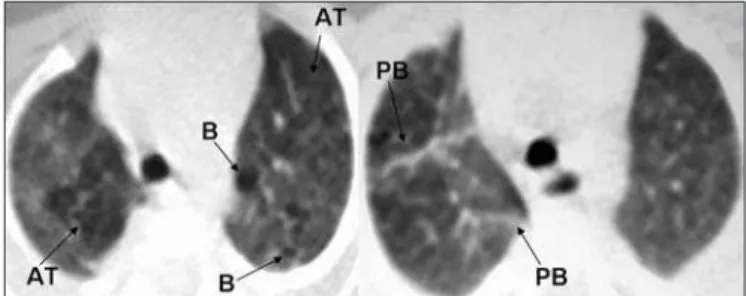
new-borns: atelectasis (opacity with reduced lung volume, secondary to alveolar collapse); consolidation (opacity, expressed as increased density of the pul-monary parenchyma, usually homogeneous, accompanied by obscuration of the underlying blood vessels); ground-glass opacity (increased density of the pulmonary parenchyma without obscuration of the vessels); air trap-ping (areas with decreased attenuation interspersed with areas of normal attenuation); and air bubbles (lesion containing air, with thin, well-deined walls, not always possible to distinguish from pulmonary cyst). Figures 1, 2, and 3 illustrate the abnormalities comprising the scoring system.
In addition to identifying the abovementioned lung abnormalities, this scoring system evaluated the topographic site (pulmonary lobes) and quantiied the afected lung area by means of a score attributed to the lobes afected by the abnormalities, with the addition of two more points in the case of bilateral lung involvement. In this study, the lingu-la was deined as a separate lobe and not as part of the left upper lobe.11
bilat-All the infants were followed up at the high-risk neonatal outpa-tient clinic by a pediatrician who was unaware of the HRCT results. Re-spiratory evolution during the irst year of life was evaluated by means of physical examination, and the presence of respiratory complications (persistent wheezing, and/or hospitalization, and/or pneumonia) dur-ing the intervals between consultations was recorded. he consultations were held monthly or according to clinical need.2 Data were collected
at each consultation, and upon completing 12 months of corrected age, it was determined whether the child had presented respiratory morbid-ity, deined as the presence of one or more of the following:persistent wheezing (presence of two or more episodes of wheezing causing respi-ratory diiculty, observed by the pediatrician by means of pulmonary auscultation, and which required bronchodilatory medication); hospi-talization due to respiratory problems (hospihospi-talization of the infant for more than 24 hours); or pneumonia (presence of tachypnea, intercostal and subcostal retraction, crackles and proven radiological abnormalities; chest X-rays were interpreted by a pediatric radiologist).2
he scores were correlated with respiratory morbidity variables. To assess the score characteristics (sensitivity and speciicity) and predictive validity (positive predictive value [PPV] and negative predictive value [NPV]) in relation to respiratory morbidity, a receiver operating char-acteristic (ROC) curve was constructed that indicated the best cutof point according to the greatest area under the curve. A statistical test was also performed (α < 0.05) to ind out whether the area under the curve
was signiicantly greater than 50%. he likelihood ratio was also cal-culated for the positive tests (score above the cutof) and negative tests (score below the cutof).
Epi Info™ version 3.32 was used to construct the database. he sta-tistical analyses used the softwares Epi Info 3.32, Stasta-tistical Package for Social Sciences (SPSS) version 12.0 and MedCalc version 9.3.8.0.
RESULTS
During the abovementioned period, 179 premature newborns were admitted to the NICU with gestational age at birth ranging from 23 to 33 weeks (mean of 28 weeks, standard deviation, SD: 2.3 weeks) and birth weight ranging from 610 to 1480 g (mean of 1101 g, SD: 235 g). Among these premature infants, 20 (11.17%) evolved to death and 58 (32.4%) were excluded (41 small for gestational age, seven with congenital malformations, seven with genetic syndrome and three with congenital infections). he parents of four infants (2.23%) refused to allow their participation in the study, and 11 patients (6.14%) failed to undergo HRCT scans due to technical problems in the CT equipment. his left 86 patients who underwent HRCT scans, with gestational ages corrected for prematurity ranging from 30 to 40 weeks (mean of 36 weeks, SD: 2 weeks) and chronological mean age of 59 days (SD: 26 days) at the time of the scan. Among these, 24 (27.9%) met the clin-ical diagnostic criteria for BPD (deined as the need for supplemen-tal oxygen for 28 days or more).32 All the premature infants who
un-derwent HRCT scans were followed up throughout their irst year of life. here was no attrition rate regarding the outcome of respiratory morbidity. Out of the 86 patients, 73 (85%) showed abnormalities on
Figure 1. High-resolution computed tomography (HRCT) scan showing air trapping (AT), air bubbles (B) and parenchymal band (PB).
Figure 2. High-resolution computed tomography (HRCT) scan showing air trapping (AT), air bubbles (B), ground-glass opacity (GG) and parenchymal band (PB).
Figure 3. High-resolution computed tomography (HRCT) scan showing atelectasis (A), consolidation (C), subpleural opacity (SPO) and ground-glass opacity (GG).
eral involvement. he maximum total score in the proposed scoring sys-tem was 40 points, i.e. eight points for each of the ive CT abnormali-ties, such that the highest values represented the most heavily altered scans and the lowest values represented the least altered scans.
Two pediatric radiologists (BM, DP) with more than 10 years’ ex-perience had evaluated the CT scans previously, when there was still no scoring system, and they now conducted the intra and interobserver re-liability study for each lesion identiied.2 For the purposes of the current
study, these same radiologists jointly identiied the target abnormali-ties and underwent training to apply the scoring system. Subsequently, the two observers independently applied the scoring system to the 86 HRCT scans, blinded to the clinical data and with no access to their own and each other’s previous readings.
he principal observer (BM) applied the scoring system to all 86 HRCT scans on two diferent occasions, with a three-month interval between the two. he intra and interobserver reliability of the scores was evaluated by means of the intraclass correlation coeicient (ICC)30 and
HRCT scans. Among the latter, 56.2% presented respiratory morbidity during the irst year of life.
A mean score was assigned to each CT by each radiologist. Table 1
shows the mean scores for the single pulmonary abnormalities, and the inal score assigned by the observers can be seen in the last column. Ac-cording to the criteria, 15% of the patients had a normal CT (mean score = zero). he mean score of the CT tests ranged from 0.5 to 12 (mean 4.2; median 3.5). he most frequent CT abnormalities were at-electasis (80.2%) and ground-glass opacity (50%).
Assessment of intraobserver reliability was based on the two evalua-tions by the principal observer, while interobserver reliability was based on the second evaluation by the principal observer and the evaluation by the second observer. Table 2 shows the results from the intraobserver and interobserver reliability evaluations for the total score and single CT abnormalities. he ICC for intraobserver reliability was very low for air trapping (ICC 0.26). For the other CT indings, the ICC ranged from 0.73 to 0.97, showing very good agreement. For interobserver reliabil-ity, the ICC ranged from 0.45 to 0.86.
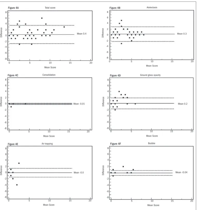
Intra and interobserver reliability values for the total score and sin-gle CT abnormalities were also evaluated in accordance with the Bland-Altman method, as shown in Figures 4 and 5. For intraobserver reliabil-ity, the total score showed a trend towards lower agreement at the higher values (Figure 4A), and no signiicant diference was observed between the results from the two evaluations in relation to consolidation (Figure 4C), ground-glass opacity (Figure 4D) or air bubbles (Figure 4F). he graphical method showed a larger diference between the irst and sec-ond evaluations for atelectasis (Figure 4B) and showed no diference for air trapping (Figure 4E).
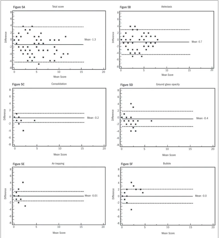
he interobserver evaluation using the Bland-Altman method showed a mean diference of -1.3 in the total score. he diferenc-es ranged from -6 to 4 and did not show any trend (Figure 5A). For consolidation, air trapping and air bubbles, the diferences between the evaluations by the two radiologists were minimal (Figures 5C, 5E and
5F, respectively). he diferences were 0.7 for atelectasis (Figure 5B) and 0.4 for ground-glass opacity (Figure 5D).
To measure the predictive validity of the scoring system in relation to respiratory morbidity over the irst year of life, a receiver operating characteristic (ROC) curve was constructed, based on the scores and the presence of respiratory morbidity. he criterion chosen for the best cutof point was a score greater than six, with an area under the curve of 0.65% (95% conidence interval, CI 0.54-0.75). his cutof (score > 6) showed the best relationship between sensitivity (39.1%; 95% CI 25.1-54.6), speciicity (90.0; 95% CI 76.3-97.1), positive likelihood ra-tio (3.91) and negative likelihood rara-tio (0.68). Based on this analysis of the score, in positive cases (score > 6) the infants had nearly a fourfold greater chance (3.91) of presenting lung disease.
Based on this analysis, the scores were divided into two categories (≤ 6 and > 6). CT scans with scores greater than six contained numer-ous abnormalities, while those with scores less than or equal to six ei-ther contained few abnormalities or were normal. Subsequent analy-sis on the scores in two categories showed that the majority of the pa-tients (64 newborns) scored from zero to six; of these, 36 papa-tients (NPV
of 56.3%) did not present respiratory morbidity during the irst year. Scores higher than six were found in 22 patients and, of these patients, 40.9% (PPV) presented wheezing, 45.5% (PPV) hospitalizations and 63.6% (PPV) pneumonia. Table 3 shows the validity measurements of the scores in relation to respiratory morbidity.
CT indings (possible range of scores) A1 Mean value (minimum-maximum) A2 Mean value (minimum-maximum) B Mean value (minimum-maximum) Mean score Mean value (minimum- maximum) Atelectasis (0-8) 2.73 (0-8) 2.42 (0-8) 3.08 (0-8) 2.75 (0-8) Ground-glass opacity (0-8) 0.67 (0-6) 0.51 (0-6) 0.89 (0-7) 0.70 (0-6.5) Air trapping (0-8) 0.14 (0-4) 0.16 (0-4) 0.22 (0-4) 0.19 (0-2.5) Air bubbles (0-8) 0.36 (0-6) 0.40 (0-5) 0.40 (0-4) 0.40 (0-4.5) Consolidation (0-8) 0.07 (0-4) 0.07 (0-4) 0.25 (0-5) 0.16 (0-4.5) Total score (0-40) 3.98 (0-15) 3.56 (0-12) 4.81 (0-14) 4.18 (0-12)
Table 1. Mean scores for separate computed tomography (CT) indings and total score, as evaluated by the two observers from high-resolution computed tomography (HRCT) scans on 86 premature newborns, and the mean score from the evaluations
A1 = Principal observer’s irst evaluation; A2 = Principal observer’s second evaluation; B = Second observer’s evaluation.
CT indings Intraobserver reliability Interobserver reliability
Atelectasis 0.87 (CI 0.81-0.91)
0.84 (CI 0.76-0.90) Ground-glass opacity 0.73 0.73
(CI 0.61-0.81) (CI 0.59-0.83)
Air trapping 0.26 0.45
(CI 0.06-0.45) (CI 0.16-0.64)
Air bubbles 0.95 0.60
(CI 0.91-0.96) (CI 0.38-0.74) Consolidation 0.97 0.68
(CI 0.95-0.98) (CI 0.50-0.79)
Total score 0.89 0.86
(CI 0.84-0.93) (CI 0.78-0.90)
Table 2. Intraclass correlation coeficient (ICC) for intraobserver and interobserver reliability in relation to total score and separate computed tomography (CT) indings
CI = 95% conidence interval.
Respiratory morbidity Sensitivity % (95% CI) Speciicity % (95% CI) PPV % (95% CI) NPV % (95% CI)
Wheezing 40.9 79.7 40.9 79.7 (20.4-61.5) (69.8-89.5) (20.4-61.5) (69.8-89.5) Pneumonia 43.8 85.2 63.6 71.9
(26.6-60.9) (75.7-94.7) (43.5-83.7) (60.9-82.9) Hospitalization 45.5 81.3 45.5 81.3
(24.6-66.3) (71.7-90.8) (24.6-66.3) (71.7-90.8) Respiratory
morbidity
39.1 90.0 81.8 56.3 (25.0-53.2) (80.7-99.3) (65.7-97.9) (44.1-68.4)
Table 3. Validity measurements for scores greater than six in relation to wheezing, pneumonia, hospitalization and respiratory morbidity during the irst year of life
Figure 4A Total score
Difference
Mean Score
Mean 0.4
0 -8 -6 -4 -2 0 2 4 6 8
5 10 15 20
Figure 4B Atelectasis
Difference
Mean Score
Mean 0.3
0 -8 -6 -4 -2 0 2 4 6 8
5 10 15 20
Figure 4C Consolidation
Difference
Mean Score
Mean -0.01
0 -8 -6 -4 -2 0 2 4 6 8
5 10 15 20
Figure 4D Ground glass opacity
Difference
Mean Score
Mean 0.2
0 -8 -6 -4 -2 0 2 4 6 8
5 10 15 20
Figure 4E Air trapping
Difference
Mean Score
Mean -0.0
0 -8 -6 -4 -2 0 2 4 6 8
5 10 15 20
Figure 4F Bubble
Difference
Mean Score
Mean -0.04
0 -8 -6 -4 -2 0 2 4 6 8
5 10 15 20
Figure 4. Bland-Altman plots showing variance in intraobserver reproducibility in relation to total computed tomography (CT) score and CT indings. Horizontal lines show the mean and ± 2 SD (standard deviation). The Y-axis shows the difference in mean score between irst and second observations.
his cutof point (> 6) was also evaluated in relation to diagnoses of BPD that were made during the neonatal period. Among the children with scores greater than six, 59.1% had BPD. Among the patients with scores less than six, 81.3% did not have BPD.
As in other hospital-based studies, a convenience sample was used. he estimators of predictive values were accompanied by their respec-tive conidence intervals, which made it possible to achieve statistical
Figure 5. Bland-Altman plots show variance in interobserver agreement in relation to total computed tomography (CT) score and CT indings. Horizontal lines show the mean and ± 2 SD (standard deviation). The Y-axis shows the difference in mean score between two observers.
found and their diferences. he power of this study was found to range between 86 and 96% for respiratory morbidity and for the separate vari-ables of each respiratory complication, except for hospitalization, which showed a test power of 51%. hus, only the estimators relating to hos-pitalization would need a larger sample (n = 106 patients).
DISCUSSION
Among the few studies in the literature on HRCT scans among pre-mature infants,2,5-11,17 only three2,3,11 analyzed CT scans performed
exclu-sively in the neonatal period. Moreover, as far as we can ascertain, only
Figure 5A Total score
Difference
Mean Score
Mean -1.3
0 -8 -6 -4 -2 0 2 4 6 8
5 10 15 20
Figure 5B Atelectasis
Difference
Mean Score
Mean -0.7
0 -8 -6 -4 -2 0 2 4 6 8
5 10 15 20
Figure 5C Consolidation
Difference
Mean Score
Mean -0.2
0 -8 -6 -4 -2 0 2 4 6 8
5 10 15 20
Figure 5D Ground glass opacity
Difference
Mean Score
Mean -0.4
0 -8 -6 -4 -2 0 2 4 6 8
5 10 15 20
Figure 5E Air trapping
Difference
Mean Score
Mean -0.01
0 -8 -6 -4 -2 0 2 4 6 8
5 10 15 20
Figure 5F Bubble
Difference
Mean Score
Mean -0.0
0 -8 -6 -4 -2 0 2 4 6 8
Ochiai et al.11 presented a scoring system to evaluate pulmonary
involve-ment based on CT abnormalities in tests performed so early in life. Studies have shown that children who developed BPD frequently presented signs of chronic lung disease. he CT indings were hyper-aeration, linear opacities, triangular subpleural opacities and broncho-vascular distortion or thickening.10,11,17 he presence of these
abnormali-ties and absence of bronchiectasis suggested that these were sequelae of BPD.10 Pulmonary hyperaeration was the most frequent abnormality
and correlated signiicantly with clinical scores, with a high interobserv-er agreement.17 HRCT scans on patients ranging in age from ive to 18
years who were born prematurely and had a history of BPD, showed a signiicant correlation with abnormal lung function.6
In 2007, Mahut et al.8 described the CT abnormalities found on
the scans of 41 very low birth weight premature patients, all with BPD. he scans were performed between 10 and 20 months after birth. hese authors concluded that despite advances in perinatal care, the CT ab-normalities currently found were still similar to those that used to be described in the pre-surfactant era and were associated with duration of oxygen therapy and mechanical ventilation, while emphasizing that they did not detect the bronchial involvement that had been described in previous studies.
Ochiai et al.11 analyzed HRCT scans on 42 premature infants, all
with a diagnosis of BPD, whose scans were performed when they were clinically stable and with an age range similar to that of our study. hey developed a scoring system based on a radiographic score for BPD with three groups of abnormalities (hyperaeration, emphysema and ibrous or interstitial abnormalities) and correlated this system with clinical scores that were assigned at the corrected ages of 28 days and 36 weeks.
he principal diference between the scoring system of Ochiai et al.11 and the system that we are proposing is that we evaluated patients
both with and without BPD, which thus allowed us to conclude that CT abnormalities like air trapping, atelectasis, consolidation, ground-glass opacity and air bubbles are not characteristic of BPD. hese CT indings are present in premature infants who are exposed to mechani-cal ventilation and oxygen therapy, with or without clinimechani-cal criteria for a diagnosis of BPD. According to our indings, these abnormalities are more prominent in patients that developed BPD and those with respi-ratory morbidity during the irst year of life.
he most frequent single pulmonary lesions demonstrated by HRCT scans in our study were atelectasis and ground-glass opacity. Other authors6-8,17 observed high frequencies of these same lesions, but
since they mostly examined older patients with images obtained under conditions of apnea and with series during inspiration and expiration, they showed a higher frequency of air trapping than seen in our study. he immaturity of the lungs of premature infants, associated with the complications caused by the oxygen therapy and mechanical ventilation needed to keep them alive, can lead to decreased pulmonary compliance and modiications to the pulmonary architecture, which are expressed on HRCT scans by these frequently identiied lesions.
One of the limitations of our study was the fact that the CT scans were performed on young, very small patients with elevated respiratory rates. Because of the young age and thus the lack of collaboration by
the patients, it was not possible to perform the test under conditions of apnea. hus, artifacts are known to occur that are secondary to the newborns’ typical movements and their respiratory pattern. Such arti-facts might be resolved if the child received general anesthesia and me-chanical ventilation, which would make it possible to obtain CT im-ages under conditions of apnea. Respiratory movements hinder the de-tailed evaluation of abnormalities like ground-glass opacity and air trap-ping. hese factors contributed towards lower interobserver agreement for these two lesions. Nonetheless, we believe that since CT is an inva-sive procedure, and given the labile clinical conditions in the majority of these newborns, performing the test under general anesthesia should be avoided.
Another possible limitation to our study was the acquisition time required for the CT images, which contributed with higher doses of radiation and increased secondary artifacts in the premature infants’ respiratory patterns. At the time when the scans were performed, the CT equipment that was used did not allow the technical settings to be changed to make the time taken and thus the radiation dose used as low as possible under the circumstances. herefore, the settings used were higher than those proposed by Lucaya et al.33 in 2000 (35 to 60 mAs),
but similar to those used recently by Mahut et al.8 in equipment
equiva-lent to ours. he latter authors performed HRCT scans on premature infants born between January 1999 and March 2001, with settings of 100 mA and 1.0 s.
Although the age of these newborns tends to decrease the quality of some CT images, we believe that this should not be viewed as an im-pediment to performing this test, which proved important for evaluat-ing the extent of pulmonary involvement. Ochiai et al.11 concluded that
their scoring system was capable of assessing the patient’s clinical status before hospital discharge and predicting the prognosis of patients with BPD, but they did not evaluate the predictive validity of the score as was done in our study.
Another important factor in performing HRCT scans on such young patients was demonstrated by the score, which showed low sensi-tivity but high speciicity, thereby indicating that a large portion of the patients without respiratory morbidity during the irst year of life pre-sented tests that were normal or only prepre-sented limited abnormalities. Meanwhile, the PPV was high, thus indicating a chance of more than 80% that the infants with abnormal scans would present respiratory morbidity during the irst year of life. Importantly, in tertiary and refer-ence centers, HRCT is a valid method for use in the late neonatal peri-od because of the very high prevalence of respiratory morbidity (56.2%) seen in the follow-ups on this high-risk population.2 here needs to be a
mechanism for indicating which patients are most likely to become ill, and therefore, investments in prevention with vaccines and other expen-sive therapies are necessary. However, radiologists need to be trained to become familiar with the types of pulmonary abnormalities identiied using HRCT on this group of very small patients that present impair-ment according to the scoring system proposed here.
predic-tive values for the CT score, i.e. good posipredic-tive predicpredic-tive value and high speciicity. he assessment on the HRCT scoring system further dem-onstrated that it should not be used for screening purpose because of the high percentage of false negatives (44%). he CT score sensitivity and speciicity showed that HRCT scans should not be indicated in hospi-tals and services where the prevalence of prematurity and respiratory morbidity could be low, because the PPV would be very low.
CONCLUSIONS
he scoring system that we propose proved to be reproducible and easy to apply. We thus conclude that this CT scoring system is important in clin-ical practice, since it allows standardization of the evaluation of CT abnor-malities in premature infants, through identifying patients with increased likelihood of respiratory morbidity during the irst year of life. Furthermore, by expanding the use of this scoring system, CT scans on premature infants with diferent risk factors for respiratory morbidity can be compared. Scores greater than six showed a capacity to predict respiratory morbidity in 81.8% of patients, but the sensitivity of the score was 39.1%, which means that out of 100 patients with respiratory morbidity during the irst year of life, only 39 patients presented scores greater than six.
REFERENCES
1. Bancalari E, Claure N, Sosenko IR. Bronchopulmonary dysplasia: changes in pathogenesis, epidemiology and deinition. Semin Neonatol. 2003;8(1):63-71.
2. de Mello RR, Dutra MV, Ramos JR, et al. Lung mechanics and high-resolution computed tomography of the chest in very low birth weight premature infants. Sao Paulo Med J. 2003;121(4):167-72.
3. Doyle LW, Faber B, Callanan C, et al. Bronchopulmonary dysplasia in very low birth weight subjects and lung function in late adolescence. Pediatrics. 2006;118(1):108-13. 4. Bhandari A, Panitch HB. Pulmonary outcomes in bronchopulmonary dysplasia. Semin
Peri-natol. 2006;30(4):219-26.
5. de Mello RR, Dutra MV, Lopes JM. Morbidade respiratória no primeiro ano de vida de pre-maturos egressos de uma unidade pública de tratamento intensivo neonatal [Respiratory morbidity in the irst year of life of preterm infants discharged from a neonatal intensive care unit]. J Pediatr (Rio J). 2004;80(6):503-10.
6. Aquino SL, Schechter MS, Chiles C, et al. High-resolution inspiratory and expiratory CT in older children and adults with bronchopulmonary dysplasia. AJR Am J Roentgenol. 1999;173(4):963-7.
7. Aukland SM, Halvorsen T, Fosse KR, Daltveit AK, Rosendahl K. High-resolution CT of the chest in children and young adults who were born prematurely: indings in a population-based study. AJR Am J Roentgenol. 2006;187(4):1012-8.
8. Mahut B, De Blic J, Emond S, et al. Chest computed tomography indings in bronchopul-monary dysplasia and correlation with lung function. Arch Dis Child Fetal Neonatal Ed. 2007;92(6):F459-64.
9. de Mello RR, Dutra MV, Ramos JR, et al. Neonatal risk factors for respiratory morbidity during the irst year of life among premature infants. Sao Paulo Med J. 2006;124(2):77-84. 10. Oppenheim C, Mamou-Mani T, Sayegh N, et al. Bronchopulmonary dysplasia: value of CT in
identifying pulmonary sequelae. AJR Am J Roentgenol. 1994;163(1):169-72.
11. Ochiai M, Hikino S, Yabuuchi H, et al. A new scoring system for computed tomography of the chest for assessing the clinical status of bronchopulmonary dysplasia. J Pediatr. 2008;152(1):90-5; 95.e1-3.
12. Edwards DK. Radiographic aspects of bronchopulmonary dysplasia. J Pediatr. 1979;95(5 Pt 2):823-9.
13. Greenough A, Kavvadia V, Johnson AH, et al. A simple chest radiograph score to predict chronic lung disease in prematurely born infants. Br J Radiol. 1999;72(858):530-3. 14. Toce SS, Farrel PM, Leavitt LA, Samuels DP, Edwards DK. Clinical and
roentgenogra-phic scoring systems for assessing bronchopulmonary dysplasia. Am J Dis Child. 1984;138(6):581-5.
15. Weinstein MR, Peters ME, Sadek M, Palta M. A new radiographic scoring system for broncho-pulmonary dysplasia. Newborn Lung Project. Pediatr Pulmonol. 1994;18(5):284-9. 16. Yuksel B, Greenough A, Karani J, Page A. Chest radiograph scoring system for use in pre-term
infants. Br J Radiol. 1991;64(767):1015-8.
17. Kubota J, Ohki Y, Inoue T, et al. Ultrafast CT scoring system for assessing bronchopulmonary dysplasia: reproducibility and clinical correlation. Radiat Med. 1998;16(3):167-74. 18. Brasield D, Hicks G, Soong S, Tiller RE. The chest roentgenogram in cystic ibrosis: a new
scoring system. Pediatrics. 1979;63(1):24-9.
19. Chrispin AR, Norman AP. The systematic evaluation of the chest radiograph in cystic ibrosis. Pediatr Radiol. 1974;2(2):101-5.
20. Koscik RE, Kosorok MR, Farrell PM, et al. Wisconsin cystic ibrosis chest radiograph scoring system: validation and standardization for application to longitudinal studies. Pediatr Pul-monol. 2000;29(6):457-67.
21. Weatherly MR, Palmer CG, Peters ME, et al. Wisconsin cystic ibrosis chest radiograph sco-ring system. Pediatrics. 1993;91(2):488-95.
22. Bhalla M, Turcios N, Aponte V, et al. Cystic ibrosis: scoring system with thin-section CT. Radiology. 1991;179(3):783-8.
23. Brody AS, Kosorok MR, Li Z, et al. Reproducibility of a scoring system for computed tomogra-phy scanning in cystic ibrosis. J Thorac Imaging. 2006;21(1):14-21.
24. Helbich TH, Heinz-Peer G, Eichler I, et al. Cystic ibrosis: CT assessment of lung involvement in children and adults. Radiology. 1999;213(2):537-44.
25. Maffessanti M, Candusso M, Brizzi F, Piovesana F. Cystic ibrosis in children: HRCT indings and distribution of disease. J Thorac Imaging. 1996;11(1):27-38.
26. Shah RM, Sexauer W, Ostrum BJ, Fiel SB, Friedman AC. High-resolution CT in the acute exa-cerbation of cystic ibrosis: evaluation of acute indings, reversibility of those indings, and clinical correlation. AJR Am J Roentgenol. 1997;169(2):375-80.
27. Webb WR, Muller NL, Naidich DP. High-resolution CT of the lung. 3rd ed. Philadelphia: Lippin-cott-Williams & Wilkins; 2001.
28. Lucaya J, Le Pointe. High-resolution CT of the lung in children - technique, indications, ana-tomy, features of lung disease, and clinical application. In: Lucaya J, Strife JL, editors. Pedia-tric chest imaging: chest imaging in infants and children. Berlin: Springer Verlag; 2002. p. 55-92.
29. Hansell DM, Bankier AA, MacMahon H, et al. Fleischner Society: glossary of terms for thora-cic imaging. Radiology. 2008;246(3):697-722.
30. Streiner DL. Learning how to differ: agreement and reliability statistics in psychiatry. Can J Psychiatry. 1995;40(2):60-6.
31. Bland JM, Altman DG. Statistical methods for assessing agreement between two methods of clinical measurement. Lancet. 1986;1(8476):307-10.
32. Jobe AH, Bancalari E. Bronchopulmonary dysplasia. Am J Respir Crit Care Med. 2001;163(7):1723-9.
33. Lucaya J, Piqueras J, García-Peña P, et al. Low-dose high-resolution CT of the chest in chil-dren and young adults: dose, cooperation, artifact incidence, and image quality. AJR Am J Roentgenol. 2000;175(4):985-92.
Conlict of interest: None
Sources of funding: None
Date of irst submission: February 26, 2010
Last received: September 18, 2010
Accepted: September 30, 2010
Address for correspondence: Márcia Boechat
Av. Rui Barbosa, 716 — 2o andar Flamengo — Rio de Janeiro (RJ) — Brasil CEP 22250-020