295
Revista da Sociedade Brasileira de Medicina Tropical 34(3): 295-297, mai-jun, 2001.
NOT NOT NOT NOT
NOTAAAAA PRÉVIA PRÉVIA PRÉVIA PRÉVIA PRÉVIA
Hepatocyte metaplasia in experimental chagasic pancreatitis:
preliminary report
Metaplasia hepatocítica em pancreatite chagásica experimental: nota prévia
V VV V
Vitorino Modesto dos Santositorino Modesto dos Santositorino Modesto dos Santositorino Modesto dos Santositorino Modesto dos Santos1, 31, 31, 31, 31, 3, Marcus Aurelho de Lima, Marcus Aurelho de Lima, Marcus Aurelho de Lima, Marcus Aurelho de Lima, Marcus Aurelho de Lima11111, Marlene Cabrine-Santos, Marlene Cabrine-Santos, Marlene Cabrine-Santos, Marlene Cabrine-Santos, Marlene Cabrine-Santos22222,,,,,
Daniela de Stefani Marquez Daniela de Stefani Marquez Daniela de Stefani Marquez Daniela de Stefani Marquez
Daniela de Stefani Marquez33333, Eliane Lages-Silva, Eliane Lages-Silva, Eliane Lages-Silva, Eliane Lages-Silva, Eliane Lages-Silva2, 32, 32, 32, 32, 3, Jaqueline Maria Matheus,, Jaqueline Maria Matheus,, Jaqueline Maria Matheus,, Jaqueline Maria Matheus,, Jaqueline Maria Matheus,
José V José V José V José V
José Vitor de Oliveira Junior and Luis Eduardo Ramírezitor de Oliveira Junior and Luis Eduardo Ramírezitor de Oliveira Junior and Luis Eduardo Ramírezitor de Oliveira Junior and Luis Eduardo Ramírezitor de Oliveira Junior and Luis Eduardo Ramírez2, 32, 32, 32, 32, 3
Abstract Abstract Abstract Abstract
Abstract Beginning the study of chronic pathologic changes in pancreas of hamsters experimentally infected with Trypanosoma cruzi Vic strain, hepatocyte metaplasia was observed in one animal from infected group. This is the first report of oncocytes in Chagas’ disease, which could be due to aberrant regenerative response to pancreas inflammatory process.
Key-words: Key-words: Key-words: Key-words:
Key-words: Hamster. Experimental Chagas’ disease. Chronic pancreatitis. Oncocyte. Hepatocyte metaplasia.
Resumo Resumo Resumo Resumo
Resumo Iniciando estudo de alterações patológicas crônicas no pâncreas de hamsters experimentalmente infectados com a cepa Vic de Trypanosoma cruzi, metaplasia oncocítica foi observada em um dos animais infectados. Este é o primeiro relato de oncocitos na doença de Chagas, que poderiam decorrer de resposta regenerativa aberrante ao processo inflamatório pancreático.
Palavras-chaves: Palavras-chaves: Palavras-chaves: Palavras-chaves:
Palavras-chaves: Hamster. Doença de Chagas experimental. Pancreatite crônica. Oncocito. Metaplasia hepatocítica.
1. Department of Internal Medicine, 2. Department of Biological Sciences and 3. Pathology Post-graduation Course of Triângulo Mineiro Medical School. Financial support: FUNEPU and CAPES/ICCTI.
Address to: Prof. Vitorino Modesto dos Santos. Depto de Medicina Interna/HE/FMTM. Praça Thomaz Ulhoa 706, 38025-050 Uberaba-MG, Brazil. Tel: 55 34 3318-5258. Fax: 55 34 3312-6640.
e-mail: [email protected] Recebido para publicação em 30/1/2001.
Hamsters infected with Trypanosoma cruzi may exhibit histopathological changes similar to human Chagas’
disease9, including pancreatitis6 7. Following injury and
regeneration, pancreas ductular cells may give rise to
metaplastic hepatocyte-like cells2 5 10. Oxyphilic, oncocytic,
ductular, hepatocytic or hepatocyte-like metaplasia has
been described in older people11, in human chronic
pancreatitis and diabetes mellitus11, and in spontaneous
or experimentally induced tumors1 3 4 8 12.
The complete study was planned in order to include the acute (first 60 days after the initial infection) and the chronic phase of experimental Chagas’ disease up to 375 days. Just at the beginning of chronic pathologic and immunohistochemical evaluation in pancreas of hamsters experimentally infected with T. cruzi, oxyphilic
metaplasia8 12 was observed in one of the infected
animals. This is the first report of pancreas hepatocyte metaplasia in Chagas’ disease.
Experimental procedures were in accordance with the Principles of Laboratory Animal Care from the National Society of Medical Search, and the Guide for
the Care and use of Laboratory Animals from the National Institute of Health. The animals were housed ten per rigid plastic cage, with hardwood chip bedding and a 12-hr light/
dark cycle1. A standard industrial pelleted diet (54%
carbohydrate, 23% protein and 4% fat) supplemented with minerals, vitamins and amino acids, and tap water given ad libitum. Ten young male non-isogenic Syrian golden hamsters, which had been infected, and reinfected 75 days after, by intraperitoneal route, with 2,000 trypomastigote
forms of the T. cruzi Vic strain were sacrificed on the 105th
day after initial infection. Ten weight and age-matched non-infected male hamsters constituted the normal control group studied.
The sacrifice and careful necropsy schedule included the 15th, 30th, 45th, 60th, 105th, 180th, 255th, 330th and 375th
day, and the reinfections were scheduled for the 75th, 150th,
225th, and 300th days of experiment. Fragments of pancreas
avidin-296
Santos VM et al
biotin-peroxidase method, antibodies anti-T. cruzi (1:3,000) and the islet cell specific antibodies: insulin (1:500), anti-glucagon (1:600), anti-somatostatin (1: 3,500), and anti-pancreatic polypeptide (1: 15,000) were employed. Statistical analysis was planned to include parametric and nonparametric methods.
At the 105th day of experiment, major evidence of
parenchyma hypotrophy, fibrosis and inflammatory infiltrate, characterizing pancreatitis, were invariably found in the infected group, while only minor changes were observed among the animals from control group.
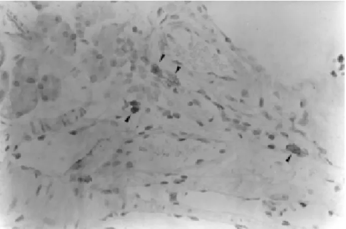
Hepatocyte metaplasia was seen in a peri-insular zone of an infected animal (Figure 1), while the phenomenon was not observed in normal control hamsters. In both groups, normal islet-cells were positive to insulin, glucagon, somatostatin and pancreatic polypeptide antibodies, while oncocytes were all negative (Figure 2). The PAP-anti-Trypanosoma cruzi test was also positive in the pancreas sample where oncocytes were observed (Figure 3).
Hamster chronic pancreatitis, due to chagasic infection, was characterized by reduction in number and
Figure 2 - On immunohistochemistry, normal islet-cells are positive to insulin (A), glucagon (B), somatostatin (C) and pancreatic polipeptide (D) antibodies, while oncocytes are negative (X400).
297
Revista da Sociedade Brasileira de Medicina Tropical 34: 295-297, mai-jun, 2001
size of acini and chronic inflammatory infiltrate around pancreas lobules and ducts, in addition to PAP anti-T. cruzi positive test in the same pancreas section. The absence of oncocytes in animals of the non-infected group could suggest that the pancreas hepatocyte metaplasia is related with an aberrant response to the inflammatory process in Chagas’
disease. Nevertheless, at the 105th day of experiment,
the number of animals already studied (n=60) was not large enough to enable such a conclusion. Moreover, since oncocytic proliferation may become a more
common event in association with animal aging11 and/
or with chagasic reinfection, the remainder (n=155) normal control as well as infected hamsters will be
appropriately evaluated after a longer period - up to 375th
day post-inoculation - as previously scheduled.
Only for the purpose of this preliminary report, the oncocytic nature of pancreas changes was based on the presence of large mitochondria, in addition to abundant, almost homogeneous, and finely granular eosinophilic cytoplasm observed under light
microscopy8 12. However, for more convincing evidence
of the phenomenon, further evaluation of the oncocytes will include liver-specific markers and ultrastructural details.
The occurrence of pancreas hepatocyte metaplasia in Chagas’ disease was not previously described. Although the authors consider it inadequate to extrapolate this very preliminary finding to human cases, the concern is an eventual relationship between Chagas’ disease and pancreas oncocytes.
Figure 3 - PAP anti-T. cruzi test positive (arrows) in the pancreas sample where oncocytes were observed (x400).
REFERENCES 1. Konishi N, Ward JM, Waalkes MP. Pancreatic hepatocytes in
Fischer and Wistar rats induced by repeated injections of cadmium chloride. Toxicology Applied Pharmacology 104:149-156, 1990.
2. Makino T, Usuda N, Rao S, Reddy JK, Scarpelli DG. Transdifferentiation of ductular cells into hepatocytes in regenerating hamster pancreas. Laboratory Investigation 62:552-561, 1990.
3. Monis B, Valentich MA, Urrutia R, Rivolta M. Multicentric focal acinar cell hyperplasia and hepatocyte-like cell metaplasia are induced by nitrosomethylurea in rat pancreas. International Journal of Pancreatology 8:119-131, 1991.
4. Pacchioni D, Papotti M, Macri L, Forte G, Bussolati G. Pancreatic oncocytic endocrine tumors. Cytologic features of two cases. Acta Cytologica 40:742-746, 1996.
5. Rao MS, Reddy JK. Hepatic transdifferentiation in the pancreas. Seminars in Cell Biology 6:151-156, 1995.
6. Ramirez LE, Lages-Silva E, Chapadeiro E. Hamster. In: Jorge TCA, Castro SL (eds) Chagas’ disease. Manual for animal experiment. FIOCRUZ, Rio de Janeiro RJ, p.145-148, 2000.
7. Ramirez LE, Lages-Silva E, Soares-Junior JM, Chapadeiro E. The hamster (Mesocricetus auratus) as experimental model in Chagas’ disease: parasitological and histopathological studies in acute and chronic phases of Trypanosoma cruzi infection. Revista da Sociedade Brasileira de Medicina Tropical 27:163-169, 1994. 8. Sadoul JL, Saint-Paul MC, Hoffman P, Plazza M, Birtwisle Y,
Freychet P, Loubiere R. Malignant pancreatic oncocytoma. An unusual cause of organic hypoglycemia. Journal of Endocrinological Investigation 15:211-217, 1992.
9. Santos VM, Teixeira VPA, Cunha DF, Cunha SFC, Monteiro JP, Santos JAM, Santos TAM, Santos LAM. Pancreatic anatomopathology changes in chronic chagasic women. Preliminary data. Arquivos de Gastroenterologia 36:127-132, 1999. 10. Sirica AE. Ductular hepatocytes. Histology and Histopathology
10:433-456, 1995.
11. Tasso F, Sarles H. Cellules canalaires et oncocytes dans le pancreas humain. Étude comparée à l’état normal et dans les pancréatites chroniques. Annales D’Anatomie Pathologique 18:277-300, 1973.