C a l d a s C a l d a s C a l d a s C a l d a s C a l d a s
Magnetic resonance imaging in staging of locoregional prostate cancer: comparison of results with analysis post-surgical histopathology 447
Rev. Col. Bras. Cir. 2010; 37(6): 447-449
Preliminary Report
Preliminary Report
Preliminary Report
Preliminary Report
Preliminary Report
Magnetic resonance imaging in staging of locoregional prostate
Magnetic resonance imaging in staging of locoregional prostate
Magnetic resonance imaging in staging of locoregional prostate
Magnetic resonance imaging in staging of locoregional prostate
Magnetic resonance imaging in staging of locoregional prostate
cancer: comparison of results with analysis post-surgical
cancer: comparison of results with analysis post-surgical
cancer: comparison of results with analysis post-surgical
cancer: comparison of results with analysis post-surgical
cancer: comparison of results with analysis post-surgical
histopathology
histopathology
histopathology
histopathology
histopathology
A ressonância magnética no estadiamento locorregional do câncer de próstata:
A ressonância magnética no estadiamento locorregional do câncer de próstata:
A ressonância magnética no estadiamento locorregional do câncer de próstata:
A ressonância magnética no estadiamento locorregional do câncer de próstata:
A ressonância magnética no estadiamento locorregional do câncer de próstata:
resultados comparados com a análise histopatológica pós-cirúrgica
resultados comparados com a análise histopatológica pós-cirúrgica
resultados comparados com a análise histopatológica pós-cirúrgica
resultados comparados com a análise histopatológica pós-cirúrgica
resultados comparados com a análise histopatológica pós-cirúrgica
M
ANOELE
DUARDOD
AUMASC
ALDAS, A
SCBC-RJ
1; L
UIZC
ARLOSD
UARTEDEM
IRANDA, ACBC-RJ
2; L
EONARDOK
AYATB
ITTENCOURT3A B S T R A C T
A B S T R A C T
A B S T R A C T
A B S T R A C T
A B S T R A C T
Conventional staging for locoregional prostate adenocarcinoma has been demonstrated as potentially underdiagnosing. Therefore, prostate MRI is emerging as an important tool for staging before surgery. Advanced techniques such as diffusion and dynamic contrast enhancement also contribute to increasing its accuracy. In this preliminary study, MRI was compared with prostate histopathology samples, reaching 78% sensitivity and 100% specificity for tumor localization; 33% sensitivity and 100% specificity for extracapsular extension; 100% sensitivity and 100% specificity for involvement of the seminal vesicles. It is possible to believe that these preliminary results are promising, and more cases will tend to confirm these data.
Key words: Key words:Key words:
Key words:Key words: Prostatic neoplasms. Neoplasm staging. Magnetic resonance spectroscopy. Histopathology.
Work done at the Departments of Urology and Radiology, Hospital Universitário Clementino Fraga Filho – Universidade Federal do Rio de Janeiro - HUCFF-UFRJ - Rio de Janeiro, Brazil.
1. Resident, Urology Service, Hospital Universitário Clementino Fraga Filho –Universidade Federal do Rio de Janeiro Rio de Janeiro, Brazil; 2. Associate Professor, Department of Surgery, Universidade Federal do Rio de Janeiro- Rio de Janeiro, Urology Service, Hospital Universitário Clementino Fraga Filho, Rio de Janeiro, Brazil; 3. M.D., Radiologist, CDPI and Multi-Imagem; Master’s Degree Graduate, Radiology Department, Hospital Universitário Clementino Fraga Filho – Universidade Federal do Rio de Janeiro Rio de Janeiro, Brazil.
INTRODUCTION
INTRODUCTION
INTRODUCTION
INTRODUCTION
INTRODUCTION
P
hysicians often face histopathology results of radical
prostatectomy specimens in patients with advanced
malignancy thaT were expected to have a prostate-confined
disease. The traditional locoregional staging with digital
exam, transrretal ultrasonography and computed
tomography is notoriously limited
1,2and the use Magnetic
Resonance Imaging (MRI) has emerged as the image
category of choice. Its advanced techniques such as diffusion,
dynamic contrast and spectroscopy have added specificity
to the findings of basic sequences
3,4.
The aim of this study is to compare MRI findings
suspicious for prostate cancer with histopathological examinationS
in patients undergoing radical prostatectomy considereing tumor
location (unilateral or bilateral), extra-capsular extension,
semi-nal vesicle invasion and obturator lymph node invasion.
METHODS
METHODS
METHODS
METHODS
METHODS
Between March and June 2009 we evaluated 11
consecutive patients in the HUCFF-UFRJ diagnosed with prostate
cancer, with an indication for radical prostatectomy according
to the criteria of the Brazilian Society of Urology guidelines. The
study was approved by the HUCFF Ethics in Research Committee
and logged in to SISNEP with CAAE number
0057.0.197.000-09. All patients signed an informed consent. We performed MRI
of the prostate with a 1.5T apparatus (Avanto, Siemens,
Germany). The suspicious appearance for neoplastic involvement
comprises nodular areas with low signal on T2-weighted
sequences (Figure 1). We also obtained advanced diffusion and
dynamic contrast enhancement sequences to confirm and
increase the specificity of T2-weighted sequences, according to
widely known criteria
3,4(Figures 2 and 3). Extra-capsular
extension was considered when we found extra-prostatic
nodules, obliteration of periprostatic fat, invasion of the
neurovascular bundle or evident trans-capsular tumor
4. Surgical
specimens were sent to standard histopathologic
examination. The results were compared using the following
criteria: location (unilateral or bilateral), extracapsular extension,
seminal vesicle invasion and obturator lymph node invasion.
RESULTS
RESULTS
RESULTS
RESULTS
RESULTS
(unila-448
Rev. Col. Bras. Cir. 2010; 37(6): 447-449
C a l d a s C a l d a s C a l d a s C a l d a s C a l d a s Magnetic resonance imaging in staging of locoregional prostate cancer: comparison of results with analysis post-surgical histopathology
teral x bilateral), MRI showed a sensitivity of 78% and
specificity of 100%, with an accuracy of 82%. The lowest
sensitivity was due to microscopic foci (<0.5 cm) of
bilate-ral disease, undetectable by MRI. In search of extracapsular
Figure 3 Figure 3Figure 3 Figure 3
Figure 3 - Dynamic contrast enhancement demonstrating early and intense uptake by suspected nodular area, suggesting neoangiogenesis (arrow).
Figure 2 Figure 2 Figure 2 Figure 2
Figure 2 - Map of apparent diffusion coefficient showing hypointense area in the same location that the image weighed in T2, suggesting high cellularity (arrow). Figure 1
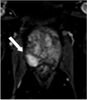
Figure 1 Figure 1 Figure 1
Figure 1 - T2-weighted image showing hypointense nodular area in the prostate peripheral zone in the middle third to the right (arrow).
extension, MRI had a sensitivity of 33%, 100% specificity
and accuracy of 82%. Only one case had seminal vesicle
invasion, which was correctly identified by MRI. There were
no cases of lymph node invasion in any of the methods.
DISCUSSION
DISCUSSION
DISCUSSION
DISCUSSION
DISCUSSION
Anticipating the results of histopathology,
thus optimizing neoplasia staging, is a big challenge in
radiology. Although we are dealing with a method
dependent on experience and training of the radiologist
and ours is a small sample, initial results are promising. The
use of magnetic resonance imaging in preoperative staging
of prostate adenocarcinoma attempts to predict events that
may influence surgical approach and the patient’s
postoperative staging
5. More cases to be aggregated in this
study can generate analyses with a higher degree of
statistical significance.
R E S U M O
R E S U M O
R E S U M O
R E S U M O
R E S U M O
Estadiamento loco-regional convencional para adenocarcinoma de próstata tem sido demonstrado um tanto quanto subdiagnosticado. Por isso, RM da próstata está emergindo como uma ferramenta importante para o estadiamento pré-cirúrgico. Técnicas avançadas, como a difusão e valorização de contraste dinâmico também contribuem para aumentar a sua acurácia. Neste estudo preliminar, a RM de próstata foi comparada com amostras de histopatologia, alcançando sensibilidade de 78% / especificidade de 100% para a localização do tumor; sensibilidade de 33% / especificidade de 100% para extensão extra-capsular; 100% de sensibilidade / especificidade e 100% da extensão das vesículas seminais. É possível acreditar que estes resultados preliminares são promissores, e mais casos tendem a confirmar estes dados.
Descritores: Descritores: Descritores: Descritores:
C a l d a s C a l d a s C a l d a s C a l d a s C a l d a s
Magnetic resonance imaging in staging of locoregional prostate cancer: comparison of results with analysis post-surgical histopathology 449
Rev. Col. Bras. Cir. 2010; 37(6): 447-449
REFERENCES
REFERENCES
REFERENCES
REFERENCES
REFERENCES
1. Smith JA Jr, Scardino PT, Resnick MI, Hernandez AD, Rose SC, Egger MJ. Transrectal ultrasound versus digital rectal examination for the staging of carcinoma of the prostate: results of a prospective, multi-institutional trial. J Urol. 1997 Mar;157(3):902-6.
2. Obek C, Louis P, Civantos F, Soloway MS. Comparison of digital rectal examination and biopsy results with the radical prostatectomy specimen. J Urol. 1999 Feb;161(2):494-8; discussion 498-9.
3. Somford DM, Futterer JJ, Hambrock T, Barentsz JO. Diffusion and perfusion MR imaging of the prostate. Magn Reson Imaging Clin N Am 2008; 16:685-695.
4. Bloch BN, Furman-Haran E, Helbich TH, et al. Prostate cancer: accurate determination of extracapsular extension with high-spatial-resolution dynamic contrast-enhanced and T2-weighted MR imaging—initial results. Radiology 2007; 245:176-185.
5. Ross R, Harisinghani M. Prostate cancer imaging—what the urologic oncologist needs to know. Radiol Clin North Am 2006; 44:711-722.
Received in 23/09/2010
Accepted for publication in 26/10/2010 Conflict of interest: None
Funding Source: None
How to cite this article: How to cite this article: How to cite this article: How to cite this article: How to cite this article:
Caldas MED, Miranda LCD, Bittencourt LK. Magnetic resonance imaging in stanging of locoregional prostate cancer: comparison of results with analysis post-surgical histopathology . Rev Col Bras Cir. [periódico na Internet] 2010; 37(6). Disponível em URL: http:// www.scielo.br/rcbc