AR
TIGO
AR
TICLE
1 Instituto de Psicologia, Universidade Federal do Rio Grande do Sul (UFRGS). Av. Paulo Gama 110, Farroupilha. 90040-060 Porto Alegre RS Brasil. [email protected] 2 Hospital Universitário Prof. Polydoro Ernani de São Thiago, Universidade Federal de Santa Catarina. Florianópolis SC Brasil. 3 Instituto de Ciências Humanas e da Informação, UFRGS. Porto Alegre RS Brasil.
4 Instituto de Ciências Biológicas, Universidade Federal do Rio Grande. Rio Grande RS Brasil.
Stress and Cognitive Reserve as independent factors
of neuropsychological performance in healthy elderly
Estresse e Reserva Cognitiva como determinantes independentes
para o desempenho neuropsicológico de idosos saudáveis
Resumo A exposição a níveis elevados de cortisol e de estresse psicológico, assim como à reserva cog-nitiva, têm sido relacionadas a sintomas da Doen-ça de Alzheimer. Contudo, não há estudos sobre a interação dessas variáveis. Objetivamos exami-nar as associações de medidas de cortisol e estresse psicológico e de reserva cognitiva com o desempe-nho neuropsicológico de idosos saudáveis, além de analisar a existência de interações entre essas variáveis. Análises transversais foram conduzidas usando dados sobre estresse, reserva cognitiva e condições clínicas em 145 idosos saudáveis. Usa-mos uma bateria neuropsicológica para medir as funções executivas, memória verbal e velocidade de processamento. Utilizamos uma medida de cortisol salivar para o nadir circadiano. Encon-tramos uma associação negativa entre diferentes medidas de estresse e o desempenho em tarefas de memória, funções executivas e velocidade de pro-cessamento. Idosos com elevada reserva cognitiva apresentaram um desempenho superior em todas as medidas neuropsicológicas. Não houve intera-ção significativa entre estresse e Reserva Cognitiva para o desempenho neuropsicológico. Estes resul-tados sugerem que idosos com níveis elevados de estresse e reduzida reserva cognitiva podem ser mais suscetíveis ao comprometimento cognitivo.
Palavras-chave Cortisol salivar, Glicocorticoides, Estresse, Reserva cognitiva
Abstract Exposure to high levels of cortisol and self-reported stress, as well as cognitive reserve, have been linked to Alzheimer’s disease pathology.
However, there are no studies on the interaction of these variables. The present study aims to assess the associations of measures of cortisol, self-re-ported stress, and cognitive reserve with neuropsy-chological performance in healthy elderly people; besides, to test the interactions between these vari-ables. Cross-sectional analyzes were conducted using data on stress, cognitive reserve and clinical conditions in 145 healthy elderly adults. A neuro-psychological battery was used to assess executive functions, verbal memory and processing speed.
Measurement of salivary cortisol at the circadian nadir was taken. A negative association between different stress measures and performance on tasks of memory, executive functions and process-ing speed was observed. Elderly people with higher cognitive reserve showed superior performance on all neuropsychological measures. No significant interaction between stress and cognitive reserve to neuropsychological performance was observed.
These results indicate that older adults with high levels of stress and reduced cognitive reserve may be more susceptible to cognitive impairment.
Key words Salivary cortisol, Glucocorticoid, Stress, Cognitive reserve
João Carlos Centurion Cabral 1
Gessyka Wanglon Veleda 2
Martina Mazzoleni 3
Elton Pinto Colares 4
Lucas Neiva-Silva 3
C
ab
ral JCC
Introduction
Social and environmental variables can have a sig-nificant effect on neuropsychological functioning, increasing vulnerability to cognitive impairment and dementia in the elderly. Alzheimer’s disease (AD), the most common type of dementia, is characterized: (a) clinically, by cognitive decline, especially of episodic memory; (b) morphologi-cally, by brain atrophy, being initially affected the hippocampal formation and entorhinal cortex; and (c) histologically, by reduction of synaptic density, presence of neurofibrillary tangles of Tau protein and aggregates of amyloid-β peptide (Aß)1,2. The accumulation of soluble Aß oligomers is considered a critical event in the pathogenesis of AD3, producing cellular changes that result in dendritic atrophy, synaptic loss, and neuronal death4,5. Late-onset AD is heterogeneous and mul-tifactorial, triggered by an interaction between ge-netic and environmental factors, as well as clinical phenotypes6. Even in healthy people, when these interactions disrupt the functioning of nervous system, there may be a reduction of brain reserves, leading to a higher susceptibility to cognitive im-pairment7. Due to the wide variability in cogni-tive abilities of older people, the factors associated with these differences remain to be elucidated.
Cognitive reserve (CR) refers to individual differences in brain or cognitive processing ca-pacity to deal with injuries to the nervous system8. This construct, often linked to educational level and intellectual experiences, has been proposed to try to explain the discrepancy between the sever-ity of disease markers and clinical manifestations in neurological disorders7. In a classic post-mor-tem study, levels of biomarkers for AD in individ-uals who did not have clinically significant symp-toms were evident, conversely these subjects had a higher amount of neurons and brain weight than those patients with the clinical manifestation of
disease9. Complementing these findings, it was
observed that subjects with a high level of edu-cation are more resistant to clinical manifestation of AD10. In addition, an in vivo study showed that education, occupation and intellectual activity are associated with measures of volume and pat-tern of brain activity11. Although the functional correlate of CR is not completely understood, the amount and interaction of specific presynaptic proteins may be some of CR components that reduce the risk of dementia12. However, little is known about the interaction between the CR and other variables that are considered risk factors for cognitive decline in the elderly.
Prolonged exposure to glucocorticoids (GC), especially cortisol in humans, and to psycholog-ical stress play a significant role in pathologpsycholog-ical cognitive impairment in elderly13-15. An increas-ing amount of evidence, includincreas-ing experimental studies in humans, demonstrated that cortisol may impair the formation of declarative memo-ry16 and may predict cognitive decline in healthy older adults17. Also, measures of executive func-tion and processing speed were negatively
asso-ciated with concentrations of GCs18. Chronic
stress and increased GC levels were also related to hippocampal neuronal loss, dendritic atrophy
and reduced hippocampal volume15,19-22.
Howev-er, these findings are still controversial because several studies have found no such associations,
nor with neuropsychological performance17,23,
nor with hippocampal measures24-26. Otherwise, different lines of evidence support the notion that chronic stress and increased GCs play a role in the risk for development of AD24. Green et al.27 found, in animal models, that GCs mediate an increase in production and reduce degradation of Aß, enhance their neuronal toxicity and en-able the formation of amyloid plaques, addition-ally increasing the accumulation of tau protein. GCs have also been associated with other patho-physiological features of AD, such as increased release of excitatory amino acids and increased expression of N-methyl-D-Aspartate (NMDA) glutamate receptor, as well as changes in calcium influx13. Furthermore, older people may be par-ticularly susceptible to the effects of stress, with reduced capacity to resist damage in neurons after prolonged exposure to GCs14. These data are still preliminary and are under intense investigation.
With the establishment of risk factors for the development of AD, the interactions between factors that may increase the likelihood of cog-nitive impairment in healthy elderly have been
increasingly studied28. Therefore, the present
aúd
e C
ole
tiv
a,
21(11):3499-3508,
2016
Method
Participants
One hundred and forty-five cognitively healthy and socially active participants, aged above or equal to 60 years, having at least 4 years of schooling, residents of southern Brazil, were re-cruited through an approach based on sampling units of time and space. For this cross-sectional study, we established units in neighborhoods of Rio Grande city, RS, Brazil, by identifying specif-ic locations and times at whspecif-ich the target popu-lation was likely to be found and constructed a sampling frame of these locations. The identified time-space units were randomly visited, allowing the target population to be systematically recruit-ed in accordance with the protocol of Muhib et al.29. Potential participants who had a history or evidence of dementia, disabling medical condi-tions or use of medicacondi-tions that affect cognitive functioning or cortisol levels (e.g., corticoste-roids) were excluded. Participants received in-structions and signed a term of informed consent before entering the study, which was approved by the Ethics Committee of Health Research of Fed-eral University of Rio Grande.
Procedure
After previous scheduling, data collection was performed by trained research assistants, at the participant’s residence. This procedure aimed to reduce the anxiety caused by testing in an atypical environment. Data collection was conducted between 10:00 a.m. and 5:00 p.m., in order to avoid the circadian variation of cortisol secretion, and was performed during one season (i.e., winter). Data were collected in the following order: interview about socio-demographic and health characteristics, and intellectual activity, measures of stress, neuropsychological testing, clinical measures, and lastly, instructions for sali-va collection procedure were provided.
Psychological and Clinical Assessments
A questionnaire was elaborated to assess health and sociodemographic characteristics, including health habits (e.g., drinking, smoking, lack of exercise), past medical history (e.g., dia-betes mellitus, cardiovascular disease, neurolog-ical disease) and social activities (e.g., traveling,
entertaining, and attending social gatherings). Moreover, to assess the mental status workup we used the Clinical Dementia Rating Scale (CDR; for more details see below) as a cognitive screen-ing instrument.
To assess stress, three scales were used and one to assess depression symptoms. The Per-ceived Stress Scale (PSS-14), which measures the perception of stress for the last month, has ade-quate psychometric qualities in its version for the Brazilian population, with internal consisten-cy and construct validity similar to the original version30. The Social Readjustment Rating Scale (SRRS) presents a list of 43 stressful events, and the respondent should indicate, among them, those which occurred during last year31. The Has-sles and Uplifts scale (HUS), in its version of 53 items, measures daily sensitivity to adverse events (only the part related to negative evaluation of events was used)32. Finally, the Geriatric Depres-sion Scale (GDS) in the verDepres-sion with 15 items was used for screening depressive symptoms. This scale showed good accuracy, sensitivity, specific-ity and reliabilspecific-ity for the Brazilian population33.
Measures of intellectual activity and educa-tion were used for the establishment of a proxy measure for CR. Data on years of formal educa-tion; school failure; weekly frequency of reading books, newspapers, magazines and other mate-rials (e.g., puzzle exercise and internet); number and fluency of foreign languages spoken (com-prehension, speaking, reading and writing) were used to quantify the proxy measures for cogni-tive reserve. Principal Component Analysis was used to combine these variables and to obtain for CR, as described in “Statistical Analysis” section. Measures of intelligence (e.g., verbal IQ) were not used for the proxy for CR, since such mea-sures are intrinsically associated with neuropsy-chological performance.
C
ab
ral JCC
Neuropsychological Assessment
The application of the neuropsychological battery lasted about 90 minutes. The instructions for each task were read aloud by a research assis-tant and made available in writing to participants before each test. The mention of the term “skills testing” was avoided to minimize anxiety related to performance of the participants. The neuro-psychological battery was assembled in order to measure the performance of executive functions, verbal memory and processing speed.
Measures of executive functions included a Verbal Fluency Test, Trail Making Test, Stroop Test, and a Digit Span task. In the Semantic Ver-bal Fluency Test, the semantic version was used, in which the participant must say as many words as possible, from a given category, during one minute. This test is highly sensitive to prefrontal cortex (PFC) dysfunction35. In order to control other sources of variations on people’s fluency and categorization (variables measured by test) two applications were carried out, one with the category “animals” and the other with the cate-gory “fruit”. The score for fluency was calculated by averaging the results of applications. The Trail Making Test B measures cognitive flexibility and attention. After demonstration, the participant must draw lines to connect the randomly spread numbers and letters in an ascending and alter-nating alphanumeric sequence, in the shortest possible time. Stroop test measures inhibitory control and attention under incongruent stimu-li conditions (i.e., words printed in an ink col-or differing from the colcol-or name of the wcol-ord). The Digit Span task measures working memory, a component of executive function that activates the Dorsolateral PFC cortex36. Only the direct or-der version was used.
The Word List Memory Task (WLMT)37
as-sesses the ability to recall ten unrelated words of everyday use. In the learning trials (immediate recall), the same words are displayed in differ-ent order in each trial. Each of the three trials has maximum score of ten. Ten minutes after the learning phase, free recall was measured through the Word List Recall Task, in which the ten words shown in the learning trials should be mentioned, without stimulus presentation. Then, the Word List Recognition Task was ap-plied, when the participant had to identify the ten words shown in previous trials, to distinguish them from other ten words of everyday use. In this task, the 20 words were randomly ordered. Each trial allowed 90 seconds for responses. After
the learning trials, an abstract task, unrelated to the WLMT (i.e., not using words in its execution) was executed, to avoid interference in the memo-ry encoding process. In this case, a Digit Symbol Substitution Task was used.
Digit Symbol Substitution Task was used to measure processing speed. In this task, a se-quence of digit-symbol pairs is used to match and complete a list of unpaired symbols. Partic-ipants were asked to make the largest number of pairings possible within 120 seconds. Moreover, Trail Making Test A (25 sequential numbers are linked with a pencil in the shortest possible time) and reading time without interference, from the Stroop Test, were used to measurement process-ing speed.
Salivary Cortisol Measurement
Salivary cortisol is a biomarker used in stress research to evaluate the activity of the hypotha-lamic-pituitary-adrenal (HPA) axis, providing a reliable measure of the biologically active (un-bound) fraction of this hormone and has good correlation with serum measurements of corti-sol38. The method of saliva collection is simple, non-invasive, and can be done in various envi-ronments, making unnecessary the presence of specialized personnel. Thus, saliva was collected by the participants in their homes, between 9:00 p.m. and 10:00 p.m. one to two days after the neu-ropsychological assessment, when they received saliva collection instructions. For this purpose, cotton swab of the Salivette sampling devices (Sarstedt, Rommeldorf, Germany) were inserted into the oral cavity. Participants were instructed to make smooth masticatory movements, with mild pressure to avoid damage to the material, until it became saturated with saliva (about 3 minutes). The samples were refrigerated until delivered to research assistant, during the next day. Then, the samples were centrifuged (1800 x g) for 20 min-utes and stored at - 20°C until the biochemical analysis. Nocturnal cortisol sampling was chosen to determine the nadir cortisol levels and, also, to avoid spontaneous fluctuations in cortisol secre-tion pattern, and its high amplitude, which are significantly lower at night17.
aúd
e C
ole
tiv
a,
21(11):3499-3508,
2016
as specified by the manufacturer’s instructions. An ELISA plate reader with a filter set at 450nm was used. The sensitivity of the kit was 1 ng/ml.
Statistical Analysis
Descriptive and exploratory analysis were performed. Tests for normality (Kolmogor-ov-Smirnov test) and homogeneity of variance (Levene test) for total sample and each group were conducted. Spearman correlation (α = 0.05) was used to measure the relationship between con-tinuous variables. Principal Component Analysis (PCA) was performed to obtain principal com-ponents and standard scores (dimensionless) for the latent variables. PCA were performed using a correlation matrix (since the variables have different units), with a varimax rotation and an extraction criterion of eigenvalue > 1. The variable loadings greater than 0.4 were used for interpretation. PCA were used to deal with the problem of multiple comparisons (Familywise Error Rate - FWER), i.e., increased the probabil-ity of type I error, reducing the dependent vari-ables. Each PCA generated a single component and a corresponding standard score. These were later named as Cognitive Reserve (CR); Execu-tive Function (EF); Verbal Memory (VM); and Processing Speed (PS). Perceived Stress score and the standardized score of cognitive reserve were categorized into two groups based on median values: High Stress (HS) / Low Stress (LS) and High Cognitive Reserve (HCR) / Low Cognitive Reserve (LCR), respectively. T tests were used to compare the means of dependent variables (EF, VM and PS) for these groups. Finally, a two-way
ANOVA was performed to determine the inter-action between the independent variables (HS / LS X HCR / LCR) on the standardized dependent variables. The Bonferroni correction was used to reduce the probability of Type I error (FWER), yielding a significance level of 0.016.
Results
Socio-demographic and health characteristics for the entire sample and for the groups of high and low level of stress, were summarized in Ta-ble 1. The salivary cortisol levels at nadir had a mean of 6.95 ng/mL (standard error (SE) = 0 , 37), and showed no significant associations with age, gender, education and socioeconomic status of the participants, nor were there significant dif-ferences between the groups classified as high or low perceived stress and those classified as high or low cognitive reserve. Each factor (for each PCA) explained approximately 60% of variance. The factor extracted for CR had an eigenvalue of 1.89. The eigenvalue for the EF factor was 2.15. While for the memory score, its eigenvalue was 3.26. Finally, the eigenvalue for PS was 2.06. Neu-ropsychological scores can be seen in Table 2.
Correlations of measures of stress and CR with performances on neuropsychological tests are shown in Table 3. All significant correlations between measures of stress and neuropsycho-logical performance were weak. The perceived stress was consistently associated with a decrease in verbal memory performance. However, this pattern was not observed in measures for adverse events. The increased level of cortisol was
sig-Variables
Age (years)a,b
Gender (female)c
Education (years)a,b
Body mass indexa
Sleep (hours per night)a,b
Regular physical activityb,c
Tobacco useb,c,d
Diabetes mellitusb,c
Cardiovascular diseaseb,c
Clinical Dementia Rating (0,5)c,e
Geriatric depressiona,f
Participants (n = 145)
69.9 (6.7) 66.2 9.8 (4.6) 26.4 (3.8)
7.4 (1.6) 53.1 10.3 23.4 58.6 17.9 2.9 (2.9)
Table 1. Sociodemographic and health variables.
Low Stress (n = 73)
70.0 (6.6) 60.3 10.9 (4.5) 26.3 (3.6) 7.4 (1.3)
61.6 11.0 19.2 56.2 6.8 1.6 (1.5)
High Stress (n = 72)
69.7 (6.8) 72.2 8.8 (4.4) 26.6 (4.0) 7.4 (1.8)
44.4 9.7 27.8 61.1 28.2 4.2 (3.4)
a Mean and standard deviation. b Self-report data. c Percentage. d Regular tobacco use in the last year. e CDR 0.5 score. f Data obtained by the Geriatric Depression Scale.
C
ab
ral JCC
nificantly correlated with a worse performance in the recognition task. Nevertheless, its associ-ation with other memory scores did not meet the significance level, even having a pattern, i.e., negative correlations with all measures. Again, the perceived stress was associated with a worse
performance on cognitive flexibility and atten-tion tasks (Trail Making Test B) and working memory (digit span), both measures of executive functions. It was not found for inhibitory con-trol and fluency measures. The two measures of processing speed were also related with perceived stress. On the other hand, the explained variance in the associations between measures of stress and neuropsychological assessment in the elderly was at most 7%. This inconsistency of results is not seen in the association between CR score and neuropsychological performances, in which all correlations were positive and highly significant. These associations reached an explained variance of up to 33%.
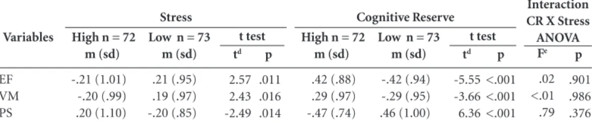
The association of high stress with reduction in neuropsychological performance in the elderly was confirmed by the comparison between High Stress and Low Stress groups (Table 4). The EF score was significantly reduced (p = 0.011) in the High Stress group. However, the mean difference (MD) of both groups was 0.41 (standard error of the difference (SED) = 0.16). The difference between stress groups was also significant (p = 0.016) for verbal memory scores, with a mean difference of 0.39 (SED = 0.16). Similar results were seen for the difference between these groups in processing speed score (p = 0.014; MD = -0.41; SED = 0.16). While these data corroborate the hypothesis that high level of stress and reduced
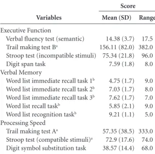
Variables
Executive Function
Verbal fluency test (semantic) Trail making test Ba
Stroop test (incompatible stimuli) Digit span task
Verbal Memory
Word list immediate recall task 1b
Word list immediate recall task 2b
Word list immediate recall task 3b
Word list recall taskb
Word list recognition taskb
Processing Speed Trail making test Aa
Stroop test (compatible stimuli)a
Digit symbol substitution task
Mean (SD)
14.38 (3.7) 156.11 (82.0) 75.34 (21.8) 7.59 (1.8)
4.75 (1.7) 7.03 (1.7) 7.62 (1.7) 5.85 (2.1) 9.21 (1.1)
57.35 (38.5) 72.9 (17.6) 38.57 (14.4)
Range
17.5 382.0 96.0 8.0
9.0 8.0 7.0 9.0 5.0
333.0 74.0 68.0
Table 2. Mean (standard deviation) and range for
neuropsychological performance in healthy elderly.
Abbreviations: SD, standard deviation. a Time. b Word list memory task.
Score
Variablesb
Stroop (IS)e
TMT-Bf
Verbal Fluency Digit Span
Immediate Recall 1 Immediate Recall 2 Immediate Recall 3 Recall
Recognition
TMT-Af Stroop (CS)g
DSST
Cortisolc
.025 (.413)d
-.083 (.231) -.102 (.183) .158 (.080)
-.170 (.064) -.069 (.271) -.118 (.147) -.140 (.107) -.208 (.031)
-.138 (.110) -.145 (.098) .216 (.027)
Table 3. Associations of measures of stress and cognitive reserve with neuropsychological performancea.
PSS14
-.100 (.116) .187 (.012) -.122 (.072) -.260 (.001)
-.201 (.008) -.189 (.012) -.151 (.035) -.181 (.015) -.244 (.002)
.212 (.005) .108 (.098) -.254 (.001)
SRRS
-.088 (.147) .120 (.076) .002 (.491) .177 (.016)
-.028 (.368) -.055 (.257) -.025 (.385) -.109 (.095) -.043 (.305)
.078 (.175) .064 (.221) -.124 (.068)
Abbreviations: PSS14, Perceived Stress Scale; SRRS, Social Readjustment Rating Scale; HUS, Hassles & Uplifts Scale; GDS15, Geriatric Depression Scale; CR, Cognitive Reserve; SCR, Standard Score for Cognitive Reserve; DSST, Digit Symbol Substitution Task; TMT, Trail Making Task; EF, Executive Function; VM, Verbal Memory; PS, Processing Speed.
a Associations by Spearman’s correlation coefficient at the 0.05 significance level. b Data were obtained from 145 participants, except where noted. c Data were obtained from 81 participants. d Correlation size effect (p-value). e Stroop test (incompatible stimuli). f Time to complete the task. gStroop test (compatible stimuli).
Stress
E F
V M
P S
HUS
-.032 (.352) .106 (.103) .082 (.164) -.050 (.274)
.070 (.201) .047 (.286) .118 (.079) .101 (.113) .075 (.184)
.123 (.071) -.004 (.480) -.097 (.122)
GDS15
-.119 (.076) .116 (.082) -.198 (.008) -.215 (.005)
-.090 (.140) -.195 (.009) -.136 (.051) -.152 (.034) -.163 (.025)
.151 (.035) .158 (.029) -.157 (.030)
SCR
.386 (<.001) -.386 (<.001) .379 (<.001) .188 (.012)
.274 (<.001) .256 (.001) .209 (.006) .242 (.002) .123 (.070)
-.444 (<.001) -.574 (<.001) .474 (<.001)
aúd
e C
ole
tiv
a,
21(11):3499-3508,
2016
neuropsychological performance are related, we realize that the size of the main effect, though ex-istent, is quite limited.
On the other hand, the means differences in standardized neuropsychological scores for the High CR and Low CR groups were highly signif-icant (Table 4). In these groups, were observed p-values of less than 0.001 for all neuropsycho-logical scores, corroborating our second hypoth-esis. In addition, the mean differences were -0.84 (SED = 0.15); -0.58 (SED = 0.15); and 0.93 (SED = 0.14); for EF, VM and PS scores, respectively.
Finally, the hypothesis of interaction effects between the independent variables was refuted (Table 4). All F-values were below 1 and η² were less than 0.001.
Discussion
Our results support the notion that healthy elder-ly adults with high level of stress have significant-ly worse scores on cognitive tests. Although this effect size was small, the relationship is consistent for all three measured variables. This consistency can be checked by the negative correlations pat-tern of perceived stress scores and cortisol levels with different measures of memory. In addition, depression scores can support these findings. Depression is largely associated with memory impairment and hippocampal atrophy and these may be due, at least in part, to hypercortisolemia, which is frequent in this pathology25. A similar pattern was seen for PS measures. Conversely, measures of EF have not shown the same con-sistency, only working memory and cognitive flexibility were impaired by perception of high stress level, both measures are associated with
the dorsolateral PFC activity36. Absence of cor-relations between adverse events or daily hassles may reflect the complexity of physiological stress, which can be modulated by several factors, such as differences in expression of GC receptor genes, differences in patterns of cortisol secretion, epi-genetic regulation, concentration of other hor-mones, reactivity to stress and resilience13,39.
Certainly, brain aging may increase the sus-ceptibility to the effects of stress. The hippo-campus and PFC, brain regions responsible for episodic memory consolidation and executive functions respectively, become increasingly sus-ceptible to deleterious effects25,40,41. In hippo-campal region, impairments can also result in persistently high levels of cortisol, since this re-gion regulates GC levels through an inhibitory
feedback loop42-44. The human hippocampus has
a high density of mineralocorticoid receptors (MR) and glucocorticoid receptors (GR), given that both mediate cortisol activity45,46. With high levels of free cortisol, MRs are fully saturated, while a large proportion of GRs are occupied, and this, via the hippocampus, inhibits the secretion of corticotropin releasing hormone (CRH) from the hypothalamus and thus reducing the cascade responsible for cortisol release from the suprare-nal glands45. Consequently, hippocampal atrophy may reduce the effectiveness of this control mech-anism, thereby increasing the susceptibility to the effects of stress. Indeed, patients with AD have a cortisol secretion pattern progressively higher47. Typically, higher evening cortisol levels are relat-ed to an impairment of the negative ferelat-edback of HPA axis48. This is consistent with our data which show an association between high evening corti-sol levels and poorer performance in the recogni-tion task, seeing as this task is less influenced by
Variables
EF VM PS
Table 4. Mean differences between stress groups stress and cognitive reserve groups for neuropsychological
standard scoresa,b,c.
Abbreviations. EF, Executive Function; VM, Verbal Memory; PS, Processing Speed; CR, Cognitive Reserve.
a T test was used to determine the neuropsychological differences between stress groups and cognitive reserve groups. b Two-way ANOVA was used to determine the interaction between independent variables (stress and cognitive reserve). c Two-tailed significance level for all tests was set at 0,0167. d Degrees of freedom = 143. e Degrees of freedom = 1; 141.
Fe
.02 <.01 .79
Interaction CR X Stress ANOVA High n = 72
m (sd)
-.21 (1.01) -.20 (.99) .20 (1.10)
Low n = 73 m (sd)
.21 (.95) .19 (.97) -.20 (.85)
Stress
t test
td
2.57 2.43 -2.49
p
.011 .016 .014
High n = 72 m (sd)
.42 (.88) .29 (.97) -.47 (.74)
Low n = 73 m (sd)
-.42 (.94) -.29 (.95) .46 (1.00)
Cognitive Reserve t test
td
-5.55 -3.66 6.36
p
<.001 <.001 <.001
p
C
ab
ral JCC
other brain structures, besides the hippocampus, than other memory tasks. Our findings on the EF impairment are also supported by neuroanatom-ical data about the distribution of MRs and GRs. Besides medial temporal structures, the PFC also has a high expression of GCs receptors, which makes this region particularly susceptible to the cortisol action13,17. The present study showed an association between stress and impaired cognitive functioning. All effect sizes found for stress were small, however, it is worth noting that the occur-rence of summation of various neurophysiolog-ical dysfunctions may further impair cognitive performance in the elderly.
On the other hand, those with higher cog-nitive reserve showed a moderately elevated performance on neuropsychological tests, when compared to those with lower CR. There was a strong consistency of the data supporting the hypothesis of an association between higher CR with better cognitive performance. CR has had a positive correlation with all measures of neuro-psychological performance49,50. This pattern of relationship occurred even with the absence of verbal intelligence measures (which were avoided because their correlation with neuropsychologi-cal measures is expected) for proxy measure of cognitive reserve. In the same line of our results, recent neuroimaging studies have supported the importance of educational level and intellectual activities for the maintenance of normal brain structures in elderly8,51. Sollé-Padullés et al.11 evidenced, through the use of functional mag-netic resonance image, that high CR is not only associated with increased brain volume, they also noted an increase in the efficacy of the neu-ral network (as reflected by a reduction in brain activity for the same performance) during the performance of cognitive tasks in healthy elders. However, these authors did not find the same for patients with Mild Cognitive Impairment (MCI) or AD. Those patients with higher CR had a smaller brain volume in both MCI and AD, and there was also a greater brain activity in patients with AD, indicating a possible anatomophysio-logical compensatory mechanism for cognitive decline11. In another cross-sectional study with elderly52, differences in the use of compensatory strategies, such as external aids, mnemonic strat-egies and increased effort investment, were also identified for the scores of CR proxies (i.e., verbal intelligence and educational level). Surprisingly, compensatory strategies were used only by older adults with a verbal intelligence level higher than their educational level. These data demonstrate
the heterogeneity in cognitive ability of older adults with differences in CR. Consequently, this highlighted the need to better understand the neurobiological and behavioral determinants of CR for both healthy elderly and those with mild cognitive impairment or neurodegenerative dis-eases as Alzheimer’s disease.
Our hypothesis of an interaction between stress and CR was refuted. High CR did not re-duce the main effect of stress on neuropsycho-logical performance in healthy elderly people. Elderly with a high level of stress (i.e., deleterious condition) and high CR (i.e., protective condi-tion) had a superior neuropsychological per-formance than those with low stress level (i.e., protective condition) and low CR (i.e., deleteri-ous condition). Accordingly, the CR had a more powerful relationship with neuropsychological performance in healthy elderly people. Never-theless, high CR did not affect the magnitude of neuropsychological impairment associated with high levels of stress in the elderly, i.e., the main effect of stress remained independently of the influence of CR. The physiological mechanisms and cognitive changes related to stress and CR are noticeably distinct and, therefore, may be ex-ercising their brain and behavioral effects with-out interacting each other.
aúd
e C
ole
tiv
a,
21(11):3499-3508,
2016
there is no evidence of a relationship between the modulating effects of CR on the impact of stress on neuropsychological performance in healthy elderly subjects. A more detailed understanding of the effect of these variables on human cogni-tion, which are recognized as risk factors for the development of AD, is important to identify pre-ventive strategies that aim to decrease cognitive decline in the healthy elderly. There have been intense researches to identify groups vulnerable
to AD prior to symptom onset6. Thus, our data
suggest that healthy elderly subjects with high stress and low CR can be especially vulnerable to cognitive impairment in old age.
Collaborations
JCC Cabral contributed to design of study, acqui-sition, analysis and interpretation of data, and in drafting the manuscript. GW Veleda contributed to design of study, acquisition, analysis and in-terpretation of data. M Mazzoleni contributed to design of study, acquisition, analysis and in-terpretation of data. EP Colares contributed to design, analysis and interpretation of hormon-al data. L Neiva-Silva contributed to design of study, mainly concerning epidemiological as-pects; contributed to analysis and interpretation of data. VT Neves was responsible for overall ori-entation as well as for the conception of the proj-ect, discussion of results, and critical analysis. All authors read and approved the final manuscript.
References
Cummings JL. Alzheimer’s disease. N Engl J Med 2004; 351(1):56-67
LaFerla FM, Green KN, Oddo S. Intracellular amyloid-β in Alzheimer’s disease. Nat Rev Neurosci 2007; 8(7):499-509. Selkoe DJ. Soluble oligomers of the amyloid beta-pro-tein impair synaptic plasticity and behavior. Behav Brain Res 2008; 192(1):106-113.
Haass C, Selkoe DJ. Soluble protein oligomers in neu-rodegeneration: lessons from the Alzheimer’s amyloid
β-peptide. Nat Rev Mol Cell Biol 2007; 8(2):101-112. Shankar GM, Li S, Mehta TH, Garcia-Munoz A, Shep-ardson NE, Smith I, Brett FM, Farrell MA, Rowan MJ, Lemere CA, Regan CM, Walsh DM, Sabatini BL, Selkoe DJ. Amyloid-β protein dimers isolated directly from Alzheimer’s brains impair synaptic plasticity and memory. Nat Med 2008; 14(8):837-842.
Barnes DE, Yaffe K. The projected effect of risk factor reduction on Alzheimer’s disease prevalence. Lancet Neurol 2011; 10(9):819-828.
Stern Y. What is cognitive reserve? Theory and research application of the reserve concept. J Int Neuropsychol Soc 2002; 8(3):448-460.
Stern Y. Cognitive reserve. Neuropsychologia 2009; 47(10):2015-2028.
Katzman R, Terry R, DeTeresa R, Brown T, Davies P, Fuld P, Renbing X, Peck A. Clinical, pathological, and neurochemical changes in dementia: a subgroup with preserved mental status and numerous neocortical plaques. Ann Neurol 1988; 23(2):138-144.
Scarmeas N, Stern Y. Cognitive reserve and lifestyle. J Clin Exp Neuropsychol 2003; 25(5):625-633.
Solé-Padullés C, Bartrés-Faz D, Junqué C, Vendrell P, Rami L, Clemente IC, Bosch B, Villar A, Bargalló N, Jurado MA, Barrios M, Molinuevo JL. Brain structure and function related to cognitive reserve variables in normal aging, mild cognitive impairment and Alzhei-mer’s disease. Neurobiol Aging 2009; 30(7):1114-1124. 1.
2. 3.
4.
5.
6.
7.
8. 9.
10. 11.
Honer WG, Barr AM, Sawada K, Thornton AE, Morris MC, Leurgans SE, Schneider JA, Bennett DA. Cognitive reserve, presynaptic proteins and dementia in the el-derly. Transl Psychiatry 2012; 2(5):e114.
de Kloet ER, Joëls M, Holsboer F. Stress and the brain: from adaptation to disease. Nat Rev Neurosci 2005; 6(6):463-475.
Lupien SJ, McEwen BS, Gunnar MR, Heim C. Effects of stress throughout the lifespan on the brain, behaviour and cognition. Nat Rev Neurosci 2009; 10(6):434-445. McEwen BS, Bowles NP, Gray JD, Hill MN, Hunter RG, Karatsoreos IN, Nasca C. Mechanisms of stress in the brain. Nat Neurosci 2015; 18(10):1353-1363.
Kirschbaum C, Wolf O, May M, Wippich W, Hellham-mer D. Stress- and treatment-induced elevations of cor-tisol levels associated with impaired declarative mem-ory in healthy adults. Life Sci 1996; 58(17):1475-1483. Li G, Cherrier MM, Tsuang DW, Petrie EC, Colasurdo EA, Craft S, Schellenberg GD, Peskind ER, Raskind MA, Wilkinson CW. Salivary cortisol and memory function in human aging. Neurobiol Aging 2006; 27(11):1705-1714. Franz CE, O’Brien RC, Hauger RL, Mendoza SP, Paniz-zon MS, Prom-Wormley E, Eaves LJ, Jacobson K, Lyons MJ, Lupien S, Hellhammer D, Xian H, Kremen WS. Cross-sectional and 35-year longitudinal assessment of salivary cortisol and cognitive functioning: the Viet-nam Era twin study of aging. Psychoneuroendocrinology
2011; 36(7):1040-1052.
Knoops AJG, Gerritsen L, van der Graaf Y, Mali WPTM, Geerlings MI. Basal hypothalamic pituitary adrenal axis activity and hippocampal volumes: the SMART-Medea study. Biol Psychiatry 2010; 67(12):1191-1198. Lupien SJ, de Leon M, de Santi S, Convit A, Tarshish C, Nair NP, Thakur M, McEwen BS, Hauger RL, Meaney MJ. Cortisol levels during human aging predict hip-pocampal atrophy and memory deficits. Nat Neurosci
1998; 1(1):69-73. 12.
13.
14.
15.
16.
17.
18.
19.
C
ab
ral JCC
McEwen BS, Sapolsky RM. Stress and cognitive func-tion. Curr Opin Neurobiol 1995; 5(2):205-216. Marcello E, Gardoni F, Di Luca M. Alzheimer’s disease and modern lifestyle: what is the role of stress? J Neuro-chem 2015; 134(5):795-798.
Peavy GM, Salmon DP, Jacobson MW, Hervey A, Gamst AC, Wolfson T, Patterson TL, Goldman S, Mills PJ, Khandrika S, Galasko D. Effects of chronic stress on memory decline in cognitively normal and mildly impaired older adults. Am J Psychiatry 2009; 166(12):1384-1391.
Csernansky JG, Dong H, Fagan AM, Wang L, Xiong C, Holtzman DM, Morris JC. Plasma cortisol and pro-gression of dementia in subjects with Alzheimer-type dementia. Am J Psychiatry 2006; 163(12):2164-2169. O’Brien JT, Lloyd A, McKeith I, Gholkar A, Ferrier N. A longitudinal study of hippocampal volume, cortisol levels, and cognition in older depressed subjects. Am J Psychiatry 2004; 161(11):2081-2090.
Tata DA, Marciano VA, Anderson BJ. Synapse loss from chronically elevated glucocorticoids: relationship to neuropil volume and cell number in hippocampal area CA3. J Comp Neurol 2006; 498(3):363-374.
Green KN, Billings LM, Roozendaal B, McGaugh JL, LaFerla FM. Glucocorticoids increase amyloid-beta and tau pathology in a mouse model of Alzheimer’s disease. J Neurosci 2006; 26(35):9047-9056.
Eshkoor SA, Hamid TA, Mun CY, Ng CK. Mild cogni-tive impairment and its management in older people.
Clin Interv Aging 2015; 10:687-693.
Muhib FB, Lin LS, Stueve A, Miller RL, Ford WL, John-son WD, Smith PJ; Community Intervention Trial for Youth Study Team. A venue-based method for sam-pling hard-to-reach populations. Public Health Rep. 2001; 116(Supl.):216-222.
Luft CDB, Sanches SDO, Mazo GZ, Andrade A. Versão brasileira da Escala de Estresse Percebido: tradução e validação para idosos. Rev Saude Publica 2007; 41(4):606-615.
Holmes TH, Rahe RH. The social readjustment rating scale. J Psychosom Res 1967; 11(2):213-218.
DeLongis A, Folkman S, Lazarus RS. The impact of daily stress on health and mood: Psychological and social resources as mediators. J Pers Soc Psychol 1988; 54(3):486-495.
Sousa RL, Medeiros JGM, Moura ACL, Souza CLM, Moreira IF. Validade e fidedignidade da Escala de Depressão Geriátrica na identificação de idosos dep-rimidos em um hospital geral. J Bras Psiquiatr 2007; 56(2):102-107.
Montaño MBM, Ramos LR. Validade da versão em português da Clinical Dementia Rating. Rev Saude Pu-blica 2005; 39(6):912-917.
Chaves MLF, Godinho CC, Porto CS, Mansur L, Carth-ery-Goulart MT, Yassuda MS, Beato R. Doença de Alz-heimer: avaliação cognitiva, comportamental e funcio-nal. Dement Neuropsychol 2011; 5(1):21-33.
Smith EE, Jonides J. Storage and Executive Processes in the Frontal Lobes. Science (80- ) 1999; 283(5408):1657-1661.
Bertolucci PH, Okamoto IH, Brucki SM, Siviero MO, Toniolo Neto J, Ramos LR. Applicability of the CERAD neuropsychological battery to Brazilian elderly. Arq Neuropsiquiatr 2001; 59(3-A):532-536.
21. 22.
23.
24.
25.
26.
27.
28.
29.
30.
31. 32.
33.
34.
35.
36.
37.
Hellhammer DH, Wüst S, Kudielka BM. Salivary corti-sol as a biomarker in stress research. Psychoneuroendo-crinology 2009; 34(2):163-171.
Lupien SJ, Maheu F, Tu M, Fiocco A, Schramek TE. The effects of stress and stress hormones on human cogni-tion: Implications for the field of brain and cognition.
Brain Cogn 2007; 65(3):209-237.
Jack CR, Petersen RC, Xu Y, O’Brien PC, Smith GE, Ivnik RJ, Tangalos EG, Kokmen E. Rate of medial tem-poral lobe atrophy in typical aging and Alzheimer’s dis-ease. Neurology 1998; 51(4):993-999.
Cerqueira JJ, Mailliet F, Almeida OFX, Jay TM, Sousa N. The prefrontal cortex as a key target of the maladaptive response to stress. J Neurosci 2007; 27(11):2781-2787. Elgh E, Lindqvist Astot A, Fagerlund M, Eriksson S, Olsson T, Näsman B. Cognitive dysfunction, hippo-campal atrophy and glucocorticoid feedback in Alzhei-mer’s disease. Biol Psychiatry 2006; 59(2):155-161. Souza-Talarico JN, Marin M, Sindi S, Lupien SJ. Ef-fects of stress hormones on the brain and cognition: evidence from normal to pathological aging. Dement Neuropsychol 2011; 5(1):8-16.
Sindi S, Fiocco AJ, Juster R-P, Lord C, Pruessner J, Lupi-en SJ. Now you see it, now you don’t: Testing Lupi- environ-ments modulate the association between hippocampal volume and cortisol levels in young and older adults.
Hippocampus 2014; 24(12):1623-1632.
de Kloet ER, Oitzl MS, Joëls M. Stress and cognition: are corticosteroids good or bad guys? Trends Neurosci
1999; 22(10):422-426.
McEwen BS, McKittrick CR, Tamashiro KLK, Sakai RR. The brain on stress: Insight from studies using the Visible Burrow System. Physiol Behav 2015; 146:47-56. Zvěřová M, Fišar Z, Jirák R, Kitzlerová E, Hroudová J, Raboch J. Plasma cortisol in Alzheimer’s disease with or without depressive symptoms. Med Sci Monit 2013; 19:681-689.
Raff H, Raff JL, Duthie EH, Wilson CR, Sasse EA, Rud-man I, Mattson D. Elevated salivary cortisol in the eve-ning in healthy elderly men and women: correlation with bone mineral density. Journals Gerontol Ser A
1999; 54(9):M479-M483.
Sobral M, Pestana MH, Paúl C. Measures of cognitive reserve in Alzheimer’s disease. Trends Psychiatry Psy-chother 2014; 36(3):160-168.
Sobral M, Pestana MH, Paúl C. The impact of cognitive reserve on neuropsychological and functional abilities in Alzheimer’s disease patients. Psychol Neurosci 2015; 8(1):39-55.
Whalley LJ, Deary IJ, Appleton CL, Starr JM. Cognitive reserve and the neurobiology of cognitive aging. Ageing Res Rev 2004; 3(4):369-382.
Garrett DD, Grady CL, Hasher L. Everyday memory compensation: the impact of cognitive reserve, subjec-tive memory, and stress. Psychol Aging 2010; 25(1):74-83.
Artigo apresentado em 07/09/2015 Aprovado em 15/12/2015
Versão final apresentada em 17/12/2015 38.
39.
40.
41.
42.
43.
44.
45.
46.
47.
48.
49.
50.
51.