BASIC RESEARCH
Exercise training prevents diastolic dysfunction induced
by metabolic syndrome in rats
Cristiano Mostarda,IIvana Cinthya Moraes-Silva,IVera Maria Cury Salemi,IIJacqueline Freire Machi,IBruno Rodrigues,I,IIIKa´tia De Angelis,IVVera de Moura Azevedo Farah,V Maria Claudia IrigoyenI
IHospital das Clinicas da Faculdade de Medicina da Universidade de Sa˜o Paulo, Hypertension Unit, Heart Institute (InCor), Sao Paulo/SP, Brazil.IIHospital
das Clinicas da Faculdade de Medicina da Universidade de Sa˜o Paulo, Cardiomiopathy Unit, Heart Institute (InCor), Sao Paulo/SP, Brazil.IIIHuman
Movement Laboratory, Sao Judas Tadeu University, Sao Paulo/SP, Brazil.IVLaboratory of Translational Physiology, Nove de Julho University, Sao Paulo Sao
Paulo/SP, Brazil.VCenter of Biological Sciences and Health, Mackenzie Presbyterian University, Sao Paulo/SP, Brazil.
OBJECTIVE:High fructose consumption contributes to the incidence of metabolic syndrome and, consequently, to cardiovascular outcomes. We investigated whether exercise training prevents high fructose diet-induced metabolic and cardiac morphofunctional alterations.
METHODS:Wistar rats receiving fructose overload (F) in drinking water (100 g/l) were concomitantly trained on a treadmill (FT) for 10 weeks or kept sedentary. These rats were compared with a control group (C). Obesity was evaluated by the Lee index, and glycemia and insulin tolerance tests constituted the metabolic evaluation. Blood pressure was measured directly (Windaq, 2 kHz), and echocardiography was performed to determine left ventricular morphology and function. Statistical significance was determined by one-way ANOVA, with significance set at
p,0.05.
RESULTS:Fructose overload induced a metabolic syndrome state, as confirmed by insulin resistance (F: 3.6¡0.2 vs. C: 4.5¡0.2 mg/dl/min), hypertension (mean blood pressure, F: 118¡3 vs. C: 104¡4 mmHg) and obesity (F: 0.31¡0.001 vs. C: 0.29¡0.001 g/mm). Interestingly, fructose overload rats also exhibited diastolic dysfunction. Exercise training performed during the period of high fructose intake eliminated all of these derangements. The improvements in metabolic parameters were correlated with the maintenance of diastolic function.
CONCLUSION:The role of exercise training in the prevention of metabolic and hemodynamic parameter alterations is of great importance in decreasing the cardiac morbidity and mortality related to metabolic syndrome.
KEYWORDS: Metabolic Syndrome; Diastolic function; Exercise Training; Insulin resistance; Cardiac hypertrophy.
Mostarda C, Moraes-Silva IC, Salemi VM, Machi JF, Rodrigues B, De Angelis K, et al. Exercise training prevents diastolic dysfunction induced by metabolic syndrome in rats. Clinics. 2012;67(7):815-820.
Received for publication onDecember 20, 2011;First review completed onFebruary 5, 2012;Accepted for publication onMarch 14, 2012 E-mail: [email protected]
Tel.: 55 11 2661-5327
INTRODUCTION
Metabolic syndrome (MS) has proven to be a major clinical challenge in this century. The presence of this syndrome increases mortality, especially due to cardiovas-cular disease (1). It is worth noting that the association of MS with cardiovascular disease increases overall and cardiovascular mortalities by 1.5- to 2.5-fold (2,3).
One of the factors that contributes to the increase in MS incidence is poor eating habits, which are mainly character-ized by a large increase in fructose consumption (4,5). Several changes are observed after chronic unhealthy eating
habits, including increased blood pressure, hyperlipidemia, obesity, glucose intolerance, insulin resistance and hyper-insulinemia.
Insulin resistance may precede and is a pathogenic factor of diabetes. Furthermore, insulin resistance is associated with left ventricular diastolic dysfunction in pre-diabetic adults, independent of blood pressure levels (6). Studies have shown that MS patients also present with left ventricular remodeling and diastolic dysfunction.
Experimentally, fructose overload in drinking water or chow has been used to study MS and promote similar metabolic and autonomic derangements. Our research group has shown that, in female rats, eight weeks of fructose overload promoted impairment in metabolic parameters and cardiac autonomic control by reducing vagal tone and increasing sympathetic modulation with additional insulin resistance in the absence of diabetes (7). However, morpho-metric and left ventricular diastolic functions in this model are seldom reported.
Copyrightß2012CLINICS– This is an Open Access article distributed under the terms of the Creative Commons Attribution Non-Commercial License (http:// creativecommons.org/licenses/by-nc/3.0/) which permits unrestricted non-commercial use, distribution, and reproduction in any medium, provided the original work is properly cited.
As a non-pharmacological treatment, aerobic physical training at moderate intensity can effectively mitigate fructose-induced hypertension in rats (8), and it has been considered a significant component in the treatment and prevention of human cardiovascular disease (9). However, little is known about the mechanisms by which exercise acts in models of high-fructose diets. Moreover, it is not known whether physical exercise of moderate intensity, performed concomitantly with a high-fructose diet, can minimize the metabolic changes, left ventricular hypertrophy and diasto-lic dysfunction that occur due to MS.
Therefore, the aim of this study was to investigate whether moderate physical exercise performed during 10 weeks of a high-fructose diet can prevent metabolic changes and morphometric and left ventricular diastolic dysfunction in Wistar rats.
MATERIALS AND METHODS
Animals
Experiments were performed on 21 male Wistar rats (251¡10 g) from the Animal Shelter at the University of
Sao Paulo, Sao Paulo, Brazil. The rats received standard laboratory chow and water ad libitum. The animals were housed in individual cages in a temperature-controlled room (22
˚
C) with a 12-h dark-light cycle. All surgical procedures and protocols used were in accordance with the Guidelines for Ethical Care of Experimental Animals from the Inter-national Animal Care and Use Committee. This study was approved by the University of Sao Paulo Medical School Ethical Committee (360/11). The rats were randomly assigned to one of three groups: control, kept sedentary (C, n = 7); sedentary, receiving a high-fructose diet (F, n = 7); and trained, receiving a high-fructose diet (FT, n = 7).High-fructose diet
The F and FT groups received an overload of D-fructose (100 g/l) in drinking water for 10 weeks. Control animals received only water during this period (7).
Exercise training
Moderate-intensity exercise training (50-70% of the max-imum running speed) was performed on a treadmill once per day, five days per week for 10 weeks, as described in detail elsewhere (10).
All animals were adapted to the procedure (10 min/day; 0.3 km/h) for one week before the beginning of the exercise training protocol. Sedentary and trained groups underwent a maximal treadmill test (10,11). The tests were performed in the first, fifth and tenth weeks of exercise training to determine aerobic capacity and adequate exercise training intensity.
Obesity parameter in rats
The Lee index for each animal was calculated after 10 weeks of fructose overload to obtain the obesity parameter. This index was calculated as the cube root of body weight (g)610/
naso-anal length (mm), and a value equal to or less than 0.300 was classified as normal at the third month of life. Rats with values higher than 0.300 were classified as obese (12).
Echocardiographic evaluation
One day after the last training session, echocardiography was performed by an observer blinded to the treatment
group, according to the guidelines of the American Society of Echocardiography. Rats were anesthetized with ketamine (50 mg/kg) and xylazine (10 mg/kg), and images were obtained using a Sequoia 512 ultrasound system (ACUSON, Mountain View, CA, USA) with a 10-14 MHz linear transdu-cer for the measurement of morphometric parameters (left ventricular (LV) mass and relative wall thickness (RWT)), systolic function (ejection fraction (EF), fractional shortening (FS) and velocity of circumferential fiber shortening (VCF)) and diastolic function (absolute and normalized LV isovolu-metric relaxation time; IVRT and nIVRT, respectively), and the ratio of maximal early diastolic peak velocity (E) and late peak velocity (A) of mitral flow (E/A ratio). The global index was quantified by the myocardial performance index (MPI). Echocardiographic parameters were measured as previously described (13,14).
Cardiovascular assessments
After the last training session, two catheters were implanted into the femoral artery and vein (PE-10) while the animals were anesthetized (ketamine 50 mg/kg+ xyla-zine 10 mg/kg) for the direct measurement of arterial pressure (AP) and drug administration, respectively.
Rats were studied one day after catheter placement; the rats were conscious and allowed to move freely during the experiments. The arterial cannula was connected to a strain-gauge transducer (P23Db, Gould-Statham, Oxnard, CA, USA), and AP signals were recorded over a 30-minute period by a microcomputer equipped with an analog-to-digital converter board (Windaq, 2-kHz sampling fre-quency; Dataq Instruments, Inc., Akron, OH). The recorded data were analyzed on a beat-to-beat basis to quantify changes in mean AP and heart rate (HR).
The rate constant for blood glucose disappearance One day after blood pressure recording, the blood glucose of all animals was measured after a 4-h fast with a glucosimeter (ACCUCHEK Advantage, Roche, Brazil). The rats were also submitted to an intravenous insulin tolerance test (ITT) after a 2-h fast. Animals were anesthetized with thiopental (40 mg/kg body weight, i.p.), and a drop of blood from the tail was collected to measure blood glucose. This procedure was performed at baseline and 4, 8, 12 and 16 min after insulin administration (0.75 U/kg). The rate constant for blood glucose disappearance (KITT) was calculated using the formula 0.693/t1/2, and the blood glucose half-time (t1/2) was calculated from the least-squares regression slope of the blood glucose concentration during the linear phase of decline (7).
Statistical analysis
Data are reported as the means ¡ SEM, and one-way
ANOVA was used to compare the groups, followed by the Student-Newman-Keulspost-hoc test. The Pearson correla-tion was used to study the associacorrela-tion between variables. Significance was established atp,0.05.
RESULTS
Hemodynamic and metabolic evaluations
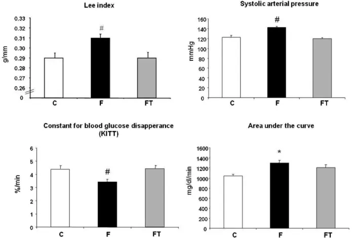
The body weight in the F group was higher than that in the C and FT groups (F = 515¡8vs. C = 412¡14 and FT = 416¡
Consequently, the Lee index was increased in the F group compared with the C and FT groups (F = 0.31¡0.001 vs.
C = 0.29¡0.001 and FT = 0.29¡0.001 g/mm). The Lee index
was similar between the C and FT groups (Figure 1). Glycemia was also increased in the F group after fructose overload compared with the control rats, but there was no difference with respect to the F and FT groups (F = 100¡4,
FT 99¡1vs. C = 88¡1 mg/dl).
In the intravenous insulin tolerance test, the rate constant for blood glucose disappearance (KITT) was reduced in F rats compared with C and FT rats (3.6¡0.2vs. 4.5¡0.2 and
4.6¡0.2 %/min, respectively). Additionally, when the area
under the curve was calculated, a higher area was obtained in the F group compared with the C group (1300¡48 vs. 1044¡30 mg/dl/min). No difference was observed between the C and FT groups for this parameter (1044¡30
vs. 1208¡38 mg/dl/min) (Figure 1).
The systolic (SBP), diastolic (DBP) and mean (MBP) blood pressure were increased in the F group compared with the C and FT groups. These parameters were similar between the C and FT groups (SBP = 142¡2vs. 122¡4 and 119¡2 mmHg, DBP = 101¡2vs. 83¡4 and 87¡4 mmHg and MAP = 118¡3 vs. 104¡4 and 101¡2 mmHg, respectively). No difference
was observed in heart rate among the studied groups (C = 330¡7, FT = 320¡6 and F = 335¡9 bpm).
Echocardiographic evaluation
The results of echocardiographic evaluations performed 10 weeks after fructose overload are shown in Table 1. The left ventricular morphometric evaluations indicated that the
left ventricular mass was higher in the F and FT groups compared with the control group. Furthermore, the RWT was increased in the F group compared with the FT and control groups. The FT and C groups exhibited similar RWT values.
Systolic function, expressed by fractional shortening, ejection fraction and VCF, was similar among the three groups. However, in the evaluation of diastolic function, lower ratios were found for IVRT, nIVRT and E/A in the F group than in the C and FT groups. The C and FT groups exhibited similar diastolic function. The myocardial perfor-mance index (MPI) was increased in the F group compared with the C and FT groups, whereas the FT and C groups exhibited similar values.
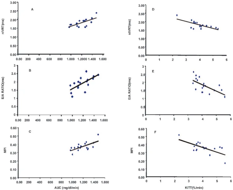
Correlation Analyses
Correlation analyses were carried out to associate meta-bolic parameters with diastolic dysfunction variables in all three groups (Figure 2). A negative correlation was found between IVRT and KITT (r = -0.67; p= 0.002). Similarly, a negative correlation between nIVRT and KITT (r = -0.70;
p= 0.0016) was demonstrated. We observed a negative correlation between the E/A ratio and KITT (r = -0.63;
p= 0.004). A negative correlation was also found between the global index MPI and KITT (r = -0.74;p= 0.004).
A positive correlation was found between IVRT, nIVRT and AUC (r = 0.67; p= 0.002 and r = 0.68;p= 0.0036, respec-tively). Furthermore, the E/A ratio and AUC were positively correlated (r = 0.66; p= 0.003), as were the MPI and AUC (r = 0.67;p= 0.003).
Figure 1 -Lee Index, systolic arterial pressure, the rate constant for blood glucose disappearance and area under the curve in control (C)
DISCUSSION
The purpose of this study was to investigate whether moderate physical exercise performed during 10 weeks of a high-fructose diet in Wistar rats can prevent metabolic changes, cardiac morphofunctional alterations and left ventricular diastolic dysfunction.
Interestingly, we found that metabolic and cardiac morphofunctional alterations promoted by a high-fructose diet can be attenuated by physical exercise when both occur in the same time period. This is the first study to demonstrate the preventive effect of physical training on the diastolic dysfunction induced by fructose overload in rats.
Metabolic and echocardiographic changes induced by fructose overload
In our study, although high fructose consumption promoted an increase in glycemia compared with control rats, the values were still within the normal range. Other studies, even those utilizing high-fructose diets over longer periods, did not find significant alterations in glycemia (7). However, we observed several changes that were character-istic of metabolic syndrome in the rats that received a high-fructose diet. For instance, obesity was defined as an increased Lee index and was associated with insulin resistance, as demonstrated by the KITT and the area under the curve. In fact, this model characterizes an increase in insulin resistance and not hyperglycemia (7). In fact, this model is characterized by an increase in systolic and diastolic blood pressure.
In humans with normal beta cell function, the secretion of insulin may be altered to accommodate different degrees of insulin sensitivity and, thus, to maintain blood glucose within the normal range. Likewise, clinical studies have also demonstrated that the consumption of fructose can induce
weight gain, dyslipidemia, reduced insulin sensitivity and high blood pressure (15,16).
Interestingly, fructose overload promoted changes in left ventricular morphometry, diastolic dysfunction and in-creased cardiac effort, as evidenced by the increase in the myocardial performance index (MPI). Other authors found similar results after submitting Wistar rats to 10% fructose overload for eight weeks (17).
Left ventricular diastolic dysfunction and morphometric alterations can result from several factors, such as increased collagen concentration and fibrosis and decreased compli-ance, which are influenced by the autonomic nervous system and the renin-angiotensin system (17). In fact, several authors showed increased blood pressure, decreased vagal tonus and increased angiotensin II in a high fructose-fed rat model (7,18,19). Furthermore, insulin resistance is a pathogenic factor and has been associated with abnormal myocardial performance in pre-diabetic adults. Additionally, as demon-strated in Figure 1, this relationship between resistance and myocardial performance reinforces the fact that the heart is a target organ in metabolic syndrome, and its evaluation should not be neglected in clinical practice.
Preventive effect of exercise training during high fructose intake
Physical exercise has been shown to be an effective, non-pharmacological treatment for cardiovascular disease (20-27). However, in the present study, it was used as a preventive tool when performed during the same period in which the rats received fructose overload in drinking water. Interestingly, the FT group presented similar values to those of the C group for several parameters, such as the Lee index, insulin resistance, and systolic and diastolic blood pressure, which confirms that exercise training was effective in the prevention of these changes induced by a high-fructose diet. The caloric expenditure due to the exercise training during the high-fructose diet period probably enhanced the energetic demand and prevented changes such as weight and fat gain, which contributed to improved insulin sensitivity and the maintenance of blood pressure in the normal range. In fact, the reduction of weight, intra-abdominal adiposity and insulin resistance by exercise training may interfere with other mechanisms of action in the treatment of hypertension, as hypertension has been associated with all of these variables (28). Indeed, regular physical exercise can promote chronic metabolic and cardiovascular adaptations. It is known that regular exercise improves glycemia, the lipid profile (29) and insulin sen-sitivity; promotes weight reduction; and improves muscle glucose uptake.
Additionally, the morphometric and functional alterations demonstrated by echocardiography in the F rats were not observed in the C and FT groups. In diabetic and hypertensive rats, physical training has been important in the treatment of metabolic and autonomic disorders by contributing to increased baroreflex sensitivity and decreased blood pressure (10,30,31). Moreover, exercise training can modulate the renin-angiotensin system and prevent the development of cardiac hypertrophy in SHR rats (32). Exercise training can increase the antioxidant capacity, increase vascular compli-ance and promote atheroprotective effects (33).
Altogether, these improvements in several systems result-ing from exercise trainresult-ing were important in the prevention of cardiovascular derangements promoted by MS. It is Table 1 -Cardiac morphometric and functional
evaluations by echocardiography in control (C), fructose-fed (F) and trained fructose-fructose-fed (FT) groups.
Variable C (7) F (7) FT (7)
morphometric
LV mass (g) 0.73¡0.03 1.3¡0.01* 1.2¡0.01*
RWT 0.33¡0.007 0.38¡0.005* 0.35¡0.004 systolic function
LVFS (%) 40¡1.5 36¡1 40¡1.6
LVEF (%) 77¡1.6 72¡1.1 76¡1.5
VCF (circ/sec) 0.0053¡0.0003 0.0046¡0.0001 0.0052¡0.0002 diastolic function
IVRT (ms) 22¡0.4 27¡0.9# 0.22¡0.6
nIVRT (ms) 1.6¡0.03 2.05¡0.07# 1.73¡0.03
E/A ratio (ms) 1.72¡0.08 2.24¡0.12# 1.72¡0.15 global index
MPI 0.32¡0.02 0.41¡0.02# 0.33¡0.01
notable that the cardiac changes observed in the F rats and absent in the FT rats due to exercise training were correlated with the indexes of insulin resistance, suggesting that these alterations play a significant role in the process of cardiac damage due to MS or diabetes.
Among the derangements caused by metabolic syndrome, cardiac morphofunctional alterations were also observed. Diastolic function seemed to be the cardiac parameter most influenced by the metabolic alterations, especially the insulin resistance.
Chronic high fructose intake favored the development of metabolic syndrome in rats. When performed concomitantly with a high-fructose diet, exercise training proved to be a vital tool for the prevention of not only metabolic but also cardiac changes promoted by fructose overload in rats. Additionally, it seems that the prevention of diastolic dysfunction was related to improved insulin resistance. Considering our results, we speculate that cardiac morphofunctional evalua-tions should also be performed in patients with metabolic
syndrome or in patients at risk for metabolic syndrome development. Nevertheless, more clinical studies that assess cardiac function in these patients should be performed.
AUTHOR CONTRIBUTIONS
Mostarda C performed and coordinated the experiments. Moraes-Silva IC and Machi JF participated in the data discussion and helped draft the manuscript. Salemi VM performed the echocardiography evaluation. Rodrigues B, De Angelis K and Farah VM participated in the design and data discussion and helped draft the manuscript. Irigoyen MC conceived the study and participated in its design and coordination.
REFERENCES
1. First Brazilian Guideline for Diagnosis and Treatment of Metabolic Syndrome. 2005.
2. Lakka HM, Laaksonen DE, Lakka TA, Niskanen LK, Kumpusalo E, Tuomilehto J, et al. The metabolic syndrome and total and cardiovas-cular disease mortality in middle-aged men. JAMA. 2002;288(21):2709-16, http://dx.doi.org/10.1001/jama.288.21.2709.
Figure 2 - Correlation analyses between insulin resistance index, diastolic index and global index. A: normalized isovolumetric
3. Girman CJ, Rhodes T, Mercuri M, Pyorala K, Kjekshus J, Pedersen TR, et al. The metabolic syndrome and risk of major coronary events in the Scandinavian Simvastatin Survival Study (4S) and the Air Force/Texas Coronary Atherosclerosis Prevention Study (AFCAPS/TexCAPS). Am J Cardiol. 2004;93(2):136-41, http://dx.doi.org/10.1016/j.amjcard. 2003.09.028.
4. Bray GA, Nielsen SJ, Popkin BM. Consumption of high-fructose corn syrup in beverages may play a role in the epidemic of obesity. Am J Clin Nutr. 2004;79(4):537-43.
5. Basciano H, Federico L, Adeli K. Fructose, insulin resistance, and metabolic dyslipidemia. Nutr Metab (Lond). 2005;2(1):5, http://dx. doi.org/10.1186/1743-7075-2-5.
6. Sliem H, Nasr G. Left ventricular structure and function in prediabetic adults: Relationship with insulin resistance. J Cardiovasc Dis Res. 2011;2(1):23-8, http://dx.doi.org/10.4103/0975-3583.78583.
7. Brito JO, Ponciano K, Figueroa D, Bernardes N, Sanches IC, Irigoyen MC, et al. Parasympathetic dysfunction is associated with insulin resistance in fructose-fed female rats. Braz J Med Biol Res. 2008;41(9):804-8, http:// dx.doi.org/10.1590/S0100-879X2008005000030.
8. Reaven GM, Ho H, Hoffman BB. Attenuation of fructose-induced hypertension in rats by exercise training. Hypertension. 198812(2):129-32, http://dx.doi.org/10.1161/01.HYP.12.2.129.
9. Carroll S, Dudfield M. What is the relationship between exercise and metabolic abnormalities?A review of the metabolic syndrome. Sports Med. 2004;34(6):371-418, http://dx.doi.org/10.2165/00007256-20043 4060-00004.
10. Moraes-Silva IC, De La Fuente RN, Mostarda C, Rosa K, Flues K, Damaceno-Rodrigues NR, et al. Baroreflex deficit blunts exercise training-induced cardiovascular and autonomic adaptations in hyper-tensive rats. Clin Exp Pharmacol Physiol. 2010,37(3):e114-20.
11. Rodrigues B, Figueroa DM, Mostarda CT, Heeren MV, Irigoyen MC, De Angelis K. Maximal exercise test is a useful method for physical capacity and oxygen consumption determination in streptozotocin-diabetic rats. Cardiovasc Diabetol. 2007;6:38, http://dx.doi.org/10.1186/1475-2840-6-38.
12. Bernardis LL, Patterson BD. Correlation between ’Lee index’ and carcass fat content in weanling and adult female rats with hypothalamic lesions. J Endocrinol. 1968;40(4):527-8, http://dx.doi.org/10.1677/joe.0.0400527. 13. Mostarda C, Rodrigues B, Vane M, Moreira ED, Rosa KT, Moraes-Silva IC, et al. Autonomic impairment after myocardial infarction: role in cardiac remodelling and mortality. Clin Exp Pharmacol Physiol. 2010;37(4):447-52, http://dx.doi.org/10.1111/j.1440-1681.2009.05327.x. 14. Wichi R, Malfitano C, Rosa K, De Souza SB, Salemi V, Mostarda C, et al.
Noninvasive and invasive evaluation of cardiac dysfunction in experi-mental diabetes in rodents. Cardiovasc Diabetol. 2007;6:14, http:// dx.doi.org/10.1186/1475-2840-6-14.
15. Elliott SS, Keim NL, Stern JS, Teff K, Havel PJ. Fructose, weight gain, and the insulin resistance syndrome. Am J Clin Nutr. 2002;76(5):911-22. 16. Ferder L, Ferder MD, Inserra F. The role of high-fructose corn syrup in
metabolic syndrome and hypertension. Curr Hypertens Rep. 2010;12(2):105-12, http://dx.doi.org/10.1007/s11906-010-0097-3. 17. Xing SS, Bi XP, Tan HW, Zhang Y, Xing QC, Zhang W. Overexpression of
interleukin-18 aggravates cardiac fibrosis and diastolic dysfunction in fructose-fed rats. Mol Med. 2010;16(11-12):465-70.
18. Farah V, Elased KM, Chen Y, Key MP, Cunha TS, Irigoyen MC, et al. Nocturnal hypertension in mice consuming a high fructose diet. Auton
Neurosci. 2006;130(1-2):41-50, http://dx.doi.org/10.1016/j.autneu. 2006.05.006.
19. Farah V, Elased KM, Morris M. Genetic and dietary interactions: role of angiotensin AT1a receptors in response to a high-fructose diet. Am J Physiol Heart Circ Physiol. 2007;293(2):H1083-9, http://dx.doi.org/ 10.1152/ajpheart.00106.2006.
20. Bacchi E, Negri C, Zanolin ME, Milanese C, Faccioli N, Trombetta M, et al. Metabolic Effects of Aerobic Training and Resistance Training in Type 2 Diabetic Subjects: A randomized controlled trial (the RAED2 study). Diabetes Care. 2012;35(4):676-82, http://dx.doi.org/10.2337/ dc11-1655.
21. Krieger EM, Da Silva GJ, Negrao CE. Effects of exercise training on baroreflex control of the cardiovascular system. Ann N Y Acad Sci. 2001;940:338-47, http://dx.doi.org/10.1111/j.1749-6632.2001.tb03689.x. 22. La Rovere MT, Bersano C, Gnemmi M, Specchia G, Schwartz PJ.
Exercise-induced increase in baroreflex sensitivity predicts improved prognosis after myocardial infarction. Circulation. 2002;106(8):945-9, http://dx.doi.org/10.1161/01.CIR.0000027565.12764.e1",-1,"xxx/ 12764.e1.
23. Ignarro LJ, Balestrieri ML, Napoli C. Nutrition, physical activity, and cardiovascular disease: an update. Cardiovasc Res. 2007;73(2):326-40, http://dx.doi.org/10.1016/j.cardiores.2006.06.030.
24. Laterza MC, de Matos LD, Trombetta IC, Braga AM, Roveda F, Alves MJ, et al. Exercise training restores baroreflex sensitivity in never-treated hypertensive patients. Hypertension. 2007;49(6):1298-306, http:// dx.doi.org/10.1161/HYPERTENSIONAHA.106.085548.
25. Legramante JM, Iellamo F, Massaro M, Sacco S, Galante A. Effects of residential exercise training on heart rate recovery in coronary artery patients. Am J Physiol Heart Circ Physiol. 2007;292(1):H510-5. 26. Senechal M, Bouchard DR, Dionne IJ, Brochu M. Lifestyle Habits and
Physical Capacity in Patients with Moderate or Severe Metabolic Syndrome. Metab Syndr Relat Disord. 2012,[Epub ahead of print]. 27. Alves AJ, Ribeiro F, Goldhammer E, Rivlin Y, Rosenschein U, Viana JL,
et al. Exercise Training Improves Diastolic Function in Heart Failure Patients. Med Sci Sports Exerc. 2011, [Epub ahead of print].
28. Jakicic JM, Clark K, Coleman E, Donnelly JE, Foreyt J, Melanson E, et al. American College of Sports Medicine position stand. Appropriate intervention strategies for weight loss and prevention of weight regain for adults. Med Sci Sports Exerc. 2001;33(12):2145-56.
29. Durstine JL, Haskell WL. Effects of exercise training on plasma lipids and lipoproteins. Exerc Sport Sci Rev. 1994;22:477-521.
30. Bertagnolli M, Schenkel PC, Campos C, Mostarda CT, Casarini DE, Bello-Klein A, et al. Exercise training reduces sympathetic modulation on cardiovascular system and cardiac oxidative stress in spontaneously hypertensive rats. Am J Hypertens. 2008;21(11):1188-93, http:// dx.doi.org/10.1038/ajh.2008.270.
31. Mostarda C, Rogow A, Silva IC, De La Fuente RN, Jorge L, Rodrigues B, et al. Benefits of exercise training in diabetic rats persist after three weeks of detraining. Auton Neurosci. 2009;28 145(1-2):11-6, http://dx.doi.org/ 10.1016/j.autneu.2008.10.010.
32. Zamo FS, Barauna VG, Chiavegatto S, Irigoyen MC, Oliveira EM. The renin-angiotensin system is modulated by swimming training depend-ing on the age of spontaneously hypertensive rats. Life Sci. 2011;18 89(3-4):93-9, http://dx.doi.org/10.1016/j.lfs.2011.05.004.