Measurement accuracy and reliability of tooth length on
conventional and CBCT reconstructed panoramic radiographs
Carlos Flores-Mir1, Mark R Rosenblatt2, Paul W. Major3, Jason P. Carey4, Giseon Heo5
Introduction: This in vivo study assessed accuracy and reliability of tooth length measurements obtained from conventional panoramic radiographs and CBCT panoramic reconstructions to that of a digital caliper (gold standard). Methods: The sample consisted of subjects who had CBCT and conventional panoramic radiographic imaging and who required maxillary premolar extraction for routine orth-odontic treatment. A total of 48 teeth extracted from 26 subjects were measured directly with digital calipers. Radiographic images were scanned and digitally measured in Dolphin 3D software. Accuracy of tooth length measurements made by CBCT panoramic reconstruc-tions, conventional panoramic radiographs and digital caliper (gold standard) were compared to each other by repeated measures one-way ANOVA with Bonferroni correction and by single measures intraclass correlation coefficient. Results: Repeated root length measures with digital calipers, panoramic radiographs and CBCT constructed panoramic-like images were all individually highly reliable. Com-pared to the caliper (gold standard), tooth measurements obtained from conventional panoramic radiographs were on average 6.3 mm (SD = 2.0 mm) longer, while tooth measurements from CBCT panoramic reconstructions were an average of 1.7 mm (SD = 1.2 mm) shorter.
Conclusions: In comparison to actual tooth lengths, conventional panoramic radiographs were relatively inaccurate, overestimating the lengths by 29%, while CBCT panoramic reconstructions underestimated the lengths by 4%.
Keywords:Reproducibility of results. Radiography. Tooth root.
How to cite this article: Flores-Mir C, Rosenblatt MR, Major PW, Carey JP, Heo G. Measurement accuracy and reliability of tooth length on conventional and CBCT reconstructed panoramic radiographs. Dental Press J Orthod. 2014 Sept-Oct;19(5):45-53. DOI: http://dx.doi.org/10.1590/2176-9451.19.5.045-053.oar
» The authors report no commercial, proprietary or financial interest in the products or companies described in this article.
Contact address: Carlos Flores-Mir 5-528 Edmonton Clinic Health Academy
Faculty of Medicine and Dentistry - University of Alberta
Edmonton, AB, Canada - T6G 1C9 - Email: [email protected] 1 Associate professor and head of the Department of Orthodontics, University of
Alberta.
2 MSc in Orthodontics, University of Alberta.
3 Chair of the Department of Dentistry, Faculty of Medicine and Dentistry, University of Alberta.
4 Associate professor, Department of Mechanical Engineering, Faculty of Engineering, University of Alberta.
5 Associate professor of Statistics, Department of Dentistry, University of Alberta, Edmonton, Alberta, Canada.
» Patients displayed in this article previously approved the use of their facial and in-traoral photographs.
Submitted: September 02, 2012 - Revised and accepted: January 08, 2013. Introdução: este estudo in vivo avaliou a precisão e a confiabilidade de medições do comprimento dentário realizadas em radiografias panorâmicas convencionais e em reconstruções panorâmicas de tomografias computadorizadas de feixe cônico (TCFC), comparando--as com medições feitas com um paquímetro digital, consideradas o padrão-ouro. Métodos: a amostra incluiu indivíduos que já tives-sem realizado tanto exames imaginológicos de TCFC quanto radiografias panorâmicas, e cujo tratamento ortodôntico exigisse a ex-tração de pré-molar superior. No total, 48 dentes extraídos, de 26 pacientes, foram mensurados diretamente com paquímetros digitais. As radiografias foram escaneadas e digitalmente avaliadas com a ajuda do software Dolphin 3D. Por meio da análise de variância simples com correção de Bonferroni e Coeficiente de Correlação Intraclasse simples, comparou-se a precisão das medições de comprimento dentário realizadas em reconstruções panorâmicas de TCFC, em radiografias panorâmicas convencionais e com paquímetro digital.
Resultados: medições repetidas de comprimento dentário feitas com o paquímetro digital, radiografias panorâmicas e recons-truções panorâmicas de TCFC foram todas consideradas, individualmente, altamente confiáveis. Em comparação ao paquímetro, as medidas obtidas por meio de radiografias panorâmicas convencionais foram, em média, 6,3 ± 2,0mm mais longas, enquanto as medidas obtidas por meio das reconstruções panorâmicas de TCFC foram, em média, 1,7 ± 1,2mm mais curtas. Conclusões: em comparação com o real comprimento dentário, as radiografias panorâmicas convencionais foram relativamente imprecisas e superesti-maram o comprimento em 29%; já as reconstruções panorâmicas de TCFC subestisuperesti-maram o comprimento em 4%.
INTRODUCTION
Panoramic radiographs are a type of tomography. The structures outside the focal trough are blurred and appear as shadows and artifacts. In order to bet-ter maintain the elliptical shape of dental structures within the focal trough, panoramic devices have a center of rotation that changes throughout the scan.1
The rotational patterns developed by the manufac-turers of these devices vary widely making the result-ing images unique to the model. Modifications in arc radius and shape as well as static versus variable cen-ters of rotation have been used to better approximate the shape of the maxillomandibular process in order to maintain patients’ dentoalveolar structures within the device’s focal trough.1 Even with standardized
head positions, the great variability in individual’s jaw dimensions and shape make achieving optimized panoramic images less predictable and repeatable.
Many reports have noted that panoramic radio-graphs do not accurately represent tooth positions, thereby requiring the clinician to supplement his ind-ings with a clinical assessment. As reviewed by Van Elslande et al,2 panoramic radiographs are fraught with
inconsistent levels of magniication and distortion er-rors. Some reports3 found vertical measurements were
± 10% diferent from direct measurements of dried
skulls, while other groups4 found the diference to be
as high as 18-21%. Diferences in magniication have been found to vary throughout panoramic images. This exacerbated the disparity between devices tested and the majority of manufacturers’ documentation which did not accurately correspond to the calculat-ed magniication in various regions of the panoramic images.2,5 While these distortions may be acceptable
for ratio calculations, they pose an unacceptable level of unreliability for linear measurements. Turp et al’s6
analysis of vertical measurements of ramus and con-dylar heights concurred with Kjellberg’s5 inding that
there was a very low correlation coeicient between the lengths recorded on the panoramic images and di-rect physical measurements.
Like many clinicians, Kaley et al7 assessed root
re-sorption by comparing pre and post orthodontic treat-ment panoramic radiographs. The study concluded that a disproportionate number of patients starting with Class III malocclusions and patients with treat-ment mechanics that positioned maxillary incisor roots
in close proximity to the lingual cortical plate had se-vere root loss. Proclination of incisors to compensate for a Class III malocclusion would have resulted in foreshortening in the panoramic images exaggerating apical resorption. By the same logic, Class II division 1 patients would have underestimated root loss.
Cone-beam computed tomography (CBCT) has ofered clinicians a radiographic technique with a high degree of resolution to identify craniofacial landmarks and a spatially accurate means of analyzing them.8,9,10
While CBCT sotware has the ability to produce pan-oramic reconstructions, the inherent inaccuracies of conventional image format have prompted only a few studies to compare the accuracy level of these recon-structions not only with conventional images, but also with true anatomy by direct measure. Ludlow et al11 scanned dried skulls with the NewTom 9000 at
a resolution of 0.5 mm slice thickness to determine vertical and horizontal length accuracy when recon-structed into panoramic projections. Researchers used metal wires of known length laid along the buccal sur-face of the ramus and mandibular body as reference knowing that while they likely did not lie in the exact plane of the panoramic reconstruction, as long as they were within 18o of the plane, the foreshortening efect
would be less than 5%. Conversely, panoramic recon-struction followed the curvature of the mandible re-sulting in linear measurements on the image that were overestimated. While operator expertise was consid-ered an important factor in measurement accuracy, the lengths recorded in the 3D volumes by landmark identiication in serial axial slices expressed levels of error in the range of 0.19 to 0.37 mm, or 0.6 to 1.7% of the measured lengths. These values were 1.5-2.5 times lower than the panoramic reconstructions of the same volumes. The present study could be considered an extension of Ludlow et al’s11 project, but instead
of using dry skulls, actual patients’ data were used. The in vivo nature of this study ofers orthodontists a clinically realistic result to apply to their diagnosis and treatment planning routines.
MATERIAL AND METHODS
The University of Alberta Health Research Eth-ics Board (Biomedical Panel) approved application #7380 on April 16th, 2008. This was a prospective
cross-sectional study. Study subjects required max-illary premolar extractions to complete their regular orthodontic treatment goals. The subjects were go-ing to undergo orthodontic treatment. The decision to get a CBCT as well as the need for premolar ex-tractions for the selected cases were generated by the treating orthodontist. Panoramic images needed to be available from previous patient’s records. They were not taken in addition to the CBCT imaging. Inclusion criteria for the study required all subjects to have also had conventional panoramic radiograph taken within the previous 24 months. All teeth in-cluded in the study were fully erupted maxillary premolars at the time conventional panoramic was taken. All the evaluated premolars appear to have closed apices. CBCT images were taken on the same day the premolars were extracted.
Sample size for the present study was set at 48 teeth. Sample size calculation was performed based on the variability of measurement differences between panoramic images and calipers. Consider-ing the 48 samples as a pilot study, the minimum sample sizes required to identify length differences of 0.5 mm would be 192, and for a 1.0 mm differ-ence, 48. The formula used for this calculation was:
�≥ �! �!∗+ �!∗ !
�
!
Where �=0.05 �=0.1 and �!∗!=1.96 �!=1.285
CBCT images were taken with the 12-bit i-CAT
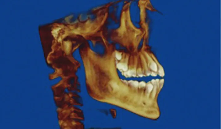
(Imaging Sciences International, Hatfield, Penn) set to a 40-second scan allowing image reconstruction with a voxel size of 0.25 mm. Standard clinical pro-tocols were used for patients’ positioning and a cot-ton roll between incisor teeth was used to stably hold the occlusion apart to improve cusp tip identification. Images were saved as DICOM files and were recon-structed in Dolphin Imaging 10.5 Premium soft-ware (Dolphin Imaging Sciences, Chatsworth, Calif, USA). Head positions in the reconstructed images were standardized anteroposteriorly by Frankfort Horizontal (Fig 1), and sagittally for maximal overlap
of bilateral structures in the maxilla, ramus and body of the mandible (Figs 1 and 2) by rotating them spa-tially. Panoramic images were reconstructed from CBCT volumes by selecting a custom focal trough that passed through the lingual cusps of the maxillary teeth and extended posterior to the condyles. Focal trough width was varied to ensure it encompassed the entire length and height of the maxillary denti-tion. Axial serial slices were reviewed to ensure the focal trough encompassed all teeth regardless of their angulation and with the center of the custom focal trough bisecting as close to the center of the long axis of the teeth as possible (Figs 3 and 4).
Conventional panoramic radiographs were pro-duced with a 17.6 second duration exposure on au-tomatic settings with an Instrumentarium Orthop-antomograph OP100 on Fuji Super HRT30 film and Kodak Lanex Regular Intensity screen. The films were developed in a Kodak M35A processor, scanned with an Epson Perfection 700 photo scanner (Epson, Long Beach, Calif) at 300 dpi and 24-bit color, and optimized for contrast and brightness with the Ep-son scanning software. JPEG images (saved at lowest compression) were imported into Dolphin Imaging for analysis (Fig 5).
Following imaging, one or two maxillary first or second premolar teeth were extracted as per the patient’s orthodontic treatment plan and stored in 95% ethanol. The 48 premolars, collected from 26 subjects, were then measured directly with a digi-tal caliper (OrthoPli, Philadelphia, Penn, USA). The minimum caliper reading was 0.013 mm and its measurement accuracy was 0.025 mm as reported by the manufacturer.
Figure 1 - Standardized volume orientation — Sagittal view.
Figure 3 - Custom focal trough selection for panoramic reconstruction from CBCT.
Figure 5 - Scanned conventional panoramic radiograph.
Figure 2 - Standardized volume orientation — Frontal View.
Figure 4 - Panoramic reconstruction from CBCT.
the appropriate root could not be determined due to the tooth’s long axis angulation or rotation. Scanned conventional panoramic radiograph measurements were standardized to measurements made on the physical films with the digital caliper. The CBCT
panoramic reconstruction measurements were cali-brated to the digital ruler produced by the Dolphin Imaging software from the 3D volume. All measure-ments were recorded to the nearest tenth of a mil-limeter and done by only one experienced examiner.
There were no premolars with clinically signifi-cant occlusal abrasion. Signifisignifi-cant root resorption was not identified in any premolar evaluated as de-termined in the panoramic image or CBCT gener-ated panoramic image. If any crown abrasion or root resorption happened between images, it was not consider clinically relevant.
Measurement error
Statistical analysis
Accuracy of tooth length measurements made by the CBCT panoramic reconstructions, conventional panoramic radiographs and the digital caliper (gold standard) were compared to each other by repeated measures one-way ANOVA with Bonferroni cor-rection and by single measures intraclass correlation coefficient using SPSS version 16.0 software (SPSS, Chicago, Ill). Statistical analysis of intra-rater reli-ability of the triplicate measurements were assessed by single measures intraclass correlation coefficient (ICC) in SPSS.
Statistical analyses for the reliability and accuracy assessments were repeated following the removal of all outlying data points. Since they were deter-mined to have no significant effect on the results, all data points were maintained for the reporting and analyses in this study. Clinically significant changes in root length were considered to be values of 1.0 mm and greater, consistent with those studies by Copeland12 and Mohandesan.13
RESULTS
Reliability
Repeated measures of root length with digi-tal calipers, conventional panoramic radiographs and CBCT panoramic reconstructions had very high reliability with ICC values of 0.999 (95% CI: 0.998, 1.000), 0.997 (95% CI: 0.993, 0.999) and 0.995 (95% CI: 0.995, 0.999) respectively. Land-mark identification and thus tooth length measure-ments were also highly repeatable (intra-observer) in the conventional panoramic images with a sin-gle measure ICC of 0.997 (95% CI: 0.993, 0.999) and for the CBCT panoramic reconstructions with a single measure ICC of 0.995 (95% CI: 0.995, 0.999). As another method to verify the degree of reliability, the mean and standard deviation for the differences between the average gold standard tooth length measurements and each corresponding con-ventional and reconstructed panoramic measure-ment were also calculated (available upon request).
Accuracy
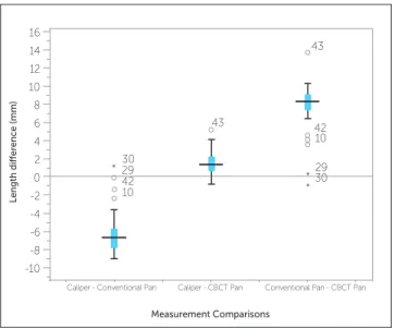
Measurements by all three techniques resulted in significantly different tooth lengths (P < 0.001), even when the Bonferroni correction was calculated. The mean tooth length for the conventional panoramic was 6.3 mm (95% C.I.: 5.6 - 7.1 mm) longer than the caliper (gold standard), while the CBCT pan-oramic mean was 1.6 mm (95% C.I.: 1.1 - 2.0 mm) shorter than the caliper (Table 1).
Box plots of tooth length differences between the three measurement techniques are depicted in Fig-ure 6. Compared to the caliper (gold standard), the conventional panoramic images resulted in tooth measurements that were generally longer and ranged from 1 mm shorter to 9 mm longer. Tooth lengths in the CBCT reconstructions, on the other hand, Figure 6 - Comparison of tooth length measurements for calipers,
conven-tional panoramic radiographs and CBCT panoramic reconstructions.
Orientation (A)
Orientation (B)
Mean diference
(A-B)
Signiicance p
95% CI for diference
Caliper
Conventional
Pan -63 < 0.001 -7.1; -5.6
CBCT 1.6 < 0.001 1.1; 2.0
Conventional
panorex CBCT Pan 7.9 < 0.001 7.0; 8.8
Table 1 - Repeated measures ANOVA for measured tooth length with Bonferroni correction.
Measurement Comparisons
Caliper - Conventional Pan
-10 -8 -6 -4 -2 0
Length difer
ence (mm)
30
43
43
42 10
29 30 29
42 10 2
4 6 8 10 12 14 16
Conventional Pan - CBCT Pan Caliper - CBCT Pan
were generally shorter than the gold standard, with a smaller measurement discrepancy. These measure-ments ranged from 1 mm longer to 5 mm shorter than that determined by the calipers.
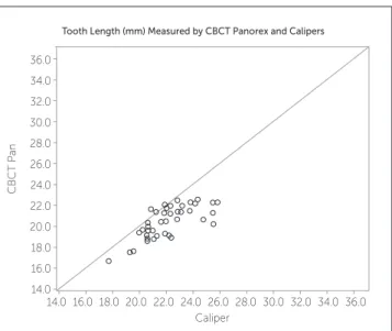
The 3D CBCT images were standardized for head position sagittally (Fig 1) and coronally (Fig 2) by the by Frankfort Horizontal, inferior or-bital rims, condylar heads and inferior border of the mandible prior to panoramic reconstruction. This standardization reduced randomness of elongation and foreshortening distortions compared to the cali-per measurements. It did not, however, account for variability in tooth angulation with respect to the standardized neutral head position. Data distribu-tion revealed in the scatter plots (Figs 7 and 8) in-dicated that tooth lengths showed relatively good measurement reliability across techniques, regard-less of actual tooth size, and a measurement bias that resulted in an overestimation of tooth lengths in conventional panoramic images and an under-estimation in CBCT panoramic reconstructions. The bias was less distinct for the CBCT reconstruc-tions, as the underestimation appeared to increase for longer teeth (Fig 8).
DISCUSSION
The average tooth length measured by the con-ventional panoramic was 6.3 mm (or 29%) longer than the calipers and the range of values was almost twice that of the other measurement techniques. The error in the conventional panoramic mea-surements in this study is greater than those found
by comparison of dry skulls: 10% by Tronje3 and
18-21% by Larheim,4 but approached the levels of
magnification (26%) found by Yitchaky’s study.14
The average tooth length measured by CBCT panoramic reconstruction was 1.6 mm shorter than the direct measurement by calipers, but the preci-sion of repeated measurements was comparably ex-tremely high for both techniques. This would imply that difference in measured tooth length was not due to misidentification of the landmarks, but to radio-graphic foreshortening or inadequate resolution of fine root apices compared to the surrounding bone.
A significant limitation of both conventional and CBCT reconstructed panoramic images lies in their inability to account for changes in tooth angulation between serial images when no other assessment means (extra imaging, clinical observation, etc.)
Figure 7 - Scatter plot of tooth length — Caliper vs. conventional panoramic. Figure 8 - Scatter plot of tooth length — Caliper vs. CBCT reconstructed panoramic.
16.0
16.0 18.0
18.0 20.0
20.0 22.0
22.0 24.0
24.0 Caliper 26.0
26.0
Film Pan
28.0
28.0 30.0
30.0 32.0
32.0 34.0
34.0 36.0
36.0 Tooth Length (mm) Measured by Film Panorex and Calipers
16.0
14.0 16.0 14.0 18.0
18.0 20.0
20.0 22.0
22.0 24.0
24.0 Caliper 26.0
26.0
CBCT Pan
28.0
28.0 30.0
30.0 32.0
32.0 34.0
34.0 36.0
are used. During orthodontic treatment, changes in tip and torque introduce elongation and foreshort-ening errors that cannot be easily accounted for.15
It is also possible to mistaken changes in root morphology for resorption as the tooth rotates dur-ing treatment and is then projected in only the
buc-colingual dimension.7 An advantage of CBCT
re-constructions over conventional panoramic is the ability to more precisely reorient the volumes with the imaging software in order to standardize the image’s anatomical planes, thus reducing the er-ror introduced by variable patient position when
radiographs are taken by several staff members.16
The point of using volumetric imaging should be to actually analyze the information 3D and not to downgrade the image potential during reconstruc-tions. The reason we used reconstructed images was because many clinicians are using them and this study and the discussion should help them better understand the drawbacks.
CBCT images created voxel sizes of 0.25 mm. This translated into a resolution limitation and thus an error of 0.25 mm at each measurement point in the image. Therefore measured tooth lengths from CBCT reconstructions would be expected to achieve accuracy within 0.5 mm of the caliper measurements. Although CBCT reconstructions resulted in measurement values that more accurately corre-sponded to direct caliper measurements compared to those of the conventional panoramic radiographs, it is interesting to note the data patterns that emerged from analysis of the scatter plots (Figs 7 and 8). Conventional panoramic appeared to result in a measurement bias that consistently overestimated tooth lengths regardless of the actual tooth size, whereas CBCT reconstructed images resulted in an underestimation bias that increased for larger tooth sizes. If this bias shows to be consistent, it would allow serial panoramic radiographs to be compared to monitor root changes during orthodontic treat-ment. Unfortunately, other studies have shown that magnification variability and inherent imaging errors throughout the panoramic images preclude the reliable use of this application.3,4,14
While the tooth length measured from CBCT panoramic reconstructions were statistically and clin-ically significantly (> 1.0 mm) different from direct
caliper measurements, these images provided im-proved clarity and accuracy compared to the mea-surements achieved by traditional film panoramic. The underestimation of measurements on CBCT reconstructions compared to direct caliper mea-surements were consistent with Ludlow’s findings11
which showed that panoramic reconstructions pro-duced measurement errors of up to 2-4%. The 1.6 mm average decrease in CBCT panoramic tooth length compared to the 22.01 mm caliper mean rep-resented a 7% decrease. With fewer confounding variables compared to conventional techniques, the differences in these measurements were likely due to buccolingual tooth angulation,15 and difficulty in
landmark identification of cusp and root tips due to tooth rotation, position and anatomy.
Study limitations
Due to ethical limitations, conventional pan-oramic radiographs were limited to historical records and, while most were taken within 12 months of the CBCT images and tooth extractions, some re-cords were taken almost 2 years prior to this study. As the patient population was in their early to mid teens, it can be expected that varying amounts of root development would have occurred during the time between conventional and CBCT imaging. While one would expect this to bias the conventional pan-oramic measurements to be shorter than the caliper and CBCT tooth lengths, the opposite was in fact the case (Fig 7). This indicated that distortion and mag-nification errors in the conventional images far out-weighed any dental growth and apical development.
imaging software prior to producing the panoramic reconstructions; and resolution loss accompany-ing the smoothaccompany-ing, compression and reconstruction algorithms that the Dolphin 3D imaging software used to store and manipulate the large data sets.
An alternative analysis of the data using single measures intra-class correlation coefficient was used to determine the reliability between the three mea-surement techniques for each tooth individually. The modest ICC value of 0.504 (95% CI: 0.334, 0.660), when calculated with the reliability defi-nition, indicated that the measurement techniques provided only fair agreement in determining if teeth measuring larger by one technique were going to measure larger by the others. Using the absolute agreement definition, however, the very low ICC value of 0.093 (95% CI: -0.016, 0.271) indicated that the magnitude of length differences recorded by one technique did not correspond to equivalent differences in tooth lengths measured by the other techniques. This should be considered when inter-preting the present results.
Aspects of the conventional panoramic radio-graphic technique that were not addressed in this study included variability in patient’s head position, imager settings, as well as technician ability and technique, as the images were obtained retrospec-tively from existing patient records. Ghost images and artifacts from overlapping anatomy are inherent to the conventional imaging process and are not re-movable to improve dental landmark identification. Additionally, indeterminate levels of magnification
and distortion intrinsic in the panoramic images were expected to produce measurement errors not easily accounted for. These may have been exacer-bated by focal trough sizes and shapes that did not adequately follow patients’ anatomy.
Clinical implications
Clinicians still must be aware of elongation/fore-shortening errors that arise from changes in tip and torque of the teeth of interest when serial panoramic images are compared during treatment. Substantial errors in linear measurement accuracy severely limit conventional panoramic radiography as a tool to identify changes in root length and as such alterna-tive methods should be considered for quantitaalterna-tively monitoring root resorption. Panoramic reconstruc-tions from CBCT volumes improve measurement accuracy over conventional imaging by reducing several sources of magnification and distortion; however, dental measurements are still significantly different from true anatomical lengths and their use diminishes the accuracy gains achieved by 3D tech-nology. While CBCT panoramic reconstructions provide more reliable representations of changes in tooth length, caution should be exercised when they are used for the diagnosis of early root resorption.
CONCLUSIONS
1. Tohnak S, Mehnert A, Crozier S, Mahoney M. Synthesizing panoramic radiographs by unwrapping dental CT data. Conf Proc IEEE Eng Med Biol Soc. 2006;1:3329-32.
2. Van Elslande DC, Russett SJ, Major PW, Flores-Mir C. Mandibular asymmetry diagnosis with panoramic imaging. Am J Orthod Dentofacial Orthop. 2008;134(2):183-92.
3. Tronje G, Eliasson S, Julin P, Welander U. Image distortion in rotational panoramic radiography. II. Vertical distances. Acta Radiol Diagn (Stockh). 1981;22(4):449-55. 4. Larheim TA, Svanaes DB. Reproducibility of rotational panoramic radiography:
mandibular linear dimensions and angles. Am J Orthod Dentofacial Orthop. 1986;90(1):45-51.
5. Kjellberg H, Ekestubbe A, Kiliaridis S, Thilander B. Condylar height on panoramic radiographs. A methodologic study with a clinical application. Acta Odontol Scand. 1994;52(1):43-50.
6. Turp JC, Vach W, Harbich K, Alt KW, Strub JR. Determining mandibular condyle and ramus height with the help of an Orthopantomogram--a valid method? J Oral Rehabil. 1996;23(6):395-400.
7. Kaley J, Phillips C. Factors related to root resorption in edgewise practice. Angle Orthod. 1991;61(2):125-32.
8. Berco M, Rigali PH Jr, Miner RM, DeLuca S, Anderson NK, Will LA. Accuracy and reliability of linear cephalometric measurements from cone-beam computed tomography scans of a dry human skull. Am J Orthod Dentofacial Orthop. 2009;136(1):17.e11-9.
REFERENCES
9. Ballrick JW, Palomo JM, Ruch E, Amberman BD, Hans MG. Image distortion and spatial resolution of a commercially available cone-beam computed tomography machine. Am J Orthod Dentofacial Orthop. 2008;134(4):573-82. 10. Lagravere MO, Carey J, Toogood RW, Major PW. Three-dimensional accuracy
of measurements made with software on cone-beam computed tomography images. Am J Orthod Dentofacial Orthop. 2008;134(1):112-6.
11. Ludlow JB, Laster WS, See M, Bailey LTJ, Hershey HG. Accuracy of measurements of mandibular anatomy in cone beam computed tomography images. Oral Surg Oral Med Oral Pathol Oral Radiol Endod. 2007;103(4):534-42. 12. Copeland S, Green LJ. Root resorption in maxillary central incisors following
active orthodontic treatment. Am J Orthod. 1986;89(1):51-5.
13. Mohandesan H, Ravanmehr H, Valaei N. A radiographic analysis of external apical root resorption of maxillary incisors during active orthodontic treatment. Eur J Orthod. 2007;29(2):134-9.
14. Yitschaky M, Haviv Y, Aframian DJ, Abed Y, Redlich M. Prediction of premolar tooth lengths based on their panoramic radiographic lengths. Dentomaxillofac Radiol. 2004;33(6):370-2.
15. Garcia-Figueroa MA, Raboud DW, Lam EW, Heo G, Major PW. Efect of buccolingual root angulation on the mesiodistal angulation shown on panoramic radiographs. Am J Orthod Dentofacial Orthop. 2008;134(1):93-9. 16. Laster WS, Ludlow JB, Bailey LJ, Hershey HG. Accuracy of measurements of