Transverse effects of rapid maxillary
expansion in Class II malocclusion patients:
A Cone-Beam Computed Tomography study
Carolina Baratieri*, Lincoln Issamu Nojima**, Matheus Alves Jr.***, Margareth Maria Gomes de Souza****, Matilde Gonçalves Nojima*****
Objective: The aim of this study was to evaluate by Cone-Beam Computed Tomography (CBCT) transversal responses, immediately and after the retention period, to rapid maxillary expansion (RME), in Class II malocclusion patients. Methods: Seventeen children (mean initial age of 10.36 years), with Class II malocclusion and skeletal constricted maxilla, underwent Haas´ protocol for RME. CBCT scans were taken before treatment (T1), at the end of the ac-tive expansion phase (T2) and after the retention period of six months (T3). The scans were managed in Dolphin software, where landmarks were marked and measured, on a coronal slice passing through the upper first molar. The paired Student´s t-test was used to identify significant differences (p<0.05) between T2 and T1, T3 and T2, and T3 and T1. Results: Immediately after RME, the mean increase in maxillary basal, alveolar and dental width was 1.95 mm, 4.30 mm and 6.89 mm, respectively. This was accompanied by buccal inclination of the right (7.31°) and left (6.46°) first molars. At the end of the retention period, the entire transverse dimension increased was maintained and the dentoalveolar inclination resumed. Conclusions: The RME therapy was an effective procedure to increase transverse maxillary dimensions, at both skeletal and dentoalveolar levels, without causing inclination on anchorage molars in Class II malocclu-sion patients with skeletal constricted maxilla.
Abstract
Keywords: Rapid maxillary expansion. Transverse effects. Cone-Beam Computed Tomography. Class II malocclusion.
IntROduCtIOn
Class II Division 1 malocclusions are strongly related to transverse problems, presenting a signif-icantly reduced maxillary width when compared to normal occlusion.2,22,25,26 However, its diagnosis is often passed unnoticed at clinical examination as transverse deficiency is camouflaged by the Class II skeletal pattern itself. The upper teeth oc-clude in a more anterior region of the mandible, showing an apparent normal transverse develop-ment, even in the presence of maxillary transverse deficiency.28 Upper molars tend to incline buccally to compensate the insufficient skeletal and alveo-lar base. For this reason, rapid maxilalveo-lary expansion (RME) may be considered before treating Class II Division 1 malocclusion patients.26
RME has been the treatment chosen by many orthodontists for correction of skeletal maxillary constriction in growing patients.10,11 The key fea-ture of RME is that the force applied to the teeth and alveolar processes by activating the expander screw promotes the opening of the midpalatal su-ture. The stability of the new transverse dimen-sion is also a fundamental part of the treatment, which turns the retention phase as important as the active phase,15 with the expander appli-ance having to remain in place for at least three months.13 The Haas expander appliance is widely used in orthodontics because its screw is covered by an acrylic block that enhances the contact with the lateral walls of palate, thus increasing the anchorage, improving the orthopedic effect, and decreasing tooth movement.11
Until recently, frontal cephalometric radio-graphs were the most precise methods for eval-uating the transverse effects of RME. However, the difficulties inherent to the technique not always allowed the precise location and iden-tification of craniofacial structures. With the use of the Cone-Beam Computed Tomography (CBCT) images, not only a three-dimensional visualization of the whole craniofacial com-plex is possible, but also precise and reliable
measurements of the changes caused by RME, since there is neither image superposition nor size distortion.8
Despite the numberless articles on rapid maxillary expansion effects,12,15,24 the literature is still scarce in studies evaluating only the re-sults from the expander appliance in Class II malocclusion patients. The objective of the present study was to evaluate, using CBCT, the dental and skeletal transverse effects of rapid maxillary expansion immediately and after a re-tention period, with Haas expander appliance in Class II malocclusion patients.
MAteRIAl And MethOds
This prospective clinical study was per-formed at the Department of Orthodontics of the Federal University of Rio de Janeiro after being approved by the research ethics commit-tee of the Institute of Collective Health Stud-ies (0052.0.239.000-09). Seventeen children (8 boys and 9 girls with mean ages of 10.67 and 10.05 years, respectively) presenting Class II, Division 1 malocclusion and skeletal transverse deficiency were selected for the study.
The inclusion criteria were: ages between 7-12 years; Class II molar (unilateral or bilater-al) and skeletal (ANB ≥ 4°)21 relationship; max-illary skeletal transverse deficiency (distance from J point to facial frontal line > 12 mm);20 and stage before pubertal growth spurt.6
Even not being an exclusion criterion, none of the patients had visible posterior crossbite. The transverse problem was first evaluated clin-ically and diagnosed as atresia, when the patient projected the mandible until a Class I relation-ship, and the posterior relationship was edge to edge or in crossbite.16
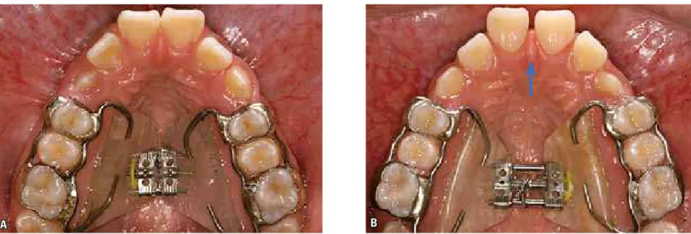
A B
(Dentaurum, Magnum model, 600.303.30) (Fig 1, A). The first screw activation was of one complete turn (0.8 mm), in the same day of appliance instal-lation, and the following activations were of two 1/4 turn per day (0.2 mm per turn, 0.4 mm daily) until the palatine surface of the upper molar con-tacted the buccal surface of the lower molar, when the patient projected the mandible to a Class I re-lationship. This active expansion treatment varied from 2-3 weeks. After this, the screw was stabilized with a 0.012-in double thread ligature (Fig 1, B) and kept in place passively for the following six months when the appliance was then removed.
CBCTs were performed before treatment (T1), immediately after screw expander stabi-lization (T2), and 1-2 days after appliance re-moval (T3). All scans were taken with the same Cone-Beam machine (i-CAT, Imaging Sciences International, Hatfield, Pennsylvania, USA), ac-cording to a standard protocol (120 KVp, 3-8 mA, FOV = 13x17 cm, voxel = 0.4 mm, and scan time = 20s). The scans performed in T1 and T2 were saved in DICOM (digital imaging and com-munication in medicine) format, and with Dol-phin Imaging software® version 11.0 (Dolphin Imaging, Charsworth, California, USA), it was possible to reconstruct 3D images for analysis.
Using specific software functions, before the measurements, it was possible to standardize
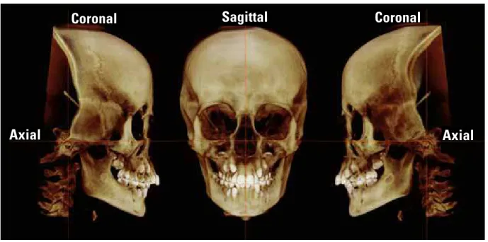
head image positions according to the axial, cor-onal, and sagittal planes4 at all studied times: The axial plane, passing through right and left orbital points as well as right porion; coronal plane, pass-ing through left and right porion, perpendicular to the chosen axial plane; and sagittal plane, pass-ing through nasion point, perpendicular to the chosen axial and coronal planes (Fig 2).
After standardization, the coronal plane and the 3D reconstructions of the images were used for determining the coronal slice and position of the landmarks (Fig 3). The most anterior coro-nal slice showing the entire palatal root of the first upper molar was chosen. All the landmarks were identified on the selected coronal slice. Landmarks and measurements were previously described by Podesser et al,18 as follows (Fig 4):
• Right and Left Maxillary (rMx and lMx): Right and left points in which the axial plane, by passing tangentially at the more inferior con-tour of nasal cavity, meets the buccal-alveolar contour of the maxilla.
• Right and Left Maxillary Alveolar (rMa and lMa): The most inferior and medial point of the buccal-alveolar process in relation to the upper first permanent molar.
rMr lMr
A B A B
FIGURE 2 - Three-dimensional image of the head position after standardization by the axial, coronal and sagittal reference planes. Dolphin Imaging® 11.0, orientation tool.
FIGURE 3 - A) Coronal slice used to identify the landmarks and measure-ments; B) 3D right lateral image, with the coronal plane passing through the right upper first molar. Dolphin Imaging® 11.0.
FIGURE 4 - Coronal slice images with the landmarks identified (rMx, lMx, rMa, lMa, rMc, lMc, rMr e lMr) and measurements: A) linear measure-ments (Maxillary base width, Maxillary alveolar width, Maxillary dental width); B) angular measurements (Right and Left molar angulation). Dol-phin Imaging® 11.0, Digitize/Measurement tool.
• Right and Left Root Molars (rMr and lMr): The most superior and medial point of the pala-tine root of the upper first permanent molar.
The Linear measurements (mm) were maxil-lary basal width (rMx-lMx), maxilmaxil-lary alveolar width (rMa-lMa), and maxillary dental width (rMc-lMc), whereas angular measurements were right (rMc.rMr.sagittal plane) and left (lMc.lMr. sagittal plane) dentoalveolar angulation.
In order to avoid possible measurement er-rors, two similar monitors were used, including the software. This allowed CBCT images to be simultaneously handled for locating planes and landmarks in all three study period of times (T1, T2, T3) for each patient, where T1 was always the reference. Measurements, regarding each period of time, were taken separately by the same examiner within a 1-week interval.
Coronal
Sagittal
Coronal
TABLE 1 - Descriptive analysis of measurements obtained in pre-treatment (T1), immediately after expansion (T2) and after 6 months retention (T3).
n = sample number; Min = minimum; Max = maximum; SD = standard deviation. error of the method
Prior to the measurements, 15 scans were randomly selected in order to determine the re-producibility of the measurement performed in the present study. The 3D position of the head image was standardized, landmarks identified and measurements were obtained in two dif-ferent periods within a 2-week interval under the same conditions. Intra-class correlation test was applied to verify the intra-observer agree-ment (95% interval confidence) for all variables. Agreement index was greater than 0.95 for all variables studied.
statistical analysis
Means, standard deviations, minimum and maximum values were calculated for each vari-able at T1, T2, and T3, as well as changes occur-ring between T1 and T2, T2 and T3, and T1 and T3 were recorded. After normal data distribu-tion was confirmed by the Kolmogorov-Smirnov non-parametric test, statistically significant dif-ferences between T2 and T1, T3 and T2, and T3 and T1 were identified using paired Student’s t test (p < 0.05). All statistical analyses were car-ried out using SPSS software version 16.0 (SPSS Inc., Chicago, IL, USA).
T1 (n=17) T2 (n=17) T3 (n=16)
Mean Min. Max. SD Mean Min. Max. SD Mean Min. Max. SD
Maxillary
Base Width 60.13 54.96 66.28 3.24 62.08 56.55 67.45 3.43 61.78 56.30 65.92 3.29
Maxillary
Alveolar Width 53.53 46.98 57.70 3.17 57.83 51.41 61.68 2.88 58.22 51.87 61.88 3.27
Maxillary
Dental Width 51.39 47.79 55.25 2.34 58.19 53.22 61.47 2.38 57.28 52.23 61.13 2.62
Right Molar
Angulation 36.23 30.96 43.81 3.80 43.54 35.07 51.74 5.44 37.82 27.51 49.40 5.53
Left Molar
Angulation 36.88 30.31 44.19 4.17 43.34 37.16 54.12 5.10 38.15 30.29 45.69 4.58
TABLE 2 - Results regarding transverse changes between pre-treatment and post-expansion (T2 – T1), post-expansion and retention (T3 – T2), and initial and retention (T3 – T1).
n = sample number; SE = standard error; SD = standard deviation; Level of significance = * p < 0.05; **p < 0.01; ***p < 0.001.
T2-T1 (n=17) T3-T2 (n=16) T3-T1 (n=16)
Mean SE SD %screw
activation Mean SE SD Mean SE SD
%screw activation
Maxillary
Base Width 1.95*** 0.18 0.74 29.10 -0.29 0.16 0.64 1.66*** .23 .92 24.97
Maxillary
Alveolar Width 4.30*** 0.30 1.20 65.38 0.39 .22 0.89 4.69*** 0.33 1.32 72.32
Maxillary
Dental Width 6.89*** 0.33 1.31 102.84 -0.91** 0.24 0.95 5.89*** 0.34 1.38 91.08
Right Molar
Angulation 7.31*** 0.85 3.40 --- -5.71*** 0.81 3.26 1.74 0.92 3.66
---Left Molar
---Results
The midpalatal suture opened in all patients. This could be clinically visualized within 3-5 days after the beginning of the expander acti-vation by the increase of inter-incisor diastema (Fig 1, B) and then confirmed in the CBCT im-age at T2 (Fig 5).
The mean screw activation was 7 mm (min. = 5.6 mm and max. = 9 mm).
During the retention period, one of the pa-tients returned without the appliance, which was replaced by a removable retention appliance, but data at T3 were not computed.
The results regarding to the descriptive analysis and Student’s t test are presented in Tables 1 and 2.
dIsCussIOn
Rapid maxillary expansion has been widely used since the mid 60’s.9,10 Numberless protocols and appliances have been proposed for correc-tion of transverse skeletal discrepancies. In 1961, Haas9 described a technique for construction of
a dental-mucous-bone-supported expansion ap-pliance and its effects have been evaluated since then.11,12 The objective of the present study was to evaluate, immediately after RME, as well as
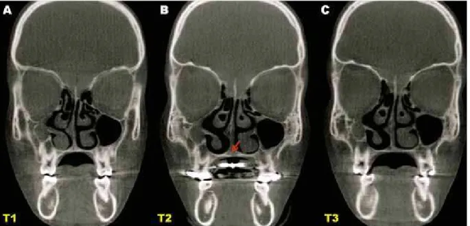
FIGURE 6 - Coronal slice used to measurements at T1, T2 and T3. A) Pre-treatment, crossbite not present in centric relation occlusion; B) Immediately after the transverse discrepancy correction, showing the palatal suture opened with slight inferior displacement (arrow) and an increase of the dentoalveolar an-gulation; C) After 6-months of retention, the transverse dimension increased, showing the buccal posterior crossbite tendency and the palatal dentoalveolar angulation. Dolphin Imaging® 11.0.
during and after the retention period, the trans-verse effects of the Haas expander in Class II malocclusion patient, since this treatment is so requested in this malocclusion.
The expansion protocol applied in this study was efficient for all patients. The opening of the midpalatal suture was easily confirmed on CBCT images realized at T2 (Fig 5), and none of the patients reported pain during the active or the retention period, just a light discomfort at the moment of the screw activation during the first 3 days. Treatment timing was an important issue to be considered, since it has been dem-onstrated that patients who underwent to RME before pubertal growth spurt exhibited greater skeletal effects, as well as greater bone stability when compared to later treatment.14 The suc-cessful results observed in our study can be at-tributed to the choice of the appliance, which provided maximum anchorage when used in the appropriate skeletal maturation period.13
Standardization of the amount of screw ex-pander activation seems to be ideal to evaluate the transverse effects. However, we thought this is ethically wrong as the patients had different orthodontic needs, i.e., some might need more expansion while for others the amount of acti-vation might not be enough. In order to make it possible to evaluate and to compare the results with previous studies, the transverse effects were proportionally analyzed according to the amount of screw activation in each patient.
Immediately after screw expander stabiliza-tion, all measurements were found to be highly significant (Table 2). Maxillary basal width in-creased, on average, 1.95 mm (29.10% of the screw activation), which was similar to what was found by Podesser et al.19 Alveolar and den-tal widths showed significantly greater results in our study, 4.3 and 6.9 mm, respectively, com-pared to 2.6 and 3.6 mm found elsewhere.19 Such difference may be related to the fact that the expander was removed at the end of
the active period for CBCT performing, which might have allowed some relapse, unlike our study, in which the expander was only removed at the end of the retention period.
Several studies reported a downward move-ment of the maxilla during the midpalatal su-ture opening following RME.1,5,9,23 This can happen because the center of resistance of the maxilla is located above the force application point, causing a buccal inclination of the dento-alveolar structures of the maxilla, with a down-ward displacement of the central region of the maxilla.17,27,29 This effect could also be observed in our study, visually, on CBCT images at T2 (Fig 6) and through the significant increase of the buccal inclination of the first upper molars (7.31°/6.46°) and the greater increase of the dental width than the total amount of screw ac-tivation (102.84%).
activation of 7 mm. Meanwhile, Garib et al7 found greater results at the basal and dental (crown) levels with the Hass appliance, re-spectively, 5.5 mm and 8.1 mm. Nevertheless, the retention period (3-months) was shorter and some relapse might be still expected.
The strong association between skeletal trans-verse deficiency and Class II, Division 1 maloc-clusions, even in the absence of posterior cross-bite, shows the importance of this discrepancy correction avoiding dental compensations.2,22,25,26 Our results showed that the RME with the Haas expander in Class II malocclusion patients did not change significantly the upper molar angulation. At the end of the retention period, dentoalveolar angulation was not found to be statistically differ-ent from that recorded at T1 despite the chang-es observed during the evaluation period. This demonstrates that the increase in dental width caused by RME had indeed promoted an effective
translation movement in the anchorage teeth. Bal-lanti et al3 also obtained the same results using Hyrax-type appliance, whereas Garib et al7 found significantly increased inclination of the molars at the end of their study. The 3-months of retention may not have been enough for molars to resume to their initial inclination.
COnClusIOns
All the Class II malocclusion patients evalu-ated had a significant increase in the skeletal and dental transverse dimension, without causing significant changes in the anchorage molars. The 6-months retention period allowed the trans-verse skeletal increase to be maintained and to return to the initial dentoalveolar inclination.
ACKnOwledgMents
The authors acknowledge the financial sup-port given by CAPES and FAPERJ.
1. Akkaya S, Lorenzon S, Uçem TT. A comparison of sagittal and vertical effects between bonded rapid and slow maxillary expansion procedures. Eur J Orthod. 1999 Apr;21(2):175-80. 2. Alarashi M, Franchi L, Marinelli A, Defraia E. Morphometric
analysis of the transverse dentoskeletal features of Class II malocclusion in the mixed dentition. Angle Orthod. 2003 Feb;73(1):21-5.
3. Ballanti F, Lione R, Fanucci E, Franchi L, Baccetti T, Cozza P. Immediate and post-retention effects of rapid maxillary expansion investigated by computed tomography in growing patients. Angle Orthod. 2009 Jan;79(1):24-9.
4. Cevidanes L, Oliveira AE, Motta A, Phillips C, Burke B, Tyndall D. Head orientation in CBCT-generated cephalograms. Angle Orthod. 2009 Sep;79(5):971-7.
RefeRenCes
5. Chung CH, Font B. Skeletal and dental changes in the sagittal, vertical, and transverse dimensions after rapid palatal expansion. Am J Orthod Dentofacial Orthop. 2004 Nov;126(5):569-75. 6. Fishman LS. Radiographic evaluation of skeletal maturation:
a clinically oriented method based on hand-wrist ilms. Angle
Orthod. 1982 Apr;52(2):88-112.
7. Garib DG, Henriques JF, Janson G, Freitas MR, Coelho RA. Rapid maxillary expansion-tooth tissue-borne versus tooth-borne expanders: a computed tomography evaluation of dentoskeletal effects. Angle Orthod. 2005 Jul;75(4):548-57. 8. Grauer D, Cevidanes LS, Styner MA, Heulfe I, Harmon ET, Zhu
20. Ricketts RM. Perspectives in the clinical application of cephalometrics. Angle Orthod. 1981 Apr;51(2):115-50. 21. Riedel RA. The relation of maxillary structures to cranium
in malocclusion and in normal occlusion. Angle Orthod. 1952;22(3):142-5.
22. Sayin MO, Turkkahraman H. Comparison of dental arch and alveolar widths of patients with Class II, division 1 malocclusion and subjects with Class I ideal occlusion. Angle Orthod. 2004 Jun;74(3):356-60.
23. Silva OG Filho, Boas MC, Capelozza L Filho. Rapid maxillary expansion in the primary and mixed dentitions: a cephalometric evaluation. Am J Orthod Dentofacial Orthop. 1991 Aug;100(2):171-9.
24. Silva OG Filho, Montes LA, Torelly LF. Rapid maxillary expansion in the deciduous and mixed dentition evaluated through posteroanterior cephalometric analysis. Am J Orthod Dentofacial Orthop. 1995 Mar;107(3):268-75.
25. Tollaro I, Baccetti T, Franchi L, Tanasescu CD. Role of posterior transverse interarch discrepancy in Class II, division 1 malocclusion during the mixed dentition phase. Am J Orthod Dentofacial Orthop. 1996 Oct;110(4):417-22. 26. Uysal T, Memili B, Usumez S, Sari Z. Dental and alveolar arch
widths in normal occlusion, Class II division 1 and Class II division 2. Angle Orthod. 2005 Nov;75(6):941-7. 27. Wertz RA. Skeletal and dental changes accompanying
rapid midpalatal suture opening. Am J Orthod. 1970 Jul;58(1):41-66.
28. Will L. Transverse maxillary deformities: diagnosis and treatment. Oral Maxillofac Surg. 1996;5:1-28.
29. Zimring JF, Isaacson RJ. Forces produced by rapid maxillary expansion. Angle Orthod. 1965 Jul;35:178-86.
Contact address Carolina Baratieri
Rua Anibal de Mendonça 16, ap. 109 CEP: 22.410-050 – Rio de Janeiro / RJ, Brazil E-mail: [email protected]
Submitted: June 2010 Revised and accepted: July 2010
9. Haas AJ. Rapid expansion of the maxillary dental arch and nasal cavity by opening the midpalatal suture. Angle Orthod. 1961 Apr;31(2):73-90.
10. Haas AJ. The treatment of maxillary deiciency by opening the
midpalatal suture. Angle Orthod. 1965 Jul;35(3):200-17. 11. Haas AJ. Palatal expansion: just the beginning of dentofacial
orthopedics. Am J Orthod. 1970 Mar;57(3):219-55. 12. Haas AJ. Long-term posttreatment evaluation of rapid palatal
expansion. Angle Orthod. 1980 Jul;50(3):189-217. 13. Haas AJ. Entrevista. Rev Dental Press Ortod Ortop Facial.
2001;6(1):1-10.
14. Lagravere MO, Major PW, Flores-Mir C. Long-term dental arch changes after rapid maxillary expansion treatment: a systematic review. Angle Orthod. 2005 Mar;75(2):155-61.
15. Lima RM Filho, Ruellas ACO. Long-term maxillary changes in patients with skeletal Class II malocclusion treated with slow and rapid palatal expansion. Am J Orthod Dentofacial Orthop. 2008 Sep;134(3):383-8.
16. Lima R, Bolognese AM, editores. Ortodontia: arte e ciência. 1ª ed. Maringá: Dental Press; 2007.
17. Majourau A, Nanda R. Biomechanical basis of vertical dimension control during rapid palatal expansion therapy. Am J Orthod Dentofacial Orthop. 1994 Sep;106(3):322-8.
18. Podesser B, Williams S, Bantleon HP, Imhof H. Quantitation of transverse maxillary dimensions using computed tomography: a methodological and reproducibility study. Eur J Orthod. 2004 Apr;26(2):209-15.