Flávia de Moraes Arantes1, Juliana Kina2, Matheus José Bueno Gonçalves3, Júlio de Araújo Gurgel4,
Omar Gabriel da Silva Filho5, Eduardo César Almada Santos6
Mini-implant and Nance button for initial retraction of maxillary
canines: A prospective study in cast models
original article
Objective: Bone anchorage is a key factor for the successful management of some malocclusions for it allows the application of continuous forces, decreases treatment time and is independent from patient compliance.
Methods: The goal of this work was to establish a dental model comparison in order to measure the anchorage loss after the initial retraction of upper canines between the two groups. Group A used mini-implants and Group B used Nance button. All patients had two models cast (M1 and M2). The first models were taken on baseline (M1) and the other models were taken after canine retraction (M2).
Results: All measurements were pooled and submitted to statistical analysis. In order to verify the inter-ex-aminer systematic error a paired t-test was performed. Dahlberg’s formula was used to estimate the casual error. For comparison purposes between Before and After stages, a paired t-test was done. For the comparison between mini-implant and Nance button groups, a Student t-test was applied. All tests adopted a 5% (p<0.05) significance level.
Conclusion: No statistically significant difference was observed between the two groups when measurements and comparisons to assess molar anchorage loss after canine initial retraction were performed. Two different anchorage systems were applied on dental models (mini-implants and Nance’s button) for each group.
Keywords: Orthodontic anchorage procedures. Dental casting technique. Orthodontics.
How to cite this article: Arantes FM, Kina J, Gonçalves MJB, Gurgel JA, Sil-va Filho OG, Santos ECA. Mini-implant and Nance button for initial retraction of maxillary canines: A prospective study in cast models. Dental Press J Orthod. 2012 July-Aug;17(4):134-9.
Submitted: February 12, 2010 - Revised and accepted: September 20, 2011
» The authors report no commercial, proprietary or financial interest in the products or companies described in this article.
» Patients displayed in this article previously approved the use of their facial and in-traoral photographs.
Contact address: Eduardo César Almada Santos
Rua José Bonifácio, 1193 – Vila Mendonça – CEP 16.015-050 – Araçatuba/SP – Brazil E-mail: [email protected]
1 Specialist in Orthodontics, UNESP. PhD in Orthodontics, FOA-UNESP. 2 Specialist in Orthodontics, UNESP. MSc and Orthodontics, UNESP.
3 Graduated in Dentistry, UNESP. Specialization student of Orthodontics, São Paulo
General Hospital .
4 Full Professor, Centro Universitário do Maranhão. Assistant Professor and PhD,
UNESP.
5 Orthodontist, Hospital for Rehabilitation of Craniofacial Anomalies, University of
São Paulo (HRAC-USP).
6 Adjunct Professor of Orthodontics, UNESP. Invited Professor at Washington
Flávia de Moraes Arantes1, Juliana Kina2, Matheus José Bueno Gonçalves3, Júlio de Araújo Gurgel4,
Omar Gabriel da Silva Filho5, Eduardo César Almada Santos6
Mini-implante e Botão de Nance para retração inicial de caninos
superiores: estudo prospectivo em modelos
Objetivo: a ancoragem óssea é fundamental para o sucesso do tratamento de algumas más oclusões, pois permite a
aplicação de forças contínuas, diminui o tempo de tratamento e independe da colaboração do paciente.
Métodos: o propósito desse trabalho foi comparar, por meio de modelos dentários, a perda de ancoragem após a
retração inicial de caninos superiores entre dois grupos. O grupo A utilizou o mini-implante enquanto o grupo B utilizou o Botão de Nance. Para todos os pacientes foram realizados dois modelos (M1 e M2). Os primeiros mode-los foram realizados ao início (M1), e os outros ao final da retração inicial de canino (M2).
Resultados: todas as medidas foram tabuladas e submetidas à análise estatística. Para verificar o erro sistemático intraexaminador foi utilizado o teste t pareado. Na determinação do erro casual utilizou-se o cálculo de erro proposto por Dahlberg. Para comparação entre as fases Início e Após, foi utilizado o teste t pareado. Para a comparação entre os grupos de mini-implante e Botão de Nance, foi utilizado o teste t de Student para medidas independentes. Em todos os testes foi adotado nível de significância de 5% (p<0,05).
Conclusão: ao se medir e comparar em modelos dentários a perda de ancoragem dos molares após a retração inicial
de canino utilizando-se dois sistemas de ancoragem distintos (Mini-implante e Botão de Nance), pôde-se observar a inexistência de diferença estatisticamente significativa entre os dois grupos.
Palavras-chave: Procedimentos de ancoragem ortodôntica. Modelos dentários. Ortodontia.
Como citar este artigo: Arantes FM, Kina J, Gonçalves MJB, Gurgel JA, Silva Fi-lho OG, Santos ECA. Miniimplant and Nance button for initial retraction of maxil-lary canines: A prospective study in cast models. Dental Press J Orthod. 2012 July--Aug;17(4):134-9.
Enviado em: 12 de fevereiro de 2010 - Revisado e aceito: 20 de setembro de 2011
» Os autores declaram não ter interesses associativos, comerciais, de propriedade ou financeiros, que representem conflito de interesse nos produtos e companhias des-critos nesse artigo.
» Os pacientes que aparecem no presente artigo autorizaram previamente a publica-ção de suas fotografias faciais e intrabucais.
Endereço para correspondência: Eduardo César Almada Santos Rua José Bonifácio 1193 – Vila Mendonça – CEP 16.015-050 – Araçatuba/SP E-mail: [email protected]
1 Especialista em Ortodontia, UNESP. Doutora em Ortodontia, FOA-UNESP.
2 Especialista em Ortodontia, UNESP. Mestre e Doutora em Odontologia, UNESP.
3 Graduado em Odontologia, UNESP. Aluno de especialização em Ortodontia,
Hospital Geral de São Paulo.
4 Professor titular do Centro Universitário do Maranhão e Professor Assistente
Doutor da Universidade Estadual Paulista Júlio de Mesquita Filho.
5 Ortodontista do Hospital de Reabilitação de Anomalias Craniofaciais da
Universidade de São Paulo (HRAC-USP).
6 Professor Adjunto da disciplina de Ortodontia, UNESP. Professor convidado da
Arantes FM, Kina J, Gonçalves MJB, Gurgel JA, Silva Filho OG, Santos ECA
INTRODUCTION
Anchorage is a key factor for a successful orth-odontic approach.19 Although orthodontic treat-ments have been quite successful, some limitations should be observed towards certain movements and much has been done to accomplish an effec-tive patient cooperation.5 Over the last two decades mini-implants have been introduced in orthodontic clinical practice with the purpose of providing an-chorage and have shown to be quite a promising op-tion.3,4,16,17 The use of a stable anchorage eliminates undesirable movements upon anchoring teeth and replaces traditional procedures such as the head-gear, what allows for continuous force application leading to a shorter treatment time.1,14
The intensity of the load may vary from one type of movement to the other. Regarding retraction movements, the load applied differs from initial canine retraction to anterior retraction. For initial retraction, the load ranges from 50 g5,11 to 100 g,5,20 whilst for anterior retraction it ranges from 150 to 200 g,5 allowing for even higher load intensi-ties such as 200 g and 300 g5,9,11,12 with good results and without any jeopardise to the root structure or the periodontium. For molar mesialization move-ments towards the gaps of prematurely lost teeth, Roberts5,15 suggests the use of 408 g for moving sec-ond and third molars.
Mini-implants may be self-piercing6 or self-tapping (with and without previous perforation procedure, respectively). Some authors7,18 state that the self piercing mini-implants are more trau-matic since this procedure induces physical pres-sure and micro fractures in the surrounding bone structure, possibly leading to periosteal or end-osteal injury. However, other professionals argue that the self-tapping mini-implant system causes larger bone trauma due to the frictional heat cre-ated by the threads during the perforation previ-ously performed.18
With regards to the time for load application there is no consensus. Many periods have been studied, varying from immediately after to 2, 4 or 6 weeks later,5,10,12 and the implant loss, in none of the works, was ever related to the waiting interval.
In an attempt to be less reliant on patient co-operation and to be able to achieve new anchorage
solutions in orthodontic treatments, mini-im-plants are recommended for adult patients in need of maximum anchorage (intrusion, extrusion, re-traction and prore-traction) who are reluctant to use extra-oral braces as well as in cases where the orthodontic anchorage cannot be accomplished due to tooth losses.
For the reasons above listed and for the indis-criminate clinical application of mini-implants the related anchorage loss there came the interest for this subject. The goal of this work was to use dental models to establish a comparison, between two groups, of the upper first molars anchorage loss after upper canines initial retraction. While Group A used mini-implants, Group B used the conventional intraoral anchorage technique. The null hypothesis to be tested is that both anchorage systems present similar results.
MATERIAL AND METHODS
In this experiment 18 patients were selected, average age 15 years old, randomly divided be-tween two groups, with 9 subjects each (A and B), out of the screening for orthodontic treatment at Araçatuba School of Dentistry - UNESP. The inclu-sion criteria for the research were:
a) Patients with a balanced facial pattern.15,16 b) Patients in the post growth spurt phase.
c) Patients presenting upper anterior crowding, demanding upper and lower pre-molars extrac-tions (Fig 1).
Orthodontic therapy for patients with upper ante-rior crowding from Group A was carried out by the use of self-tapping 1,6 x 10mm titanium mini-implants from SIN (Sistema de Implante, São Paulo, Brazil).
Squeff et al,18 after determining topography, arche-type and torque test of some SIN mini-implants, stated that all tested mini-implants were adequate for clinical application as an adjunct to orthodon-tic anchorage.
Implants were inserted both sides into the up-per alveolar bone, between the second pre-molar and first molar, over attached gingiva. In order to prevent root lesions, they were inserted in the in-terseptal areas of these teeth, guided by the periapi-cal radiographs, taken under the parallelism tech-nique. After implant insertion, the first pre-molars were extracted, with an immediate onset of 150 cN load applied over the canines. In the lower arch, the lingual arch was used as anchorage. Alignment and levelling were performed with Edgewise fixed ap-pliance, according to Capelozza prescription, Stan-dard I (Abzil 3M, São José do Rio Preto, Brazil).
Group B received orthodontic treatment using Nance button as the anchorage system for the up-per arch and Nance lingual arch for the lower teeth, both manufactured using a 0.9 mm wire (Morelli). The anchorage systems were first incorporated fol-lowed by Edgewise appliance fixation, according to Capelozza prescription, Pattern I (Abzil 3M), and subsequent extraction of the first pre-molars. Ini-tial canine retraction under 150cN load per side was immediately started.
All patients had two impressions taken: M1 mod-el (taken at basmod-eline) and M2 modmod-el (taken after the
initial canine retraction). This initial canine re-traction phase was chosen due to the great anchor-age loss that usually takes place during this treat-ment stage4 and also for the clinically satisfactory incisors alignment. This work had no aim to assess anterior retraction, since during that treatment stage some anchorage loss was needed concomi-tantly to the group retraction
Plaster casts were obtained after an impression was taken with Kromopan alginate, batch number 0155300130.103 514 (Lascod SpA Florence Italy) and water, under a 1/1 proportion. Impressions were then poured with Durone V stone, batch 539589 (Dentsply Indústria e Comércio Ltda Petrópolis, RJ Brazil) and water under the proportion of 19 mm of water for each 100 g of plaster, in order to reproduce the dental structures as precisely as possible.
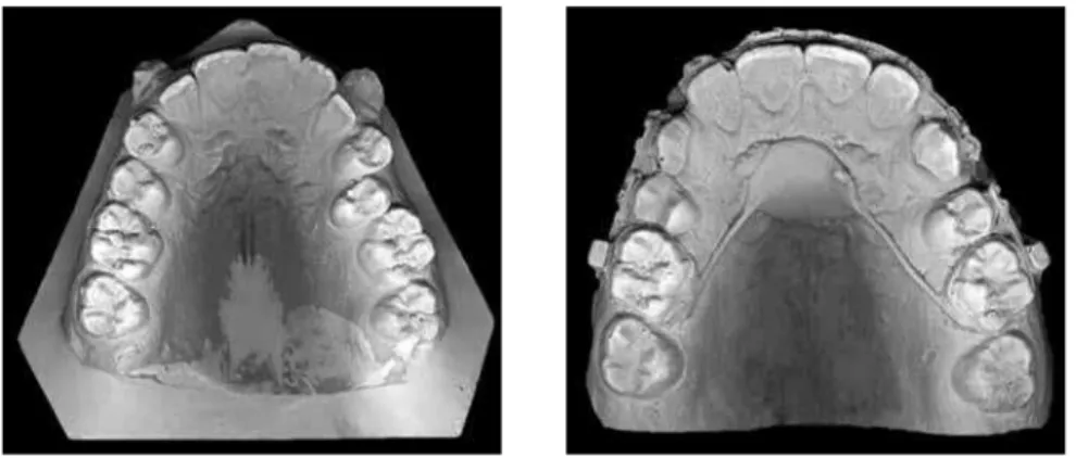
Aiming optimize the measurement of the dental models, which are tridimensional by nature, all upper models underwent a photographic scanning with an HP scanner (Scanjet G4050 China), being transferred to a two-dimensional and flat image. No models need-ed to be cut or trimmneed-ed since only the occlusal surface touched the scanner avoiding undesired tilting.
Both upper models (baseline and post canine initial retraction) from each patient were scanned together in order to avoid possible dimensional changes due to different scanning process. More-over, models from each patient were scanned one at a time (Fig 2).
Arantes FM, Kina J, Gonçalves MJB, Gurgel JA, Silva Filho OG, Santos ECA
The images obtained from the scanned models were transferred in the format of a Microsoft Of-fice PowerPoint 2003 file to achieve better organi-zation and standardiorgani-zation of the image selection process. Each patient was illustrated by two upper model images (baseline and post canine initial re-traction), which were cut out in a standardized size, without dimensional alterations. Subsequently, this file was saved as a JPEG image and transferred to an AutoCad 2007 Autodesk software.
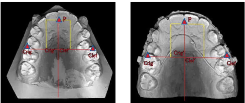
Measurements were performed based on the following reference points: point P, located at the centre of the incisive papillae, from which a line was drawn along the median palatal raphe; two other points, Crig and Clef, located at the centre of the mesiobuccal cusp of each permanent upper first molar, right and left sides, respectively. A line was traced from each of these points, perpendicu-larly to the median palatal raphe line, crossing it in two distinct locations, generating two other points: Clef’ (representing the intersection be-tween the line drawn from the palatal raphe and Clef point) and intersection point Crig’ (between the palatal raphe and the line stemming from Crig point) (Fig 3).
The distances between the points Clef’ to P, and points Crig’ to P, crossing the median palatal ra-phe line are the measurements compared in both sides between the images of the models obtained at baseline and after the canine initial retraction.
Therefore, these measurements represent the po-sition of the permanent upper first molar on both left and right sides, relative to the incisive papilla, at baseline and post canine initial retraction stag-es. As these distances decrease, after canine initial retraction, it indicates the amount of mesializa-tion of the permanent upper first molar on each side (anchorage loss).
All measurements were completed and submit-ted to statistic analysis. In order to verify the in-tra-examiner systematic error a paired t test was performed. For the casual error determination, Dahlberg’s (Houston, 1983) formula was utilized.
d = difference between the first and second mea-surements.
n = number of repetitions.
The results of the systematic error, measured by the paired t test and the casual error assess-ments are displayed on Table 1. Data were scribed according to the average and standard de-viation shown on tables (Tables 2, 3, 4). In order to compare Before and After stages a paired t test was used. In order to compare Mini-implant and Nance button groups a student t test was applied for in-dependent measurements. All tests considered the 5% significance level (p<0.05). All statistic calcula-tions were performed by the 5.1 Statistics for Win-dows software (StatSoft Inc, Tulsa, USA).
Figure 3 - Upper models (baseline and inter-mediate) with lines and points traced. Crig
Clef Crig’ Clef’
Crig Clef
P
Clef’ Crig’
Table 2 - Average, standard deviation and comparison between Before and After measurements obtained from the models for the mini-implant group. Right Side (distance between the centre of the mesiobuccal cusp of the right permanent upper first molar to point P). Left Side (distance between the centre of the mesiobuccal cusp of the left permanent upper first molar to point P).
ns: no statistically significant difference.
Measure Beggining After Difference t p
Mean SD Mean SD Mean
Right side 53.06 8.29 50.41 5.86 -2.65 1.684 0.131 ns
Left side 52.96 5.70 51.06 7.21 -1.90 1.080 0.312 ns
Table 3 - Average, standard deviation and comparison between Before and After measurements obtained from the models for the Nance button group. Right Side (distance between the centre of the mesiobuccal cusp of the right permanent upper first molar to point P). Left Side (distance between the centre of the mesiobuccal cusp of the left permanent upper first molar to point P).
ns: no statistically significant difference *statistically significant difference (p < 0.05).
Measure Beginning After Difference t p
Mean SD Mean SD Mean
Right side 53.01 7.18 50.16 7.05 -2.85 3.555 0.007 *
Left side 53.60 7.46 50.87 10.17 -2.73 1.576 0.154 ns
Table 4 - Average and standard deviation of the variations between Before and After, and comparison between mini-implant and Nance button groups. Right Side (distance between the centre of the mesiobuccal cusp of the right permanent upper first molar to point P). Left Side (distance between the centre of the mesiobuccal cusp of the left permanent upper first molar to point P).
ns: no statistically significant difference.
Measure Mini-implant Nance button Difference t p
Mean SD Mean SD Mean
Right side -2.65 4.73 -2.85 2.41 0.20 0.109 0.914 ns
Left side -1.90 5.29 -2.73 5.19 0.83 0.333 0.743 ns
Measure First measurement Second measurement t p Error
Mean SD Mean SD
Right side 55.57 7.13 55.08 6.86 1.041 0.325 ns 1.07
Left side 56.76 6.37 57.59 6.62 1.358 0.207 ns 1.43
Table 1 - Average, standard deviation for both measurements, paired t test, and Dahlberg’s systematic and casual errors. Right side (distance between the centre of the mesiobuccal cusp of the upper permanent right first molar to point P); Left side (distance between the centre of the mesiobuccal cusp of the left permanent upper first molar to point P).
ns: no statistically significant difference.
The choice for assessing standardized stone dental models is justified by the existence of specific software capable of estimating molars anchorage loss. Canine initial retraction is an ordinary procedure within the orthodontic treatment. Many systems (appliances) are used to retract canines during space closure, which depends on many factors: type of system used, load ap-plied, technique and periodontal condition.20
In this study, for Group A, mini-implants were in-serted between the roots of permanent upper second pre-molars and first molars. Group B Nance button was employed as an anchorage system.
RESULTS
Results are shown on tables 1 - 4.
DISCUSSION
Studies based on mini-implants positioned between the roots of second pre-molars and first molars have proved to be successful for mass retraction of the anteri-or segment as well as during canine initial retraction.19,20
Arantes FM, Kina J, Gonçalves MJB, Gurgel JA, Silva Filho OG, Santos ECA
The results revealed an average anchorage loss of 2,85 mm on the right side and 2.73 mm on the left side for the group with Nance button, even though the left side did not present any statistically significant dif-ference (Table 3). These results were expected since load application is performed directly over the molars, confirming thus the null hypothesis.
It was also possible to observe by that even when bone anchorage is used, anchorage loss was detected on the upper molars for the mini-implant group (Table 2). These results demonstrate that bone anchorage cannot be ensured, since the mechanics, the intensity and con-trol of the loads applied are still very relevant factors to be considered.20 One hypothesis to explain the anchor-age loss in the mini-implant group can be possible attrib-uted to the levelling and alignment stage, since the fric-tion between the wire and the molar tubes might have caused the molars to rotate, as no stabilization procedure was ever done. At the end of the canine retraction molars were assessed but no conclusion was drown whether the molars had gone through rotation or tilting, as it would usually happen in any orthodontic movement.
Table 4 shows no statistically significant difference between the two groups, mini-implant and Nance but-ton), what doesn’t necessarily mean there are no dif-ferences between, but rather the absence of evidence
that could support that difference. Clinically, it could be observed that there was a higher anchorage loss in the group wearing Nance button and that the interval for canine initial retraction was also longer.
Eighteen patients is quite a shy value for statistic analysis, although it could be considered quite a good number for a clinically based work. In addition, other scientific papers published used much smaller sam-ples or even a single clinical case.
According to the present study, mini-implants can-not be considered as an absolute anchorage method. Nonetheless, studies that advocate it as the ultimate anchorage option, are actually based on clinical reports, what hinders the possibility of taking a conclusion, in other words, there are no scientific evidences. Such an argument encourages the development of standardised methodology studies. Therefore, studies that verify the effectiveness of the anchorage and the biological cost of the mini-implants are to be further developed.
CONCLUSION
After measuring and comparing models for the as-sessment of molars anchorage loss after canine initial retraction by means of two systems (mini-implants and Nance button) no statistically significant differ-ence could be observed between the two groups.
1. Costa A, Raffaini M, Melsen B. Microscrews as orthodontic anchorage. In J Adult Orthod Orthognath Surg. 1998;13:201-9.
2. Capelozza Filho L. Diagnóstico em Ortodontia. Maringá (PR): Dental Press; 2004.
3. Goodacre CJ, Brown DT, Roberts WE, Jeiroudi MT. Prosthodontic considerations
when using implants for orthodontic anchorage. J Prosthet Dent. 1997 Feb;77(2):162-70.
4. Hong RK, Heo JM, Ha YK. Lever-arm and mini-implant system for anterior torque
control during retraction in lingual orthodontic treatment Angle Orthod. 2005 Jan;75(1):129-41.
5. Janson M, Sant’Ana E, Vasconcelos W. Ancoragem esquelética com minimplantes:
incorporação rotineira da técnica na prática ortodôntica. Rev Dental Press Ortodon Ortop Facial. 2006;5:85-100.
6. Kyung HM, Park HS, Bae SM, Sung JH, Kim IB. Development of orthodontic
micro-implant for intraoral anchorage. J Clin Orthod. 2003 Jun;37(6):321-8; quiz 314. 7. Lee JS, Kim JK, Park YC, Vanarsdall RL Jr. Application of orthodontic mini-implants.
Hanover Park (IL): Quintessence Books; 2007.
8. Mah J, Bergstrand F. Temporary anchorage devices: a status report. J Clin Orthod. 2005;39:132-36.
9. Park HS, Bae SM, Kyung HM, Sung JH. Simultaneous incisor retraction and distal
molar movement with micro-implant anchorage. World J Orthod. 2004;5:164-71. 10. Park HS, Bae SM, Kyung HN, Sung JH. Micro-implant anchorage for treatment of
skeletal Class I bialveolar protrusion. J Clin Orthod. 2001;35:417-22.
11. Park HS, Know TG, Sung JH. Microscrew implant anchorage sliding mechanics.
World J Orthod. 2005;6:265-74.
REFERENCES
12. Park HS, Know TG, Sung JH. Sliding mechanics with microscrew implant
anchorage. Angle Orthod. 2004;74:703-10.
13. Reis SAB, Capelozza LF, Cardoso MA. Características cefalométricas dos indivíduos padrão I. Rev Dental Press Ortodon Ortop Facial. 2005;10:67-78.
14. Roberts WE, Arbuckle GR, Analoui M. Rate of mesial translation of mandibular molars using implant-anchored mechanics. Angle Orthod. 1996;66:331-8.
15. Roberts WE, Marshall KJ, Mozzary PG. Rapid endosseous implant utilizes as
anchorage to protract molars and close an atrophic extraction site. Angle Orthod. 1990;60:135-52.
16. Roberts WE, Smith RK, Zilberman Y, Mozsary PG, Smith RS. Osseous adaptation to
continuous loading of rigid endosseous implant. Am J Orthod Dentofacial Orthop. 1984;86:95-111.
17. Shapiro PA, Kokich VG. Uses of implants in orthodontics. Dent Clin North Am. 1988;32:539-50.
18. Squeff LR, Simonson MBA, Elias CN, Nojima LI. Caracterização de mini-implantes
utilizados na ancoragem ortodôntica. R Dental Press Ortodon Ortop Facial. 2008;13:49-56.
19. Thiruvenkatachari B, Ammayappan P, Kandaswamy R. Comparison of rate of
canine retraction with conventional molar anchorage and titanium implant Anchorage. Am J Orthod Dentofacial Orthop. 2008;123:30-5.