Frictionless segmented mechanics for
controlled space closure
Ildeu Andrade Jr1
Extraction spaces may be needed to achieve specific orthodontic goals of positioning the dentition in harmony with the craniofa-cial complex. However, the fundamental reality that determines the occlusion final position is the control exerted by the ortho-dontist while closing the extraction spaces. A specific treatment objective may require the posterior teeth to remain in a constant position anteroposteriorly as well as vertically, while the anterior teeth occupy the entire extraction site. Another treatment objective may require the opposite, or any number of intentional alternatives of extraction site closure. The present case report describes a simple controlled segmented mechanic system that permitted definable and predictable force systems to be applied and allowed to predict the treatment outcome with confidence. This case was presented to the Brazilian Board of Orthodontics and Dentofacial Orthopedics (BBO) in partial fulfillment of the requirements for Diplomate certification.
Keywords: Angle Class II malocclusion. Dental extraction. Corrective orthodontics.
How to cite this article: Andrade Jr I. Frictionless segmented mechanics for controlled space closure. Dental Press J Orthod. 2017 Jan-Feb;22(1):98-109. DOI: http://dx.doi.org/10.1590/2176-9451.22.1.098-109.bbo
» The author reports no commercial, proprietary or financial interest in the products or companies described in this article.
Contact address: Ildeu Andrade Jr.
Rua Padre Rolim, 815, conj. 406 - Santa Efigênia CEP: 30.130-090 – Belo Horizonte/MG Brasil E-mail: [email protected]
» Patients displayed in this article previously approved the use of their facial and in-traoral photographs.
Submitted: December 06, 2016 - Revised and accepted: December 16, 2016 1 Adjunct Professor, Pontifícia Universidade Católica, Minas.
DOI: http://dx.doi.org/10.1590/2176-9451.22.1.098-109.bbo
INTRODUCTION
This patient was a healthy 31-year-old woman with a chief complaint of “protruded teeth afecting my smile”. Her medical history showed no contraindica-tion to orthodontic treatment. There was no history of dental trauma or oral habits. The patient had good
oral hygiene. Regarding function, the patient pre-sented lateral disocclusion through molar guidance on the right side. Mouth opening and closure movements were performed without deviation. No signs or symp-toms of temporomandibular dysfunction were noted. Os espaços gerados pelas extrações de pré-molares podem ser necessários para que objetivos ortodônticos específicos sejam alcançados dentro do complexo craniofacial. No entanto, a realidade fundamental que determina a posição final da oclusão é o controle biomecânico exercido pelo ortodontista no fechamento dos espaços das extrações. Um objetivo específico do tratamento pode exigir que os dentes posteriores permaneçam estáveis em uma posição sagital e vertical, enquanto os dentes anteriores são retraídos e ocupam todo o espaço da extração. Outro objetivo de tratamento pode exigir o inverso, ou quaisquer alternativas intencionais de fechamento de espaços de ex-tração. O presente relato de caso descreve um sistema simples de mecânica segmentada que permite, de maneira controlada, que sistemas com forças definidas e previsíveis sejam aplicados, permitindo também predizer o resultado do tratamento com segurança. Esse caso foi apresentado ao Board Brasileiro de Ortodontia e Ortopedia Facial (BBO), em cumprimento parcial dos requisitos para certificação.
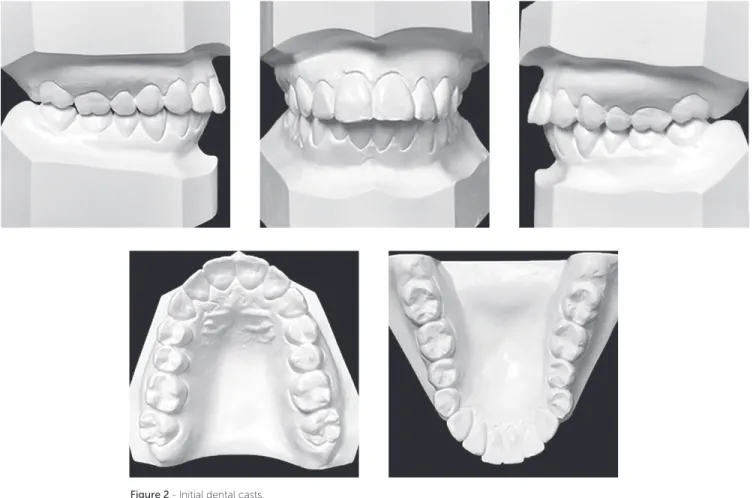
Figure 1 - Initial facial and intraoral photographs. causing tension in the orbicularis oris muscles. Moreover, the lower lip was procumbent, 4.5 mm ahead of the Steiner’s S-line (Table 1). Intraorally, she presented a bilateral end-to-end Class II, with a severe bimaxillary protrusion, an overjet of 5 mm and a pronounced curve of Spee (Fig 1). The Bolton analysis was equal to 75.3% in the anterior teeth and equal to 87.1% of total (Fig 2). Both arches were constricted in the posterior region, which contributed to the creation of the “black corridors”.
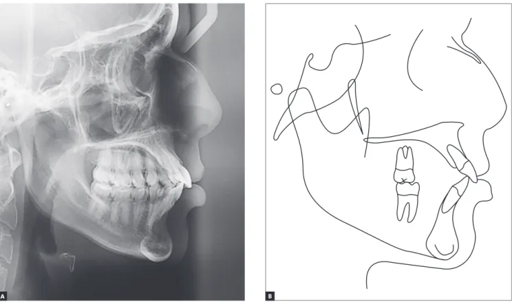
reveals an ANB angle of 3.5o and an Angle of
Convex-ity equal to 2o. The FMA, Y-axis and SN-GoGn were
equal to 18o, 52o and 30o, respectively. The maxillary
and mandibular incisors were signiicantly lared, with a maxillary central incisor to SN angle of 117o, 1.NA
of 32o; mandibular central incisor to MP angle of 105o,
and 1.NB of 34o. The maxillary and mandibular
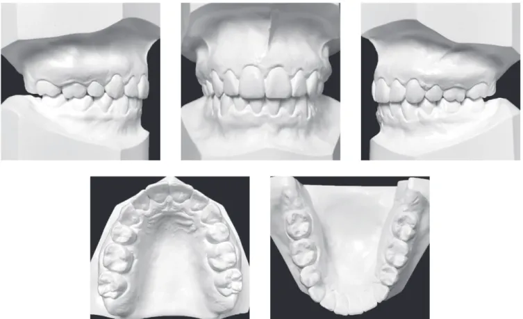
Figure 2 - Initial dental casts.
TREATMENT PLAN
The treatment objectives were to: (1) reduce the excessive protrusion of both dental arches; (2) obtain normal canine and incisal guidance; (3) correct lip in-competence and lip strain on closure; (4) correct the Class II dental relationship; (5) level the curve of Spee; (6) achieve optimal overjet and overbite, and (7) im-prove the facial balance.
Based on these objectives, two treatment options were proposed, both requiring the extraction of all irst premolars. The irst alternative was to perform an en-masse retraction by using TADs. The pros and cons of absolute anchorage were explained. The sec-ond was a two-step space closure (beginning with canine retraction and followed by incisor retraction) with Frictionless controlled segmented mechanics and anchorage control without TADs. The patient refused any surgical treatment other than the extraction itself and requested the second option.
TREATMENT PROGRESS
A transpalatal bar and a lower lingual arch were placed for anchorage considerations, such as molar
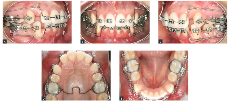
rotations and undesirable transverse changes, and the maxillary and mandibular first premolars were ex-tracted two weeks after these appliance installation (Fig 5). Standard edgewise brackets and tubes were passively bonded to the maxillary and mandibular ca-nines, second premolars and second molars (0.022-in American Orthodontics, Sheboygan, WI, EUA). The maxillary and mandibular incisors were also bonded for alignment and levelling. The distalization of the canines in both arches was initiated one week after extraction with segmented arch mechanics, using a Ricketts cuspid retractor made by a 0.016 x 0.016-in Elgiloy (Rocky Mountain, Denver, CO, EUA), up to closure of 2/3 of the spaces (Fig 5). Seven months lat-er, the maxillary and mandibular incisors were than retracted simultaneously with closed 12-mm length (150 g) NiTi closed coil springs (G&H Orthodon-tics, Franklin, IN, EUA) adapted to two power arms on each quadrant (Fig 6A). The posterior power arms were located in the distal part of the auxiliary tubes of the first molars. The anterior ones were part of a wire passing through the slots of the incisors, creating an incisor segment. The posterior and anterior power Figure 4 - Initial cephalogram (A) and cephalometric tracing (B).
arms were constructed to be as close as possible to the center of rotation of the molar and incisor segments, respectively (Fig 6). As the objective was to close the extraction spaces and at the same time upright the maxillary and mandibular incisors, the height of the power arms were adjusted throughout the treatment as needed (eg. shortening of the anterior power arms provides more lingual/palatal crown torque).
Once the incisors were fully retracted (6 months), the patient was debonded and them rebonded for fi-nal alignment and levelling. Once the arches were
aligned and leveled, continuous 0.017 x 0.025-in stainless steel (SS) arch wires were inserted in both arches and the patient was instructed to wear 3/8 ounces Class II elastics. Three months later, 0.019 x 0.025-in SS wires were placed for torquing control. Root paralleling and vertical elastics were used to detail and settle down the occlusion.
The appliances were removed ater 24 months of treatment, when a maxillary wraparound and a man-dibular bonded premolar-to-premolar ixed retainer were installed on the patient.
RESULTS
The posttreatment facial and intraoral photo-graphs (Fig 7) illustrate the improvement in the patient’s proile. The proile line to nose relationship improved and no mentalis strain was noted. The lips were compe-tent at rest and the upper lip to S-plane changed from 1 to -2.5 mm; the lower lip to S-plane changed from 4.5 to 0 mm. Frontal analysis revealed a balanced face, with pro-portional facial thirds and an estheti cally pleasant smile.
The posttreatment dental casts (Fig 8) and intraoral photos (Fig 7) show an Angle Class I occlusion with normal overjet, overbite, and canine and incisal guid-ance. There was a slight increase in the mandibular arch transverse dimension, as needed.
The posttreatment panoramic radiograph (Fig 9) shows that all spaces were closed without alveolar bone loss and root resorption ater treatment. The posttreat-ment cephalometric radiograph and its tracing (Fig 10) illustrate the changes achieved with treatment. As planned, the maxillary incisors were uprighted 17o
and 7 mm over the basal bone (to a inal 1.NA of 14.5o
and 1-NA of 3 mm). Mandibular incisors were retract-ed 16.5o and 6 mm from the mandibular plane (to a inal
IMPA angle of 88.5o and 1-NB of 4 mm). The
interin-cisal angle was signiicantly increased (32o). The FMA
angle increased 2o (from 18o to 20o) and the SN-GoGn
reduced 1o (from 30o to 29o) (Table 1).
The cephalometric tracings confirmed that the maxillary and mandibular incisors were significantly uprighted and the lower lip soft tissue protrusion was reduced (Figs 4 and 10). Cephalometric tracings su-perimposition also revealed no extrusion in the max-illary and mandibular molars. As pre dicted and de-sired, the sagittal position of the maxillary molars re-mained almost unchanged, with minimal anchorage loss; and the mandibular ones slightly came forward to achieve a Class I relationship.
The case was retained by means of a maxillary wraparound retainer and a bonded premolar-to-pre-molar ixed retainer in the mandibular arch. The pa-tient was instructed to only remove the retainer dur-ing periods of eatdur-ing and toothbrushdur-ing. The appoint-ments were set in 2 to 3 months intervals in the irst year and 6 months intervals in the second year.
Treatment objectives were achieved with excellent esthetic and functional results. The patient became very happy with the results of her treatment, which was accomplished in 24 months.
Figure 6 - Progress records (incisors’ retraction).
A B C
Figure 8 - Final dental casts.
Figure 10 - Final profile cephalometric radiograph (A) and cephalometric tracing (B).
Figure 11 - Total (A) and partial (B) superimpositions of initial (black) and final (red) cephalometric tracings.
A B
Skeletal pattern
Wits (Jacobson) ♀ 0 ±2 mm
♂ 1 ±2 mm 4 mm 1 mm 3
Angle of convexity (Downs) 0o 2o 3o 1
Y-axis (Downs) 59o 52o 53o 1
Facial angle (Downs) 87o 95o 94.5o 0.5
SN-GoGn (Steiner) 32o 30o 29o 1
FMA (Tweed) 25o 18o 20o 2
Dental pattern
IMPA (Tweed) 90o 105o 88.5o 16.5
1.NA (degrees) (Steiner) 22o 31.5o 14.5o 17
1-NA (mm) (Steiner) 4 mm 10 mm 3 mm 7
1.NB (degrees) (Steiner) 25o 34o 20o 14
1-NB (mm) (Steiner) 4 mm 10 mm 4 mm 6
1
1- Interincisal angle (Downs) 130o 110o 142o 32
1-APo (Ricketts) 1 mm 11 mm 4 mm 7
Profile
Upper lip — S-line (Steiner) 0 mm 1 mm -2.5 3.5
Lower lip — S-line (Steiner) 0 mm 4.5 mm 0 mm 4.5
FINAL CONSIDERATIONS
Controlling Newton’s third law and the tip-ping and torque of anterior and posterior segments is critical to successfully closing extraction spaces. Two methods have been used for extraction space closure: (1) En-masse retraction of the incisors and canines;1,2 (2) A two-step retraction, beginning with
the distalization of the canines and followed by in-cisor retraction.3,4 The first method has two types
of mechanics. The first is segmented mechanics, in which the anterior teeth are retracted directly with a
spring such as a T-loop space closure spring,3 which
needs a complicated design to achieve bodily tooth
movement.4 The second type is sliding mechanics,
where incisors and canines are retracted with an archwire guided by the brackets placed on the pos-terior teeth.3,4 However, in sliding mechanics,
fric-tion occurs at the wire-bracket interface, dissipating some of the applied force and decreasing the rate of tooth movement.5-8 To overcome the friction, a high
To achieve controlled extraction space closure, the appliance used must deliver determinable force sys-tems regulated by the orthodontist, and not produce closure in an indeterminate way. Only when force systems are deinable, dental movements are predict-able and treatment outcomes can be predicted with
conidence.11 In addition, the force systems should
move teeth with an optimal velocity and an extended range of activation, while producing a relatively con-stant force system, which will reduce the tissue injury and the number of appointments, while yielding tooth movement with a nearly constant center of rotation. This case report described a mechanical system that met these goals without using temporary anchorage devices (TADs). TADs have expanded the horizons of orthodontic treatment, because they allow treatment to proceed successfully with virtually no anchor-age loss and minimal patient cooperation. However, sometimes we are faced with patients that, for difer-ent reasons, do not want invasive methods, such as TADs, and/or extraoral force for anchorage matters.
Therefore, precise control of tooth movement dur-ing closure of extraction spaces in three dimensions is of paramount importance in meeting treatment goals. This includes control of the anchorage units, vertical forces, root positions, and rotations. Many methods for controlling the posterior anchorage movement in extraction space closure have been described.2-5,10,11,13
Regulation of the space closure is ultimately deter-mined by the biomechanical forces applied to the an-terior and posan-terior teeth. Variation in the force and moment magnitude and the moment-to-force ratio are important determinants of the orthodontic tooth movement. Tweed tip-back bends, Begg or tip-edge mechanics, intermaxillary elastics, and headgear can produce diferences in the moment-to-force ratios (and the moment diferential) between the anterior teeth and posterior teeth. This diference in the moment-to-force ratio acting on the anterior versus the posterior segments is produced by either applying unequal mo-ments (a moment diferential) or unequal forces (i.e., use of a headgear or intermaxillary elastics).
The advantage of using a segmented mechanics is that it is possible to develop a precise and predict-able force system between an anterior and a posterior segments, enabling sagittal, vertical and axial control of the anterior and posterior teeth. The mechanism
described in this case report enables the magnitude of the moments and forces delivered to be well con-trolled.12 Consequently, constant levels of force can be
maintained, and the moment to force ratio (M/F) at the centers of resistance is easily regulated to produce the desired tooth movements. If sliding mechanics or closing loops archwires techniques were used for re-traction, the posterior dental anchorage would be sig-niicantly afected. Consequently, the treatment time would probably be longer. Furthermore, retracting the canines without prior alignment and leveling save treatment times and allows you to use the window of opportunity created by the corticotomy during the extraction of the irst premolars.
To design this segmented mechanics optimally to obtain the desired force system, the position of the center of resistance of the anterior teeth may be esti-mated on a lateral cephalometric X-ray ilm. In clini-cal situations such as the one reported in this paper, where incisors are proclined, the center of resistance of the anterior segment lies further lingual to the inci-sors crowns.13 It is important to monitor the anterior
and posterior segments and alter the force system if in-dicated, especially the axial inclination of the anterior teeth.14 The resulting force system can be modiied by
changing the magnitudes and points of application of the distal forces with respect to the center of resistance of the anterior segment.
In: Nand R, editor. Biomechanics in clinical orthodontics. Philadelphia: Saunders; 1997. p. 156-87.
4. Burstone CJ. The segmented arch approach to space closure. Am J Orthod.
1982 Nov;82(5):361-78.
5. Nanda R, Ghosh J. Biomechanical considerations in sliding mechanics. In: Nanda R, editor. Biomechanics in clinical orthodontics. Philadelphia: Saunders; 1997. p. 188-217.
6. Kusy RP, Whitley JQ, Prewitt MJ. Comparison of the frictional coeicients for selected archwire-bracket slot combinations in the dry and wet states. Angle Orthod. 1991 Winter;61(4):293-302.
7. Noda T, Okamoto Y, Hamanaka H. Friction property of orthodontic wires: evaluation by static frictional coeicients. J Jpn Orthod Soc. 1993;52:154-60.
e1-6; discussion 702-4.
11. Braun S, Sjursen RC Jr, Legan HL. On the management of extraction sites. Am J Orthod Dentofacial Orthop. 1997 Dec;112(6):645-55.
12. Burstone CJ, Koenig HA. Optimizing anterior and canine retraction. Am J Orthod. 1976 July;70(1):1-19.
13. Shrof B, Lindauer SJ, Burstone CJ, Leiss JB. Segmented approach to simultaneous intrusion and space closure: biomechanics of the three-piece base arch appliance. Am J Orthod Dentofacial Orthop. 1995 Feb;107(2):136-43.