Distalization of impacted mandibular second
molar using miniplates for skeletal anchorage:
Case report
Belini Freire-Maia*, Tarcísio Junqueira Pereira**, Marina Parreira Ribeiro***
This study describes a case with an impacted right mandibular second molar which was distalized using miniplates for skeletal anchorage. Uprighting impacted mandibular second molars has been a great challenge for orthodontists and oral surgeons because of the scarcity of anchorage options. Skeletal anchorage was first used in clinical ortho-dontics in the middle of the 1980s. Since then, several devices have been developed for that purpose, such as mini-screws, tooth implants and, lately, miniplates, which have been tested and showed encouraging results. This topic is relevant for orthodontists and oral surgeons because the use of miniplates may significantly change the treatment of impacted mandibular molars.
Abstract
Keywords: Skeletal anchorage. Tooth impaction. Molar distalization.
* MSc in Oral and Maxillofacial Surgery and Trauma (OMFST), Unicastelo. Head of the Specialization Course in OMFST, Pontifícia Universi-dade Católica de Minas Gerais (PUC-Minas), Brazil.
** MSc in Orthodontics, PUC-Minas. Professor, Specialization Course and Master’s Program in Orthodontics, PUC-Minas, Brazil. *** Senior Undergraduate Student, School of Dentistry, PUC-Minas, Brazil.
introduction
Impacted mandibular second molars are a rather uncommon problem with an incidence of 3 in 1,000 and often pose a challenge for orthodontists and oral surgeons.4,8,10
Unilater-al impaction is a more common problem and more frequently affects the right side of the mandible of male patients.10
The possible causes of second molar impac-tion are the late erupimpac-tion of second premolars, premature extractions or ankylosis of first mo-lars, dentigerous cysts or odontomas and, finally,
the competition for space by the third molar.8
Iatrogenic factors, such as bands and orthodontic loops fixed to the mandibular first molar, may also lead to impactions.12
Treatment options depend on tooth inclina-tion, the position of third molars, and the type of movement desired, which may be performed sur-gically or orthodontically.12 The best age for
treat-ment is between 11-14 years, when the develop-ment of the root of the permanent second molar is still incomplete. Several options have been ad-opted to treat mandibular molar impaction, and
» The authors report no commercial, proprietary, or inancial interest in the
products or companies described in this article. How to cite this article: Freire-Maia B, Pereira TJ, Ribeiro MP. Distalization
one of them is skeletal anchorage, successfully used in orthodontics.
Skeletal anchorage is not a recent procedure. It was first used by Creekmore and Eklund in 1983, who placed screws below the anterior nasal spine for incisor intrusion.9
After skeletal anchorage became a regular procedure in orthodontics, several fixation meth-ods and rigid devices have been used for tooth movement,5 such as tooth implants,3,11,13,17
mini-screws2,3,6,7,11,13 and titanium miniplates.1,2,3,6,7,11,13
Miniplates are made of commercially pure titanium, which is biocompatible and adapts to bones.1,2,6 Miniplates have been used to treat
fa-cial fractures for many years1 and have recently
achieved a prominent place among orthodontic anchorage methods due to its high stability.
Kuroda et al6 reported that miniplates
pro-vide rigid anchorage for several types of tooth movement, but require patient cooperation af-ter implantation, particularly for oral hygiene. Although infections are rare, they occur in 10% of the cases and are only controlled by strict oral hygiene and, in more severe cases, the use of antibiotics.15 In addition to infections, other
complications such as plate fracture and loosen-ing of screws may occur.
cAse report
A 16-year-old boy sought dental care at an oral surgery service with partial impaction of tooth 47.
A panoramic radiograph confirmed mesial impaction of tooth 47, unerupted maxillary third molars and absence of mandibular third molars (Fig 1). Clinical examination revealed that the crown of the tooth under evaluation was partially exposed in the oral cavity.
The suggestion for treatment was skeletal anchorage using a rigid device placed in the region of the retromolar trigone/mandibular ramus to move the impacted tooth 47 and achieve good occlusion and intercuspation. A 1.0 mm thick straight miniplate with four holes (MDT System 2.0 Ø, MDT®, Rio Claro,
Brazil) and two 2.0 mm Ø and 5.0 mm and 7.0 mm long screws.
Surgery for miniplate fixation was per-formed after the extraction of the maxillary third molars under local anesthesia. A flap was raised by making an incision in the right ret-romolar region which extended buccally and along the gingival crevice of teeth 47, 46 and 45 to expose the cortical bone (Fig 2).
The selected straight miniplate was previ-ously molded to adapt better to the retromolar region/mandibular ramus, and the screws were fixed after a 1.5-mm bur was used to make the holes in the cortical bone (Fig 3).
The end of the miniplate to be used for orth-odontic anchorage was exposed in the oral cavity. Immediately after the surgery, an orthodontic de-vice was placed on the distal face of tooth 47 and
traction with an elastic band was initiated. The incision was closed with 4.0-silk su-ture, which was removed seven days after the surgery. After the miniplate was fixed, another radiograph was obtained (Fig 4).
For two months, tooth 47 underwent gradu-al distgradu-alization and uprighting produced by the
FIGURE 4 - Radiograph obtained immediately after miniplate fixation and before orthodontic movement was initiated.
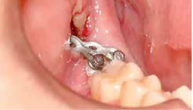
FIGURE 5 - Elastic chain for traction placed from the end of the miniplate to two orthodontic devices bonded to tooth 47.
FIGURE 6 - Tooth 47 in upright position after 3 months of treatment. FIGURE 3 - Fixation of miniplate and screws in retromolar
region/man-dibular ramus.
FIGURE 7 - Miniplate and screws surgically removed after 3-months treatment.
FIGURE 8 - Panoramic radiograph shows tooth 47 in correct position af-ter 3-months treatment.
discussion
According to Miyahira et al,10 mandibular
sec-ond molar impaction is statistically more frequent in the right side of male patients, which was con-firmed in the case reported in the present article.
The treatment for second molar impaction, no matter what technique is chosen, should be initiated immediately after the diagnosis because this abnor-mality may cause caries and periodontal problems, as well as root resorption of the adjacent tooth.
Different treatment options have been dis-cussed in the literature when orthodontic treat-ment is contraindicated, such as surgical repo-sitioning of the impacted tooth, an option that poses greater risks of complications, such as pulp necrosis, ankylosis and root resorption.12
Skeletal anchorage has been evaluated in numerous recent studies and discussions. It provides absolute anchorage, facilitates tooth movement, and is a valuable alternative to orth-odontic treatment. Mini-screws,2,3,6,11,13 tooth
implants3,11,13,17 and miniplates1,2,3,6,7,11,13 have
been used for that purpose.
Mini-screws used in orthodontic anchorage have the advantage of fewer adverse effects and lower operational costs than tooth implants,11
which, according to Miyahira et al,10 require a
lon-ger time for osseointegration, have a higher cost and are difficult to remove.
Mini-screws, however, are not free of compli-cations. Substantially high fracture rates, including fractures that result from their placement, have been reported.2
Choi et al1 investigated complications after the
placement of miniplates for orthodontic anchor-age and found high postoperative infection rates. Of the 69 miniplates used in mandibles and max-illa, five led to infection and had to be removed.
Other reasons may also explain miniplate failure, such as the surgical techniques used for insertion, the amount of force, the patient oral hygiene hab-its and the thickness of the cortical bone, which may contribute to the loss of implanted material.
Sugawara et al14 showed that skeletal
anchor-age using miniplates was successful for molar intrusion, distalization and protrusion, which are hardly achieved when using conventional mechanical techniques. Sugawara and Nishimu-ra15 used the same technique as in our case and
achieved success in about 85% of their cases, with plate loosening in only 1% of the cases.
In a recent study, miniplates had a success rate of 96.4% because they resisted reciprocal forces of several traction movements.6 Miyawaki et al11
found similar success rates when fixing miniplates with screws longer than 5.0 mm and with a di-ameter greater than 2.0 mm, a screw size that en-sured stability. Similar results have been reported by several authors, who found that miniplates were stable after fixation.7,10,13
conclusion
Miniplates, due to their high stability, may be used for the uprighting of impacted, partially im-pacted or mesially positioned molars.
In the case reported here, orthodontic treatment was successfully completed after 3 months, and the clinical result was excellent. Based on this experi-ence, we believe that the use of miniplates is a pre-cise, safe and simple method of skeletal anchorage.
references
contact address
Belini Freire-Maia
Avenida Contorno, 4747 conjunto 1011 – Serra CEP: 30.113-921 – Belo Horizonte/MG, Brazil E-mail: [email protected]
Submitted: September 19, 2008 Revised and accepted: November 24, 2008 1. Choi BH, Zhu SJ, Kim YH. A clinical evaluation of titanium
miniplates as anchors for orthodontic treatment. Am J Orthod Dentofacial Orthop. 2005:128(3):382-4.
2. Cornelis MA, Schefler NR, Nyssen-Behets C, De Clerck HJ,
Tulloch JF. Patients’ and orthodontists’ perceptions of miniplates used for temporary anchorage: a prospective study. Am J Orthod Dentofacial Orthop. 2008;133(1):18-24.
3. Eziroglu FV, Uckan S, Ozden UA, Arman A. Stability of zygomatic plate-screw orthodontic anchorage system. Angle Orthod. 2007;78(5):902-7.
4. Giancotti A, Arcuri C, Barlattani A. Treatment of ectopic mandibular second molar with titanium miniscrews. Am J Orthod Dentofacial Orthop. 2004;126(1):113-7.
5. Kim S, Herring S, Wang IC, Alcalde R, Mak V, Fu I, et al. A comparison of miniplates and teeth for orthodontic anchorage. Am J Orthod Dentofacial Orthop. 2008;133(2):189-97. 6. Kuroda S, Sugawara Y, Deguchi T, Kyung HM, Takano-Yamamoto
T. Clinical use of miniscrew implants as orthodontic anchorage: success rates and postoperative discomfort. Am J Orthod Dentofacial Orthop. 2007;131(1):9-15.
7. Leung MTC, Rabie ABM, Wong RWK. Stability of connected mini-implants and miniplates for skeletal anchorage in orthodontics. Eur J Orthod. 2008;30(5): 483-9.
8. McAboy CP, Grumet JT, Siegel EB, Iacopino AM. Surgical uprighting and repositioning of severely impacted mandibular second molars. J Am Dent Assoc. 2003;134(11):1459-62. 9. McNamara JA. Microimplants as temporary orthodontic
anchorage. Michigan: Ann Arbor; 2007.
10. Miyahira YI, Maltagliati LA, Siqueira DF, Romano R. Miniplates as skeletal anchorage for treating mandibular second molar impactions. Am J Orthod Dentofacial Orthop. 2008;134(1):145-8. 11. Miyawaki S, Koyama I, Inoue M, Mishima K, Sugahara T,
Takano-Yamamoto T. Factors associated with the stability of titanium screws placed in the posterior region for orthodontic anchorage. Am J Orthod Dentofacial Orthop. 2003;124(4):373-8. 12. Sawicka M, Racka-Pilszak B, Rosnowska-Mazurkiewicz A.
Uprighting partially impacted permanent second molars. Angle Orthod. 2007;77(1):148-54.
13. Sherwood KH, Burch JG, Thompson WJ. Closing anterior open bites by intruding molars with titanium miniplate anchorage. Am J Orthod Dentofacial Orthop. 2002;122(6):593-600.
14. Sugawara J, Baik UB, Umemori M, Takahashi I, Nagasaka H, Kawamura H, et al. Treatment and posttreatment dentoalveolar changes following intrusion of mandibular molars with application of a skeletal anchorage system (SAS) for open bite correction. Int J Adult Orthodon Orthognath Surg. 2002;17(4):243-53.
15. Sugawara J, Nishimura M. Minibone plates: the skeletal anchorage system. Semin Orthod. 2005;11(1):47-56. 16. Tseng YC, Chen CM, Chang HP. Use of a miniplate for skeletal
anchorage in the treatment of a severely impacted mandibular second molar. Br J Oral Maxillofac Surg. 2008;46(5):406-7. 17. Yanosky MR, Holmes JD. Mini-implant temporary anchorage