Pleura Laboratory, Pulmonary Division, Heart Institute (InCor), São Paulo University Medical School - São Paulo/SP, Brazil.
Email: [email protected]
Received for publication on May 26, 2006. Accepted for publication on October 23, 2006.
BASIC RESEARCH
EXPERIMENTAL PLEURODESIS INDUCED BY
ANTIBIOTICS (MACROLIDES OR QUINOLONES)
Lisete R. Teixeira, Francisco S. Vargas, Milena M. P. Acencio, Renan U. M. Bumlai, Leila Antonangelo, Evaldo Marchi
Teixeira LR, Vargas FS, Acencio MMP, Bumlai RUM, Antonangelo L, Marchi E. Experimental pleurodesis induced by antibiotics (macrolides or quinolones). Clinics. 2006;61(6):559-64.
PURPOSE: Chemical pleurodesis is a therapeutic tool for the treatment of recurrent pleural effusions, mainly those of neoplastic etiology. In the past, tetracycline was the sclerosant agent of choice in clinical practice, but presently, there is no consensus about an ideal agent. The aim of this study was to evaluate the effectiveness of macrolides (azithromycin and clarithromycin) or quinolones (levofloxacin and gatifloxacin) in inducing experimental pleurodesis in rabbits.
METHOD: Forty New Zealand rabbits randomized into groups of 10 received (at a total volume of 2 mL for each animal) 1 of the 4 drugs by intrapleural injection. After 28 days, the animals were euthanized and the pleural cavity was evaluated macroscopically and microscopically.
RESULTS: The intensity of the macroscopic adhesions was mild in all groups. On microscopic analysis, minimal pleural fibrosis and inflammation were observed in all animals.
CONCLUSION: The macrolides (azithromycin or clarithromycin) and the quinolones (levofloxacin or gatifloxacin) when injected into the normal pleural space of rabbits are not effective in promoting pleurodesis. Additional research is required to identify sclerosing agents capable of inducing pleurodesis.
KEYWORDS: Pleural effusion. Pleurodesis. Macrolides. Quinolones.
INTRODUCTION
Chemical pleurodesis is a method used to treat recur-ring pleural effusions, especially those with malignant etiology in which symphysis between the pleural layers is needed.1 Intrapleural instillation of a sclerosing agent that acts on the mesothelial layer leads to an inflammation that progresses to a fibrotic process and coalesces the parietal and visceral layers, obliterating the pleural space and pro-ducing pleurodesis.1
The choice of a sclerosing agent is still controversial. An ideal sclerosant would be low cost, easily available,
easily manipulated and sterilized; it would produce low morbidity (eg, pain should be easily controlled), and no mortality.2 To date, no sclerosant agent with all these char-acteristics has been identified.
The most frequently used sclerosants are antibiotics (doxycycline), antineoplastic agents (bleomycin), immunostimulants (C. parvum), and irritants (talc).2 The effectiveness of antibiotics varies between 60% and 80%, of antineoplastic agents between 60% and 70%, and for ir-ritants, it is generally greater than 90%.2,3 Although talc might be considered an ideal agent, several cases of acute respiratory distress syndrome have been reported in asso-ciation with talc pleurodesis.2,4,5
Macrolides have been used to induce pleurodesis. Eryth-romycin was used in an experimental model as a sclerosing agent, with inflammation and fibroblast proliferation be-ing evident after 8 days.9 In a clinical trial evaluating 15 patients with malignant pleural effusion, erythromycin was effective inducing pleurodesis in 84% of cases.10
Quinolones are widely used broad-spectrum antibiotics. Nevertheless, as far as we know, there have been no clini-cal or experimental studies evaluating pleural sclerosis pro-duction with this drug.
This study aimed to evaluate, using an experimental model in rabbits, the effectiveness of intrapleural instilla-tion of macrolides (azithromycin or clarithromycin) or quinolones (levofloxacin or gatifloxacin) in inducing pleurodesis.
METHODS
This study was carried out at the Pleura Laboratory and the Research Division of the Heart Institute, Hospital das Clínicas, São Paulo University Medical School [HC-FMUSP], having been previously approved by the Institu-tional Ethics Committee.
Forty New Zealand rabbits weighing 2.0 to 3.0 kg, randomized into 4 groups (with 10 animals each), received (in a total volume of 2 mL) by intrapleural injection, 15 mg/kg of azithromycin or clarithromycin or 10 mg/kg of levofloxacin or gatifloxacin.
The surgical procedure followed a previously described methodology.8,11,12 After anesthesia by intramuscular injection (35 mg/kg of ketamine HCl and 5 mg/kg of xylazine hydro-chloride), the animals were prepared for aseptic surgery. Af-ter an incision (0.5 cm) in the right hemithorax, the parietal pleura was exposed and the sclerosing agent was injected into the pleural space through a drain (6F); the surgical wound was closed by sutures. The drain, aspirated daily, was removed after 48 hours if the daily fluid outflow was less than 1 mL/day. The left hemithorax served as a control and did not receive intrapleural instillation of any substance or drain.
According to the protocol, animals exhibiting apparent distress were immediately euthanized.
After 28 days, the surviving animals were euthanized by an IV injection of 40 mg/kg of pentobarbital into the marginal ear vein. The thorax was removed en bloc, and the lungs were expanded with 60 mL of 10% formalin and immerged in this solution for at least 48 h. After this pe-riod, each pleural cavity was exposed using the method pre-viously described by our group.13-16 Then, with the thorax opened, we evaluated the presence of macroscopic pleural adhesions and collected fragments from the pleura and pul-monary parenchyma for later microscopic analysis.
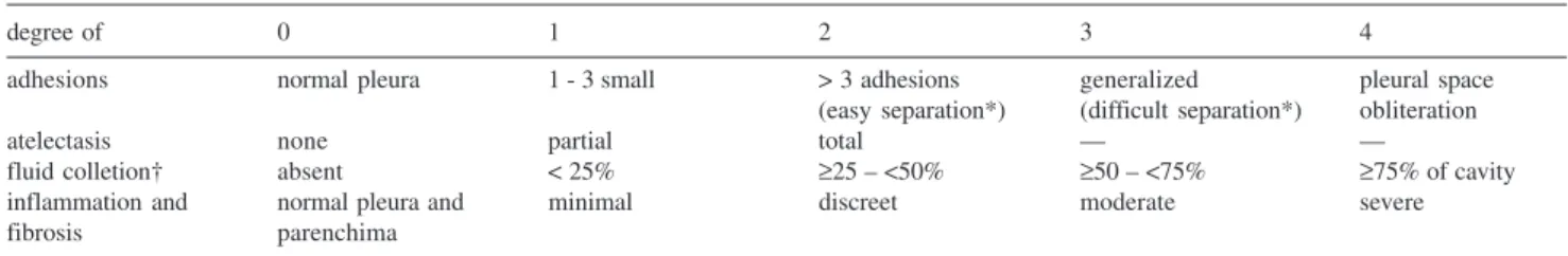
Table 1 summarizes scoring scheme for adhesion, atel-ectasis, and fluid collection. Such scores were established in order to macroscopically quantify the degree of pleural adhesion (0-4), atelectasis (0-2), and fluid collection (0-4). Pleurodesis was considered effective when the degree of pleural adhesions was equal to or greater than a score of 311-16 and no presence of atelectasis or fluid collection. Ta-ble 1 also shows the scores used to quantify collected frag-ments of pleura and parenchyma; these were processed and stained with hematoxylin and eosin. Similarly to the mac-roscopic assessment, we based our micmac-roscopic analysis on previously validated scores,13-16 quantifying the degree of inflammation and fibrosis (0 to 4).
The descriptive statistics developed were mean and standard deviation. Group means were tested for signifi-cant differences using the t test to compare the 2 groups of antibiotics (macrolides vs quinolones) and ANOVA fol-lowed by multiple comparison tests (Tukey or Dunn) to compare the groups among themselves. Statistical signifi-cance was considered when the difference was less than or equal to 0.05.
RESULTS
Only 1 death was observed, and it occurred on the sec-ond postoperative day. Necropsy revealed no significant al-terations in the pleural space that could have contributed to the lethal outcome (this case was excluded from the sta-tistical analysis). The side effects of the drugs injected were acute episodes of diarrhea 3 hours after the intrapleural in-jection in more than 75% of the animals that received
Table 1 - Scoring scheme for the macroscopic and microscopic evaluations of the pleura
degree of 0 1 2 3 4
adhesions normal pleura 1 - 3 small > 3 adhesions generalized pleural space
(easy separation*) (difficult separation*) obliteration
atelectasis none partial total — —
fluid colletion† absent < 25% ≥25 – <50% ≥50 – <75% ≥75% of cavity
inflammation and normal pleura and minimal discreet moderate severe
fibrosis parenchima
azithromycin or clarithromycin. After 24 hours, the animals returned to normalcy, and no drug intervention was neces-sary. These episodes of diarrhea were not observed with the intrapleural use of quinolones.
No animal presented pleural fluid during the 48 hours after injection of the sclerosing agent (the period in which the drain remained in the animals), and this made sampling for further biochemical or cytological fluid analysis impos-sible.
Macroscopic Analysis
The intensity of the adhesions (Table 2) was discreet in the groups that received macrolides (1.25 ± 0.75) and in those receiving quinolones (0.64 ± 0.63), and no sig-nificant difference (P = 0.057) was observed between them. The scores attributed to the adhesions in the animals that received azithromycin (1.25 ± 0.50), clarithromycin (1.25 ± 1.04), levofloxacin (1.00 ± 0.53), or gatifloxacin (0.16 ± 0.41) were significantly different (P = 0.033), although significant differences were not found from the multiple comparisons test.
In all animals, the left hemithorax, used as a control because it had not been injected with the agent, received a score of 0.
Microscopic Analysis
The microscopic changes observed in the visceral pleura resulting from the intrapleural injection of the agents in-vestigated were minimal on the right and absent (score = 0) on the left. These results (Table 3) were both observed in the group dosed with macrolides (fibrosis: 0.94 ± 0.73, inflammation: 0.37 ± 0.52) and in the group dosed with quinolones (fibrosis: 0.43 + 0.51, inflammation: 0.36 ± 0.50), with no significant differences (fibrosis: P = 0.108, inflammation: P = 0.973).
The pleural fibrosis scores resulting from injections of azithromycin (0.37 ± 0.48), clarithromycin (1.50 ± 0.41), levofloxacin (0.62 ± 0.52), or gatifloxacin (0.17 ± 0.41)
were significantly different one from the other (P = 0.002). Tukey’s test showed that this difference was a result of the comparison between clarithromycin and the other drugs.
Regarding pleural inflammation, there was no signifi-cant difference among the groups (P = 0.553). The scores attributed were 0.25 ± 0.50; 0.50 ± 0.58; 0.50 ± 0.54, and 0.17 ± 0.41 for azithromycin, clarithromycin, levofloxacin, and gatifloxacin, respectively.
The pulmonary parenchyma was not affected by the in-trapleural injection of these agents (score = 0).
DISCUSSION
This study demonstrates that the intrapleural injection of azithromycin, clarithromycin, levofloxacin, or gati-floxacin is not effective in inducing pleurodesis in an ex-perimental rabbit model. The degree of pleural adhesion after the intrapleural injection of these drugs was notably inferior to that observed in a prior study after the pleural instillation of talc (2.2 ± 0.7) or silver nitrate (3.2 ± 1.1).12 The choice of an ideal pleural sclerosing agent is still object of research and is based on efficacy, morbidity-mor-tality, ease in manipulation, availability, and cost. It is note worthy that although the century have elapsed since pleurodesis was first performed (1901)2,7, the ideal agent only have far been identified. The nonexistence of a sub-stance that satisfies all the basic prerequisites and clinical needs explains the ongoing research, including the revival of studies on agents that had been used in the past.17,18
The interest in antibiotics as pleural sclerosing agents dates back to the early phases of studies on pleurodesis. The first antibiotic used in clinical practice was tetracy-cline, which was used until the 1980s when production of the injectable form was discontinued. Tetracycline was im-mediately replaced, although without the same impact, by its natural derivatives, doxycycline, and minocycline.2,3 Tetracycline, employed as a pleural sclerosing agent in hu-mans for more than 20 years, was effective in inducing pleurodesis in approximately 70% of cases, surpassing the currently achieved results obtained with doxycycline and minocycline that experimentally produce pleurodesis in about 80% of injected animals, but can result in hemothorax and death.8
In addition to tetracycline and its derivatives, macrolides, represented by erythromycin, were evaluated for sclerosing effects both in experimental models and in humans. In animals, after a period of 8 days, the intrap-leural instillation of erythromycin produces marked inflam-mation, allowing recognition of its irritant action on pleu-ral layers and suggesting a potential sclerosing effect.9 This effect was later confirmed in humans (26 patients) in whom Table 2 - Scores of pleural adhesions observed
macros-copically in the injected hemithorax
ANTIBIOTICS PLEURAL ADHESIONS p* p§
Macrolides 1.25 ± 0.75 0.057
Quinolones 0.64 ± 0.63
Azithromycin 1.25 ± 0.50 0.033
Clarithromycin 1.25 ± 1.04 Levofloxacin 1.00 ± 0.53 Gatifloxacin 0.17 ± 0.41
effectiveness was observed in 85% of patients with neoplastic pleural effusion, with a complete response in 15 (58%). In these 85% (22) of patients, the formation of nu-merous pleural adhesions and control of the pleural effu-sion was noted.10
It is important to note that antibiotics, provided that they are inexpensive, effective, and cause few side effects, could be considered ideal agents due to their wide availability. The established sclerosing effectiveness of tetracycline and erythromycin (macrolides), justifies further investigation with other antibiotics.
The mechanisms responsible for producing pleurodesis are not completely clear. Apparently, the instillation of a sclerosing agent causes damage to the mesothelial layer with the subsequent formation of an inflammatory pro-cess, characterized by the production of exudative fluid.1,2
In spite of prior reports9,10 describing the effectiveness of erythromycin, other macrolides such as azithromycin and clarithromycin were not capable of producing a pleural in-flammatory process intense enough to induce the forma-tion of pleurodesis. No exudaforma-tion, evidenced by the pres-ence of pleural effusion, was seen during the first 48 hours of observation. This absence of pleural fluid correlates with the data noted both in the macroscopic and microscopic evaluations.
The nonexistence of fluid suggests a mild effect of these drugs on the pleura. The pleural fluid, characteris-tically an exudate, expresses acute inflammatory altera-tions that are the beginning of a succession of events lead-ing to the development of pleural adhesions and the re-sulting pleurodesis formation. Factors capable of interfer-ing in this progress include the intensity of the injury, the capacity of the mesothelial cells and fibroblasts to secrete collagen, the relative equilibrium between the metalo-proteinases and their inhibitors, and the balance between the activators and inhibitors of plasminogen, in addition to the relationship between the inflammation and the pro-inflammatory cytokines with fibrinolysis mecha-nisms.19,20
Earlier studies9,10 showing the sclerosing action of eryth-romycin led to the hypothesis that other macrolides (azithromycin or clarithromycin) might also be capable of promoting mesothelial layer damage sufficient to produce pleural effusion and subsequent pleural adhesions. This was not observed in this study, which might be explained by pharmacokinetic characteristics. In theory, these antibiot-ics would be rapidly absorbed and might not remain in the pleural cavity long enough to cause mesothelial damage and initiate the necessary inflammatory process. The ob-servation of diarrhea 3 hours after intrapleural dosing of the macrolides suggests precocious systemic manifestations resulting from the rapid absorption and distribution of these antibiotics.
With the objective of finding sclerosing agents with an effect similar to that of tetracycline, and considering the current ample use and wide distribution of quinolones, we chose to evaluate the possible effects of inducing pleurodesis with levofloxacin and gatifloxacin, even though there are no past reports (as far as we know) on the use of these antibiotics.
The pleural adhesions observed after instillation of these agents were not encouraging: there was no acute evidence of pleural fluid formation, and the degree of inflammation noted at microscopic assessment after 28 days was irre-levant, because the degree of pleural adhesion resulting from the experimental treatment was not considered ad-equate. Therefore, the quinolones analyzed (levofloxacin and gatifloxacin) produced minimal damage to the mes-othelium. Thus, the succession of events that might cul-minate in macroscopically visible pleural adhesions , and microscopically recognizable fibrosis was not established. The statistical difference observed among the 4 groups analyzed in the macroscopic evaluation of adhesions and the microscopic evaluation of pleural fibrosis was not sub-stantial and has no concrete significance since those scores are irrelevant as noted above.
Although the sclerosing effect of erythromycin had pre-viously been demonstrated in clinical and experimental
Table 3 - Scores for pleural fibrosis and inflammation observed microscopically in the injected hemithorax
ANTIBIOTICS FIBROSIS INFLAMMATION
X ± SD p* pP§ X + SD p* p§
Macrolides 0.94 ± 0.73 0.108 0.37 ± 0.52 0.973
Quinolones 0.43 ± 0.51 0.36 ± 0.50
Azithromycin 0.37 ± 0.48 0.002 0.25 ± 0.50 0.553
Clarithromycin 1.50 ± 0.41 0.50 ± 0.58
Levofloxacin 0.62 ± 0.52 0.50 ± 0.54
Gatifloxacin 0.17 ± 0.41 0.17 ± 0.41
studies,9,10 no significant pleural adhesion was noted in the present experiments, after the intrapleural injections of azithromycin or clarithromycin, also belonging to the mac-rolide class. Similarly, the studied quinolones (levofloxacin and gatifloxacin) showed no pleural sclerosing effect in our experimental model and therefore should not be considered for use in humans.
These negative results are deliberately collected and offered as a finished study because we firmly believe that
they add important data to the field, indicating that addi-tional drugs must be tested with the purpose of identify-ing the ideal pleural sclerosant.
ACKNOWLEDGEMENTS
We thank Carlos S. R. Silva, laboratory technician, and Gabriela G. Carnevale, pharmacist, for their help during surgical procedures and care for the animals.
RESUMO
Teixeira LR, Vargas FS, Acencio MMP, Bumlai RUM, Antonangelo L, Marchi E. Pleurodese experimental induzida por antibióticos (macrolídeos e quinolonas). Clin-ics. 2006;61(6):559-64.
OBJETIVO: A pleurodese química representa uma ferramenta terapêutica utilizada no tratamento dos processos pleurais recidivantes, principalmente nos derrames neoplásicos. A escolha do melhor esclerosante pleural é ainda motivo de controvérsia, não havendo consenso com relação ao agente considerado ideal. O objetivo deste estudo é avaliar a efetividade dos macrolídeos (azitromicina e claritromicina) e das quinolonas (levofloxacina e gatifloxacina) na indução de pleurodese experimental em coelhos.
MÉTODOS: Quarenta animais randomizados em grupos de 10, receberam, em volume total de 2 mL, estas drogas
através de injeção intrapleural.
RESULTADOS: Após 28 dias, os animais foram sacrificados sendo avaliada a cavidade pleural. A intensidade das aderências macroscópicas assim como da fibrose e da inflamação observadas à microscopia foi discreta tanto no grupo que recebeu macrolídeos quanto naquele que recebeu quinolonas.
CONCLUSÃO: Azitromicina, Claritromicina, Levofloxacina e Gatifloxacina quando injetados na cavidade pleural de coelhos, não são eficazes na indução de pleurodese. Novas pesquisas devem ser realizadas com o intuito de identificar agentes esclerosantes capazes de produzir sínfise pleural.
UNITERMOS: Pleurodese. Macrolídeos. Quinolonas. Azitromicina. Claritromicina. Levofloxacina. Gatifloxacina.
REFERENCES
1. Light RW, Vargas FS. Pleural sclerosis for the treatment of pneumothorax and pleural effusion. Lung. 1997;175:213-23.
2. Pleurodesis, Vargas FS, Teixeira LR in Pleural Effusion, Vargas FS, Teixeira LR, Marchi E. 1st Ed – Ed Roca, S Paulo - SP, Brazil, 2004. 3. Walker-Renard PB, Vaughan LM, Sahn SA. Chemical pleurodesis for
malignant pleural effusions. Ann Intern Med. 1994;120:56-64. 4. Rehse DH, Aye RW, Florence MG. Respiratory failure following talc
pleurodesis. Am J Surg. 1999;177:437-40.
5. Brant A, Eaton T. Serious complications with talc slurry pleurodesis. Respirology. 2001;6:181-85.
6. Light RW, Wang NS, Sassoon CS, Gruer SE, Vargas FS. Comparison of the effectiveness of tetracycline and minocycline as pleural sclerosing agents in rabbits. Chest. 1994;106:577-82.
7. Marchi E, Teixeira LR, Vargas FS. Management of malignancy-associated pleural effusion: current and future treatment strategies. Am J Respir Med. 2003;2:261-73.
8. Wu W, Teixeira LR, Light RW. Doxycycline pleurodesis in rabbits. Comparison of results with and without chest tube. Chest. 1998;114:563-68.
9. Carvalho P, Knight LL, Olson RD, Crowley JJ, Hawk PA, Charan NB. Effects of erythromycin on the rabbit pleura: its potential role as a pleural sclerosant. Am J Respir Crit Care Med. 1995;151:1228-32.
10. Tang X, Cheng D. The value of erythromycin pleurodesis in the treatment of malignant pleural effusions. Hua Xi Yi Ke Da Xue Xue Bao. 1997;28:437-39.
11. Teixeira LR, Wu W, Cheng DS, Light RW. The effect of corticosteroids on pleurodesis induced by doxycycline in rabbits. Chest. 2002;121:216-19.
13. Teixeira LR, Vargas FS, Antonangelo L, Mattos VC, Vaz MAC, Marchi E. Low concentration silver nitrate pleurodesis in rabbits: optimal concentration for rapid and complete sclerosing effect. Lung. 2003;181:353-59.
14. Vargas FS, Carmo AO, Marchi E, Vaz MAC, Ramos KP, Teixeira LR. Effectiveness of silver nitrate compared to talc slurry as pleural sclerosing agent in rabbits. Influence of concomitant intrapleural lidocaine. Rev. Hosp Clin Fac Med S Paulo. 1999;54:199-208. 15. Vargas FS, Antonangelo L, Capelozzi V, Vaz MAC, Genofre EH, Marchi
E , Teixeira LR. Lung damage in experimental pleurodesis induced by silver nitrate or talc. 1-year follow-up. Chest. 2002;122:2122-26. 16. Vargas FS, Teixeira LR, Antonangelo L, Vaz MAC, Carmo AO, Marchi
E, Light RW. Experimental pleurodesis in rabbits induced by silver nitrate or talc. 1-year follow-up. Chest. 2001;119:516-20.
17. Vargas FS, Carmo AO, Teixeira LR. A new look at old agents for pleurodesis: nitrogen mustard, sodium hydroxide, and silver nitrate. Curr Opin Pulm Med. 2000;6:281-86.
18. Dikensoy O, Light RW. Alternative widely available, inexpensive agents for pleurodesis. Curr Opin Pulm Med. 2005;11:340-44.
19. Lee YC, Lane KB. The many faces of transforming growth factor-beta in pleural diseases. Curr Opin Pulm Med. 2001;7:173-79.