284 Radiol Bras. 2013 Set/Out;46(5):284–289
Suggestion of a national diagnostic reference level for
18
F-FDG/PET scans in adult cancer patients in Brazil
*
Sugestão de nível de referência em diagnóstico nacional para 18F-FDG/PET em procedimentos oncológicos adultos no Brasil
Cássio Miri Oliveira1, Lidia Vasconcellos de Sá2, Thêssa Cristina Alonso3, Teógenes Augusto da Silva4
Objective: To suggest a national value for the diagnostic reference level (DRL) in terms of activity in MBq.kg–1
, for nuclear medicine procedures with fluorodeoxyglucose (18
F-FDG) in whole body positron emission tomography (PET) scans of adult patients. Materials and Methods: A survey on values of 18
F-FDG activity administered in Brazilian clinics was undertaken by means of a questionnaire including questions about number and manufacturer of the installed equipment, model and detector type. The suggested DRL value was based on the calculation of the third quartile of the activity values distribution reported by the clinics. Results: Among the surveyed Brazilian clinics, 58% responded completely or partially the questionnaire; and the results demonstrated variation of up to 100% in the reported radiopharmaceutical activity. The suggested DRL for 18
F-FDG/PET activity was 5.54 MBq.kg–1
(0.149 mCi.kg–1
). Conclusion: The present study has demonstrated the lack of standardization in administered radiopharmaceutical activities for PET procedures in Brazil, corroborating the necessity of an official DRL value to be adopted in the country. The suggested DLR value demonstrates that there is room for optimization of the procedures and 18
F-FDG/PET activities administered in Brazilian clinics to reduce the doses delivered to patients. It is important to highlight that this value should be continually revised and optimized at least every five years. Keywords: Nuclear medicine; Positron emission tomography; Diagnostic reference level; Radiopharmaceutical 18
F-FDG.
Objetivo: Sugerir um valor nacional de “nível de referência em diagnóstico” (NRD), em termos de atividade em MBq.kg–1
, para procedimentos de medicina nuclear com fluordeoxiglicose (18
F-FDG) em exames de corpo inteiro de pacientes adultos, por meio da tomografia por emissão de pósitron (PET). Materiais e Métodos: Um levantamento dos valores das ativi-dades do 18
F-FDG administradas nas clínicas brasileiras foi realizado por meio de um questionário, que incluiu informa-ções sobre o número de equipamentos instalados, fabricante, modelo e detector utilizados. O valor sugerido para o NRD foi baseado no cálculo do terceiro quartil da distribuição das atividades reportadas pelas clínicas. Resultados: Das clínicas consultadas, 58% responderam totalmente ou parcialmente ao questionário; os resultados demonstraram variações de até 100% nas atividades do radiofármaco administradas. O NRD para a atividade do 18
F-FDG/PET sugerido foi de 5,54 MBq.kg–1
(0,149 mCi.kg–1
). Conclusão: O estudo demonstrou falta de padronização das atividades administradas nas clínicas brasileiras, justificando a necessidade de oficializar um valor de NRD a ser adotado no País. O valor do NRD sugerido neste trabalho demonstrou que há espaço para a otimização dos procedimentos e das atividades administra-das nas clínicas brasileiras, com a intenção em reduzir a dose aos pacientes. Ressalta-se que tal valor deve ser conti-nuamente revisto e otimizado em períodos de pelo menos cinco anos.
Unitermos: Medicina nuclear; Tomografia por emissão de pósitron; Nível de referência em diagnóstico; Radiofármaco
18
F-FDG. Abstract
Resumo
* Study developed at Centro de Desenvolvimento da Tec-nologia Nuclear – Comissão Nacional de Energia Nuclear (CDTN/ CNEN) and at Escola de Engenharia – Departamento de Enge-nharia Nuclear da Universidade Federal de Minas Gerais (UFMG), Belo Horizonte, MG, Brazil.
1. PhD, Collaborator of Centro de Desenvolvimento da Tec-nologia Nuclear – Comissão Nacional de Energia Nuclear (CDTN/ CNEN), Post-Graduation student in Sciences and Nuclear Tech-niques, Universidade Federal de Minas Gerais (UFMG), Belo Horizonte, MG, Brazil.
2. PhD, Technologist, Comissão Nacional de Energia Nuclear (CNEN), Professor of Post-Graduation at Instituto de Radiopro-teção e Dosimetria (IRD/CNEN), Rio de Janeiro, RJ, Brazil.
3. Fellow PhD degree, Technologist, Comissão Nacional de
Oliveira CM, Sá LV, Alonso TC, Silva TA. Suggestion of a national diagnostic reference level for 18F-FDG/PET scans in adult cancer pa-tients in Brazil. Radiol Bras. 2013 Set/Out;46(5):284–289.
INTRODUCTION
Medical radiation is currently the most relevant source of radiation exposure of ar-tificial origin to humans, and nuclear medi-cine is responsible for 1% of the annual collective dose as the world population is considered(1). For this reason, it is essential
that radiological protection methods be continuously optimized in order to ensure Energia Nuclear (CNEN), Head of Serviço das Radiações
Aplica-das à Saúde (SERAS) do Centro de Desenvolvimento da Tecno-logia Nuclear – Comissão Nacional de Energia Nuclear (CDTN/ CNEN), Belo Horizonte, MG, Brasil.
4. PhD, Researcher and Professor, Centro de Desenvolvi-mento da Tecnologia Nuclear – Comissão Nacional de Energia Nuclear (CDTN/CNEN), Professor of Post-Graduation in Sciences and Nuclear Techniques, Universidade Federal de Minas Gerais (UFMG), Belo Horizonte, MG, Brazil.
Mailing Address: Dr. Cássio Miri Oliveira. Avenida Antônio Carlos, 6627, Campus UFMG, Pampulha. Belo Horizonte, MG, Brazil, 31270-901. E-mail: [email protected].
protection to patients. The radiological pro-tection to patients is based on the funda-mental principles of radioprotection –
jus-tification and optimization – as the
prin-ciple of individual dose limitation is not applicable to medical exposures, consider-ing that the diagnostic value of the image shall not be restricted by a dose limit. How-ever, there are values named “diagnostic reference levels” (DRLs), which serve as a reference to identify atypical operations with the purpose of promoting the optimi-zation of radiodiagnosis procedures and the protection of patients by means of doses reduction.
The concept of DRLs was introduced by the International Commission on Radio-logical Protection (ICRP) in its 73rd is-sue(2). It is important to highlight that the DRLs do not constitute a boundary be-tween good and bad diagnostic procedures. However, such levels must be reviewed and investigated as they are systematically ex-ceeded in standard procedures(2). Thus, DRLs should be established for each coun-try or region and should be jointly imple-mented by governments, national regula-tory authorities and professional associa-tions(3).
In the conventional diagnostic radiol-ogy, DRLs must be based upon dose val-ues measured at several hospitals and clin-ics, either well equipped or not, and deter-mined by the calculation of the third quartile of the evaluated doses distribu-tion(4). In nuclear medicine, DRLs are
sug-gested and based on the administered ac-tivity necessary to obtain a good image quality required for a given procedure. Thus, an “optimum” value must be utilized for a DRL, instead of a percentile: the ref-erence level for administration of radionu-clide activities sufficient to obtain data for specific patient groups. However, the ICRP in its 103rd issue(3), asserts that, in practice,
the DRLs in nuclear medicine can be de-termined by means of the calculation of the third quartile of the distribution of activi-ties administered to patients. Such condi-tion is based on the assumpcondi-tion that the activities administered in the clinics pro-duce studies with satisfactory image qual-ity. Therefore, it is important to highlight that the DRLs in nuclear medicine do not constitute reference levels which should
not be exceeded, but rather a guidance level for administered activities(4).
Currently, there is an increase in the number of positron emission tomography (PET) apparatuses to meet the increasing clinical demand for exams with fluoro-deoxyglucose (18F-FDG) in Brazil. A simi-lar increase was also observed in developed countries, such in United States of America, where 18F-FDG is utilized in more than 1.5 million exams per year(5), and is
one of the most widely produced radiophar-maceuticals in the world(6). However, the
PET technique is relatively new in Brazil, where the first dedicated PET apparatus was installed in the early 2000s(7) and, for that reason, specific Brazilian standards related to quality control procedures and medical exam protocols are yet to be estab-lished , such as the case of recommended “doses” or activities to be administered to patients.
The Agência Nacional de Vigilância Sanitária (Anvisa), through Resolution RDC No. 38 of 2008, establishes rules for “Installation and Operation of In Vivo Nuclear Medicine Services”(8), but it only provides some routine tests and their fre-quencies. Therefore, the PET clinics cur-rently follow the equipment manufacturer’s recommendations or different international recommendations which suggest, for ex-ample, the activity to be administered to the patient. Thus, this situation can lead to variations in administered activities and protocols of the exams performed in the Brazilian 18F-FDG/PET clinics. In the Eu-ropean Community (EC), the administered activities present a high variability from country to country(1), as there are no
recom-mended DRLs in nuclear medicine within the EC (4). The nationwide recommendation
on the activity to be administered to pa-tients is an important parameter to be con-sidered, as it directly interferes in the qual-ity of the exams and, mainly, in the well-being of the patient who might otherwise be exposed to unjustifiable ionizing radia-tion. Therefore, the establishment of a prac-tical method for surveying, estimating and defining DRLs is of paramount importance. The present study is aimed at surveying the activities administered to adult cancer patient in order to estimate and suggest the first national DRL for 18F-FDG/PET
proce-dures in Brazil. Additionally, the estimation of DRL is aimed at providing a numeric value to serve as a parameter for standard-ization and resulting optimstandard-ization of ad-ministered activities in Brazilian clinics. It must be highlight the fact that the DRLs must be continuously reviewed in order to assure the quality of the procedures, ac-cording to the development of the tech-nique whose utilization is expanding in Brazil.
MATERIALS AND METHODS
Some European countries have differ-ent methods and guidelines for the estab-lishment of DRLs. In Greece, for example, the establishment and application of DRLs are based on data collection during inspec-tions carried out in the nuclear medicine services, as a part of a licensing program accomplished every two years. The DRLs, both in conventional radiology (dose) and in nuclear medicine (activity), are based on the calculation of the third quartile of the distribution of collected administered activities, which are updated every 5 years(9,10).
In Germany, the DRLs are also based on nationwide surveys, with a revaluation pe-riod of two to three years(10,11). However,
Italy has established its values on the basis of literature reviews, in particular on the European Community Directives. For all procedures with existing DRLs in Italy, the hospitals or clinics are responsible for per-forming the survey of doses or activities and compare the found values with the national reference levels. The DRLs were standardized in the Decree-Law No. 187 dated May 26th, 2000, which implemented the European Directive 97/43/Euratom in Italian regulation. According to that decree, every nuclear medicine or radiology depart-ment must impledepart-ment a quality control pro-gram aimed at optimizing the procedures. Additionally, the doses delivered to patients at each procedure must undergo evaluation every two years, in order to verify the com-pliance with the DRLs(10).
In Belgium, a questionnaire was devel-oped for the survey of the administered ac-tivities in the different nuclear medicine centers. The 25 most frequently performed exams were selected for the establishment of the DRLs(13).
Swedish hospitals and clinics, likewise in Italy, have the responsibility of evaluat-ing the administered activities and compar-ing the values with the respective DRLs. The survey of the administered activities in Sweden is carried out on an annual basis. However, the determination of adminis-tered doses and activities is mandatory and must be established every two years(10). In Switzerland, the DRLs are also suggested on the basis of nationwide surveys on ad-ministered activities, with provisions for their updating every five or ten years(10).
Other countries, such as Ireland, are yet to publish data on the DRLs studied in the last years. However, for convenience, the DRLs from the United Kingdom are adopted as parameters for the evaluation of radiodiagnosis procedures(14).
As regards to DRLs to procedures in-volving 18F-FDG/PET in adult cancer pa-tients, there are only a few countries which have already published their data or estab-lished their DRLs. Among such countries are Germany(11), Australia and New
Zealand(15), Finland(16), France(12), United
Kingdom(17), Sweden and Switzerland(10).
The Table 1 presents the DRLs established in each of those countries.
For better understanding of the current 18F-FDG/PET scenario in Brazil, a
ques-tionnaire was developed with simple and direct questions related to the evaluation of number of apparatuses and type of PET detectors, number of procedures per-formed, adopted recommendations for
ac-Table 1 DRLs for 18F-FDG/PET scans in adult cancer patients.
DRLs 18F-FDG
Country
Germany (2003) Australia and New Zealand (2009)
Finland (2009) France (2012) United Kingdom (2006)
Switzerland (2007) Sweden (2006)
MBq
370 (2D) 200 (3D) 385 370 350 400 – tumor and heart
350 350
MBq.kg–1
2.86 (3D) 5.5 5.3 5.0 5.7 5.0 5.0
mCi.kg–1
0.08 (3D) 0.148 0.143 0.135 0.154 0.135 0.135 tivity administration and, mainly, the ad-ministered activities in different clinics.
After obtaining the data, the DRL for oncologic exams with 18F-FDG/PET in adults was estimated taking into consider-ation the calculconsider-ation of the third quartile, as done in some European countries. Such a manner to obtain the DRL is conservative, but in the absence of an established DRL for 18F-FDG/PET in Brazil, that is an appro-priate way to obtain and recommend an initial value.
Once the DRL value is suggested, it is necessary to make it known by the inter-ested public, besides establishing the fre-quency for its review. The continuous evaluation of the DRL is extremely impor-tant for the complementation of a quality assurance program in the services.
The frequency in review of DRLs was based upon the developments in appara-tuses and related techniques in Brazil, as new methods, detector materials and pro-cedures are constantly being introduced, thus inducing the implementation of a con-tinuous optimization process.
RESULTS
The results from the survey carried out by means of the questionnaire are presented below. The investigation started in August 2011 and was completed in August of 2012. Out of the 72 18F-FDG/PET clinics cur-rently registered at Comissão Nacional de Energia Nuclear (CNEN), 42 totally or par-tially responded to the questionnaire (58% of the clinics). In only one of the clinics, the PET equipment was undergoing mainte-nance. Two of the 42 clinics participating in the survey have two PET apparatuses each, thus the respondent clinics comprise
44 PET apparatuses, one of them, a hybrid gamma camera with a coincidence detec-tion system. Out of the 42 respondent clin-ics, 41 reported the activities administered in the 18F-FDG/PET oncology procedures. As regards geographical distribution of the respondent clinics, 19 are located in the Southeastern region, 10 clinics in the Northeastern region, 5 in the Southern re-gion, six in the Mid-western rere-gion, and two clinics in the Northern region.
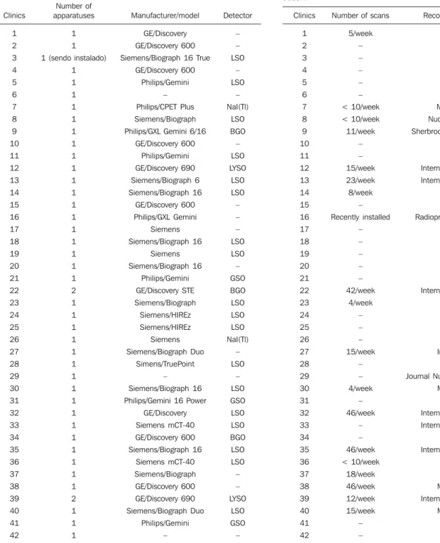
It is important to observe that the data were supplied by the workers and/or heads of the 18F-FDG/PET clinics, and no change or correction was made on the question-naires answers. Table 2 presents the results related to the number of PET and/or PET/ CT apparatuses with their respective manu-facturers/models and detector types.
On Table 2, one observes that 14 clin-ics did not provide the manufacturer/model of their equipment and/or could not de-scribe the type of utilized detectors. One observed that the most PET apparatuses utilized in Brazil are manufactured by Si-emens, with 20 registered. General Electric (GE) manufactured 11 apparatuses, fol-lowed by Philips with 8 apparatuses. As regards detectors, 18 of the 28 clinics which provided such information utilize lutetium orthosilicate (LSO) crystals, and in all such cases the apparatuses were made by Siemens, except for one, made by GE. Only one of the Siemens apparatuses uti-lized sodium iodide NaI(Tl) crystals, which consists of a single photon emission com-puted tomography (SPECT) hybrid gamma camera. Besides that equipment, another apparatus manufactured by Philips also uti-lizes the NaI(Tl) crystals. Three of the clin-ics utilize bismuth germanate (BGO) crys-tals. The remaining three apparatuses uti-lize gadolinium orthosilicate (GSO) detec-tors patented by Philips.
It is important to highlight that the ap-paratuses equipped with BGO detectors were some of the first acquired by Brazil in the early 2000’s. In 2011, at least two ap-paratuses with the time-of-flight (TOF) technology with lutetium yttrium ortho-silicate (LYSO) crystals were acquired.
As regards number of scans, some of the clinics provided an approximate interval for number of performed procedures; for example, clinics 32, 35 and 38 presented the interval of 41 up to 50 scans per week. Thus, in order to facilitate the interpretation and description, the mean values of such intervals were calculated. It is important to highlight that according to the obtained
data, the total number of scans per week in the 17 clinics was 337. In a simplified es-timation, at the end of a month such num-ber of scans would reach 1,350. In one year, such number would reach 16,000 18F-FDG/ PET procedures. Considering the other 55 clinics which did not provide data on this particular question or which did not re-spond to the questionnaire, and by
assum-ing that, proportionally, such clinics per-formed approximately the same number of procedures as the others which provided the data, the total number of 18F-FDG/PET scans performed in one year in Brazil would reach 68,000.
The recommendations followed for ad-ministration of “doses” were provided by 16 clinics, of which nine informed that they Table 2 Number of PET apparatuses, manufacturer/model and detectors.
Clinics 1 2 3 4 5 6 7 8 9 10 11 12 13 14 15 16 17 18 19 20 21 22 23 24 25 26 27 28 29 30 31 32 33 34 35 36 37 38 39 40 41 42 Number of apparatuses 1 1 1 (sendo instalado)
1 1 1 1 1 1 1 1 1 1 1 1 1 1 1 1 1 1 2 1 1 1 1 1 1 1 1 1 1 1 1 1 1 1 1 2 1 1 1 Manufacturer/model GE/Discovery GE/Discovery 600 Siemens/Biograph 16 True
GE/Discovery 600 Philips/Gemini
– Philips/CPET Plus Siemens/Biograph Philips/GXL Gemini 6/16
GE/Discovery 600 Philips/Gemini GE/Discovery 690 Siemens/Biograph 6 Siemens/Biograph 16 GE/Discovery 600 Philips/GXL Gemini Siemens Siemens/Biograph 16 Siemens Siemens/Biograph 16 Philips/Gemini GE/Discovery STE Siemens/Biograph Siemens/HIREz Siemens/HIREz Siemens Siemens/Biograph Duo Simens/TruePoint – Siemens/Biograph 16 Philips/Gemini 16 Power
GE/Discovery Siemens mCT-40 GE/Discovery 600 Siemens/Biograph 16 Siemens mCT-40 Siemens/Biograph GE/Discovery 600 GE/Discovery 690 Siemens/Biograph Duo Philips/Gemini – Detector – – LSO – LSO – NaI(Tl) LSO BGO – LSO LYSO LSO LSO – – – LSO LSO – GSO BGO LSO LSO LSO NaI(Tl) – LSO – LSO GSO LSO LSO BGO LSO LSO – – LYSO LSO GSO –
Table 3 Number of scans and recommendations for activity adminis-tration. Clinics 1 2 3 4 5 6 7 8 9 10 11 12 13 14 15 16 17 18 19 20 21 22 23 24 25 26 27 28 29 30 31 32 33 34 35 36 37 38 39 40 41 42
Number of scans
5/week – – – – – < 10/week < 10/week 11/week – – 15/week 23/week 8/week – Recently installed – – – – – 42/week 4/week – – – 15/week – – 4/week – 46/week – – 46/week < 10/week 18/week 46/week 12/week 15/week – – Recommendations by – – – – – – Manufactures Nuclear physicians Sherbrook University/Canada – – International standards International standards – – Radioprotection supervisor – – – – – International standards – – – – Image quality –
followed international recommendations, while four clinics reported that they fol-lowed the equipment manufacturer’s rec-ommendations. The clinics 8 and 16 in-formed that they followed recommenda-tions provided by their nuclear physicians and radioprotection supervisor, respec-tively. The clinic 27 informed that their administered activity was calculated with basis on image quality tests. Such way to evaluate the “correct” activity to be admin-istered is appropriate, besides being recom-mended for the establishment of DRLs. However, the clinic 27 did not present the lowest administered activity per weight.
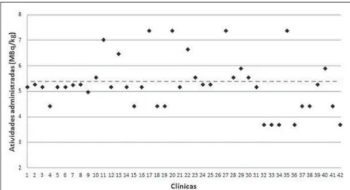
Figure 1 presents the reported adminis-tered activity values in MBq.kg–1 for each one of the 41 clinics, except for clinic 26, which did not provide such value.
With basis on the data of the 41 clinics, representing 57% of the PET clinics in Brazil, the suggested national DRL18 F-FDG/PET suggested for adult cancer pa-tients, represented on Figure 1 by the hori-zontal dotted line, is 5.54 MBq.kg–1 or 0.149 mCi.kg–1, corresponding to 387.7 MBq or 10.48 mCi, considering a standard patient with a body weight of 70 kg.
On Figure 1, it was possible to observe that the activities administered to adult can-cer patients present significant differences, reaching variations of up to 100%. It is in-teresting to highlight that the variations in administered activities occur in clinics which operate apparatuses from a same manufacturer and same detector type, as in the case of clinics 33 and 36, which
oper-ate Siemens apparatuses with LSO detec-tors, and where the administered activities are 3.7 MBq.kg–1 and 7.4 MBq.kg–1, respec-tively. Such a fact demonstrates the lack of standardization and optimization of the procedures. A smaller but no less significant variation was observed for GE apparatuses with BGO detectors in clinics 34 and 22, where differences of up to 80% between administered activities were observed. The clinics operating Philips apparatuses dem-onstrated the smallest variations in reported administered activities, 18.3% between clinics 41 and 7 with LSO detectors.
As the renovation of the different types of apparatuses acquired by Brazilian clin-ics since the first decade of the 2000’s was evaluated, it was possible to suggest a fre-quency for DRL revaluation. The first dedi-cated PET equipment with BGO crystals was installed at the beginning of 2000(7) and, after 5 years, apparatuses equipped with more effective detector crystals such as LSO were installed, and again, five years later, apparatuses equipped with LYSO detectors. By comparing the renovation of apparatuses acquired by Brazilian clinics and the frequency of revaluation of the DRLs in some European countries, the five-year frequency for revaluation has shown to be convenient.
DISCUSSION
The “standard” administered activity in a clinic should be a function of image qual-ity and, for that reason the scan protocols
must be optimized and established accord-ing to such parameter. A method utilized for such optimization consists of balancing the administered activity and the acquisition times per case. Thus, the aim of such a proposition is decreasing the administered activity, compensating such decrease by means of increased acquisition time. Such balancing may contribute for the decrease in doses delivered to patients, promoting savings in radioactive materials and even improving the image quality. This can be verified by means of specific simulators which evaluate the quality of the images as a function of the acquisition time.
Another important fact observed in some clinics is that the responsible person-nel informed that the administered activity is “a function of the supplied doses avail-ability”, and not a function of image qual-ity to produce a satisfactory exam. Such type of approach in the performance of a scan will cause unnecessary doses to pa-tients and should not be encouraged.
In comparison with the DRLs in Euro-pean countries, the suggested DRL of 5.54 MBq.kg–1 is a value 93.7% higher than the DRL in Germany. However, this values is in accordance with the values (5.0 to 5.5 MBq.kg-1) presented by other countries as listed in Table 1. s. It is important to high-light that the higher DRLs than those from other countries does not indicate that the scans are being erroneously carried out, but only that the procedures can and should be optimized.
CONCLUSIONS
When the present study was initiated, Brazil had 64 clinics registered at CNEN. In less than one year, such number in-creased to 72 clinics, a fact which demon-strates the increasing acquisition of equip-ment to meet the demand for 18F-FDG/PET scans in the country. However, the lack of standardization of administered activities observed in the Brazilian clinics is a factor which requires attention; although 12 clin-ics informed that their administered activi-ties were lower than the lowest DRL of the European countries, with the exception of Germany.
FDG/PET scenario in Brazil and also may serve to increase awareness by demonstrat-ing that procedures in clinics should be continuously optimized and that updating and technical training of involved profes-sionals are of utmost importance.
The suggestion of a first national DRL 18F-FDG/PET does not make such value an
official “diagnostic reference level” how-ever it serves the purposes of being a start-ing reference value to encourage respon-sible professionals to optimize scan proto-cols.
Acknowledgements
The author Cássio Miri Oliveira thanks the Coordenação de Apoio ao Pessoal de Nível Superior (Capes) for the Doctoral scholarship, and MRA Indústria de Equi-pamentos Eletrônicos Ltda., for the finan-cial support. The authors thank the clinics which anonymously collaborated with them providing information for the present study. The present study is a part of the project from Instituto Nacional de Ciência e Tecnologia (INCT) – Metrology of Ion-izing Radiations in Medicine.
REFERENCES
1. Council Directive 97/43. Euratom on health pro-tection of individuals against the dangers of ion-izing radiation in relation to medical exposure,
and repealing. Directive 84/466/Euratom. Official Journal of the European Communities. 1997;180: 22–7.
2. International Commission on Radiological Protec-tion. Radiological protection and safety in medi-cine. ICRP Publication 73. Ann ICRP. 1996;26(2). 3. International Commission on Radiological Protec-tion. The 2007 Recommendations of the Interna-tional Commission on Radiological Protection. ICRP Publication 103. Ann ICRP. 2007;37:1– 332.
4. European Commission. Radiation Protection 109. Guidance on diagnostic reference levels (DRLs) for medical exposures. Luxembourg: European Comission; 1999.
5. Zigler SS. Instrumentation and radiopharmaceu-tical validation. Q J Nucl Med Mol Imaging. 2009;53:402–10.
6. International Atomic Energy Agency. Nuclear Technology Review 2006. Vienna, Austria: IAEA; 2006.
7. Robilotta CC. A tomografia por emissão de pósi-trons: uma nova modalidade na medicina nuclear brasileira. Rev Panam Salud Publica. 2006;20: 134–42.
8. Brasil. Ministério da Saúde. Agência Nacional de Vigilância Sanitária. Resolução Nº. 38, de 4 de junho de 2008. Dispões sobre a instalação e fun-cionamento de serviços de medicina nuclear in
vivo. Brasília, DF: Diário Oficial da União, 18 de
dezembro de 2008. Sec. 1, p. 175.
9. Vogiatzi S, Kipouros P, Chobis M. Establishment of dose reference levels for nuclear medicine in Greece. Radiat Prot Dosimetry. 2011;147:237–9. 10. European ALARA Network Survey. The diagnos-tic reference levels (DRLs) in Europe, 2007. [acessado em 1º de junho de 2012]. Disponível em: http://www.eu-alara.net/index.php?option= com_content&task=view&id=156&Itemid=53.
11. Nosske D, Minkov V, Brix G. Establishment and application of diagnostic reference levels for nuclear medicine procedures in Germany. Nuklear-medizin. 2004;43:79–84.
12. Institute de Radioprotection et de Sûreté Nu-cléaire. Niveaux de référence diagnostiques en radiologie et en médecine nucléaire. Journal Of-ficiel de la République Française. 2012. [acessado em 24 de julho de 2012]. Disponível em: http:// nrd.irsn.fr/document/site_49/fckfiles/File/Arrete-NRD-24102011.pdf.
13. De Geest E, Jacobs F, Dierckx RA. A multicenter study of the administered activity in nuclear medi-cine departments in Belgium [Abstract]. In: XI In-ternational Congress of the InIn-ternational Radia-tion ProtecRadia-tion AssociaRadia-tion; 2004 May 23–28; Madri, Espanha.
14. The Medical Council Regulates the Medical Pro-fession in Ireland. Diagnostic reference levels, 2004. [acessado em 12 de junho de 2012]. Dispo-nível em: http://www.medicalcouncil.ie/About- Us/Legislation/Medical-Ionising-Radiation/Diag-nostic-Referance-Levels-03-12-2004.pdf. 15. Botros GM, Smart RC, Towson JE. Diagnostic
reference activities for nuclear medicine proce-dures in Australia and New Zealand derived from the 2008 survey. ANZ Nuclear Medicine. 2009; 40:2–11.
16. Korpela H, Bly R, Vassileva J, et al. Recently re-vised diagnostic reference levels in nuclear medi-cine in Bulgaria and in Finland. Radiat Prot Do-simetry. 2010;139:317–20.