CASE REPORT
A combined minimally invasive approach for the
treatment of aortoesophageal fistula caused by the
ingestion of a chicken bone: case report and
literature review
Xiaodong Chen,IJianwen Li,IJie Chen,IIYu Zhou,IIIYuanqi Zhang,I,IVHongfei Ding,IShuichuan Huang,IZhi ZhangI
IAffiliated Hospital of Guangdong Medical College, Department of Vascular Surgery, Zhanjiang, Guangdong province, People
9s Republic of China. IIAffiliated Hospital of Guangdong Medical College, Department of Cardiothoracic Surgery, Zhanjiang, Guangdong province, People9s Republic of China. IIIAffiliated Hospital of Guangdong Medical College, Department of Gastroenterology, Zhanjiang, Guangdong province, People’s Republic of China. IVFirst Affiliated Hospital of Sun Yat-sen University, Research Center of Vascular Surgery, Department of Vascular Surgery, the Guangzhou, People’s Republic of China, Guangdong, China.
Email: [email protected] Tel.: 86 759-2369322
INTRODUCTION
Aortoesophageal fistula (AEF) is a rare, catastrophic complication that can be caused by the ingestion of a foreign body (1,2). In the past, AEF was treated with surgical repair using thoracic esophagectomy and esophagogastrostomy. However, the mortality rate for patients with AEF remains extremely high (2-4). Recently, we successfully treated a patient with AEF that was caused by the ingestion of a chicken bone. We placed an aortic endovascular stent graft followed by thoracoscopic mediastinal debridement and drainage. A retrievable esophageal stent was placed to isolate the fistula, which was removed after 80 days. In this report, we present our experience using a combined minimally invasive therapeutic approach for the treatment of AEF.
CASE DESCRIPTION
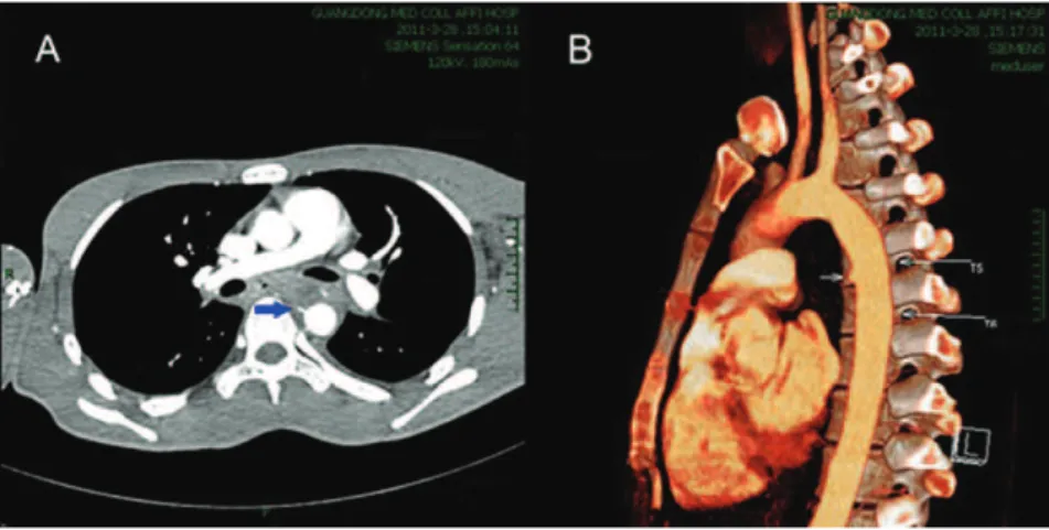
A 22-year-old man was admitted to our hospital on March 28, 2011 complaining of seven days of retrosternal discomfort and a single episode of vomiting of approximately 50 mL of fresh red blood. The patient had swallowed a chicken bone prior to the onset of the discomfort. His vital signs were stable during physical examination, and there was no fever on arrival at the hospital. Laboratory findings revealed a white blood cell (WBC) count of 19.96109/L and hemoglobin (Hb) levels of 84 g/L. He vomited approximately 300 ml of bright red blood and produced a large amount of tarry stool on March 29, 2011. His Hb levels subsequently decreased to 73 g/L. CT angiography (CTA) demonstrated a mediastinal esophageal fistula with inflammatory exudate surrounding the descending aorta. An aortic pseudoaneurysm at the anterior wall of the descending aorta was also observed at the
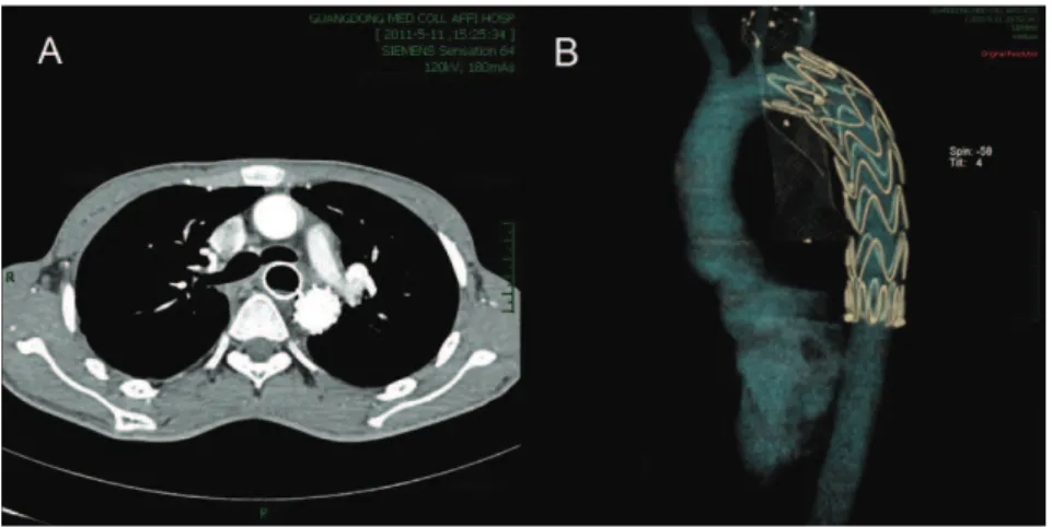
lower edge of thoracic vertebrate 5 (Figure 1). A diagnosis of AEF was highly suspected. Angiography was performed on March 31, 2011 and showed a 0.7 cm60.4 cm pseudoaneur-ysm of the descending aorta approximately 4.5 cm below the origin of the left subclavian artery, which was consistent with the results of the CTA. This finding confirmed the diagnosis of AEF. After an immediate multidisciplinary discussion, a plan for definitive treatment was established. An endovas-cular stent-graft (catalog number: TF2424C200X, Medtronic, Inc., USA) was placed inside the descending aorta to isolate the pseudoaneurysm. Postoperative angiography showed satisfactory exclusion results, and the patient was transferred to the operating room for thoracoscopic exploration. Swelling in the esophageal bed and inflammatory exudates were observed under the thoracoscope. Immediate mediastinal debridement and irrigation was performed using the thoracoscope. Two F16 silicone tubes were placed in the pleural cavity and the mediastinum to facilitate postoperative drainage. The patient underwent an esophageal endoscopy, and an esophageal fistula approximately 1.0 cm in diameter was found 25 cm away from the incisors (Figure 2A). A single-use retrievable esophageal stent-graft (Micro-Tech Co., Ltd. Nanjing China, length 8.0 cm, diameter 2.0 cm) was implanted to isolate the esophageal perforation followed by the placement of a three-lumen gastrojejunal tube for additional enteral nutrition. After spending three days in the intensive care unit (ICU), the patient was transferred back to our department. He had no melena and no fever or vomiting, and the mediastinal and pleural drainage gradually became clear. On the 7thday after the operation, esophageal imaging with meglumine diatrizoate showed no leakage, and the three-lumen gastrojejunal tube was removed. The patient was provided with a liquid diet. Both of the drainage tubes in the pleural cavity and the mediastinum were removed 25 days after the operation. Repeat CTA confirmed successful tamponade of the fistula 40 days after the operation (Figure 3). Antibiotic treatment was administered during hospitaliza-tion. Laboratory findings showed that the patient’s WBC count was 23.06109/mL with 79.1% neutrophils (NEs) on the second day after the operation. The WBC count and Copyrightß2012CLINICS– This is an Open Access article distributed under
the terms of the Creative Commons Attribution Non-Commercial License (http:// creativecommons.org/licenses/by-nc/3.0/) which permits unrestricted non-commercial use, distribution, and reproduction in any medium, provided the original work is properly cited.
No potential conflict of interest was reported.
CLINICS 2012;67(2):195-197 DOI:10.6061/clinics/2012(02)19
NE returned to normal after 14 consecutive days of administration of the antibiotic meropenem based on cultures of the mediastinal exudates, which were positive for
Pseudomonas aeruginosaseven days after the operation. The
patient was switched to oral antibiotics and discharged on May 18, 2011. He was re-admitted to remove the esophageal stent 80 days after the operation. Esophageal endoscopy revealed a complete healing of the original fistula after removal of the esophageal stent (Figure 2B). No fever was observed during the treatment follow-up. After continuous administration of oral antibiotics for three months, the patient stopped the medication when blood laboratory findings and chest X-ray results were normal. The patient lived a normal life during six months of follow-up after the operation.
DISCUSSION
The widely accepted approach for treating AEFs caused by foreign bodies is a thoracotomy with aortic stent-graft interposition followed by reconstruction of the esophagus. However, this procedure has extremely high mortality and morbidity rates (4,5).
The formidable challenges in treating these injuries are rupture of the AEF, mediastinal infection caused by esopha-geal perforation and endovascular stent-graft infection.
Although several successful cases have been documented since the first AEF operation reported by Ctercteko (6) in 1980, the survival rate of patients with AEF has remained dismal. Zhang et al. (2) reported 32 cases of esophageal foreign body and/or aortoesophageal fistula between 1963 and 2010. Of 19 patients who underwent surgical interventions, three patients were completely cured and 16 patients died of fatal hemorrhage. The other 13 patients who were treated nonsurgically all died as a result of various treatments. The optimal management of AEFs remains controversial.
With the rapid development of new endovascular techniques, endovascular aortic stent grafts have shown significant advantages over traditional open surgery for the treatment of aorta fistulas (1). Endovascular stent placement for AEF has been described with good results (3,7). Verhoeven et al. (1) suggested that endovascular aortic stent grafts should be considered as a first-choice lifesaving procedure. These grafts prevent fatal exsanguination and provide the valuable time necessary to optimize the patient’s condition before proceeding with additional treatments.
Postoperative infections due to the placement of aortic stents depends on the treatment of the esophageal fistula, mediastinal drainage and anti-infection treatments. Esoph-ageal exclusion with mediastinal drainage followed by
Figure 1 -CT angiography images.A) Mediastinal esophageal fistula with inflammatory exudate surrounding the descending aorta (arrow).B) An aortic pseudoaneurysm at the anterior wall of the descending aorta at the lower edge of thoracic vertebrae 5 (arrow).
Figure 2 -Esophageal endoscopy.A) An esophageal fistula approximately 1.0 cm in diameter was found 25 cm away from the incisors prior to implantation of the esophageal stent (arrow).B) Complete healing of the original fistula after the removal of the esophageal stent (arrow).
An approach for treating aortoesophageal fistula
Chen X et al. CLINICS 2012;67(2):195-197
secondary esophageal reconstruction was previously a standard surgical approach for the treatment of esophageal fistulas. However, this procedure is time consuming and can cause significant surgical trauma. Thoracoscopy is a mini-mally invasive technique for managing mediastinal and thoracic diseases (8,9). Esophageal stents are universally applied to treat stenosis of the esophagus and esophageal perforation (10,11). We used thoracoscopy instead of med-iastinal debridement, irrigation and drainage. We implanted retrievable esophageal stents and placed a three-lumen gastrojejunal tube. Esophageal stents avoid saliva and/or exudates passing through the esophageal fistula into the mediastinum to prevent mediastinal infection. In this case, we chose a removable esophageal stent because the patient was a young man with benign esophageal disease. The three-lumen gastrojejunal tube ensured early postoperative intake of nutrients. After removal of the three-lumen gastrojejunal tube on postoperative day 7, the patient was provided with a liquid diet for nutritional support.
This report represents the first case in which the minimally invasive approaches described were combined to successfully treat a patient with AEF. Based on our limited experiences, we suggest performing aortic endovas-cular repair by stent-graft to ultimately reduce the risk of aortic rupture, followed by thoracoscopy to treat mediast-inal infections and placement of a retrievable esophageal stent in patients with AEF who have slight inflammation or perforation of the esophageal region and surrounding tissues. The combined approach resulted in less trauma and a more rapid recovery compared to conventional thoracotomy. This minimally invasive approach may be a safe and feasible method for treating patients with AEF and has the potential for improved treatment options for AEFs. A larger sample size and long-term follow-up of patients with AEFs will be required to further evaluate the feasibility of this novel combined approach.
ACKNOWLEDGMENTS
We would like to thank Prof. Wenjian Wang and Xun Hou, PhD of the First Affiliated Hospital of Sun Yat-sen University for editing the manuscript.
AUTHOR CONTRIBUTIONS
Chen XD and Li JW inserted the endovascular stent, drafted the document, reviewed the literature, revised and critically edited the drafts and contributed equally to this work. Zhang YQ assisted in drafting the document and participated as a surgeon in the case. Ding HF, Huang SC, and Zhang Z participated as surgeons in the case. Chen J performed the thoracoscopic operation. Zhou Y inserted and removed the esophageal stent.
REFERENCES
1. Verhoeven EL, Vourliotakis G. Thoracic endovascular aortic repair for aortobronchial or aortoesophageal fistulas: permanent or temporary salvage or not an option at all? J Endovasc Ther. 2009;16(4):441–42, http://dx.doi.org/10.1583/09-2741C.1.
2. Zhang X, Liu J, Li J, Hu J, Yu F, Li S, et al. Diagnosis and treatment of 32 cases with aortoesophageal fistula due to esophageal foreign body. Laryngoscope. 2011;121(2):267-72, http://dx.doi.org/10.1002/lary.21366. 3. Jonker FH, Heijmen R, Trimarchi S, Verhagen HJ, Moll FL, Muhs BE. Acute management of aortobronchial and aortoesophageal fistulas using thoracic endovascular aortic repair. J Vasc Surg. 2009;50(5):999-1004, http://dx.doi.org/10.1016/j.jvs.2009.04.043.
4. Flores J, Shiiya N, Kunihara T, Yoshimoto K, Yasuda K. Aortoesophageal fistula: alternatives of treatment case report and literature review. Ann Thorac Cardiovasc Surg. 2004;10(4):241–46.
5. da Silva ES, Tozzi FL, Otochi JP, de Tolosa EM, Neves CR, Fortes F. Aortoesophageal fistula caused by aneurysm of the thoracic aorta: successful surgical treatment, case report and literature review. J Vasc Surg. 1999;30(6):1150–57, http://dx.doi.org/10.1016/S0741-5214(99) 70056-X.
6. Ctercteko G, Mok CK. Aorta-esophageal fistula induced by a foreign body: the first recorded survival. J Thorac Cardiovasc Surg. 1980;80(2): 233-35.
7. Metz R, Kimmings AN, Verhagen HJ, Rinkes IH, van Hillegersberg R. Aortoesophageal fistula successfully treated by endovascular stent-graft. Ann Thorac Surg. 2006;82(3):1117–19, http://dx.doi.org/10.1016/ j.athoracsur.2006.01.091.
8. Kozuki A, Shinozaki H, Tajima A, Kase K. Successful treatment for descending necrotizing mediastinitis with severe thoracic emphysema using video-assisted thoracoscopic surgery. Gen Thorac Cardiovasc Surg. 2010;58(11):584-87, http://dx.doi.org/10.1007/s11748-009-0553-7. 9. Melo CB, Sarmento PA, Imaeda CJ, Daud DF, Hasimoto FN, Lea˜o LE.
Descending necrotizing mediastinitis: minimally invasive thoracic surgical treatment. J Bras Pneumol. 2010;36(6):812-18, http:// dx.doi.org/10.1590/S1806-37132010000600019.
10. Bakken JC, Wong Kee Song LM, de Groen PC, Baron TH. Ues of a fully covered self-expandable metal stent for the treatment of benign esophageal diseases.Gastrointest Endosc. 2010;72(4):712-20, http:// dx.doi.org/10.1016/j.gie.2010.06.028.
11. Petersen JM. The use of a self-expandable plastic stent for an iatrogenic esophageal perforation. Gastroenterol Hepatol. 2010;6(6):389-91. Figure 3A and B -CT angiography confirmed successful tamponade of the fistula.
CLINICS 2012;67(2):195-197 An approach for treating aortoesophageal fistula Chen X et al.