RADIOLOGY PAGE
137
Proximal Bulbar Periurethral Abscess
Sarah D. Blaschko, Dana A. Weiss, Anobel Y. Odisho, Kirsten L. Greene, Matthew R. Cooperberg
Department of Urology, University of California San Francisco, CA, USA
_______________________________________________________________________________
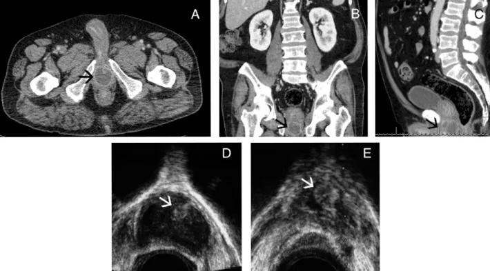
A 67 year-old male with poorly controlled diabetes and persistent leukocytosis despite appro-priate antibiotic treatment for pneumonia underwent computer-tomography (CT) scanning to evaluate for additional sources of infection. He was noted to have a 3.5 centimeter rim enhancing fluid collection at the level of his bulbar urethra (Figure-1, Panel A, B, C). Upon questioning, the patient recalled an aching testicular pain that had resolved one week prior. He
denied any difficulty voiding, and post-void resi-dual measurements were zero. Digital rectal exam, penile, scrotal, and perineal examination were nor-mal. Transrectal ultrasound demonstrated an abscess surrounding the bulbar urethra (Figure-1, Panel D). Transrectal ultrasound-guided needle aspiration was performed with return of 30 milliliters of frank pus and visible resolution of the abscess (Figure-1, Pa-nel E). The patient had subsequent rapid clinical
Vol. 39 (1): 137-138, January - February, 2013
Figure 1 - Panel A) Axial CT-scan of the pelvis demonstrating a rim-enhancing fluid collection (black arrow) at the level of the proximal bulbar urethra; Panel B) Coronal CT-scan showing the periurethral abscess (black arrow) adjacent to the prostate, which is distinct from the abscess; Panel C) Sagittal CT-scan showing the periurethral abscess (black arrow) adjacent to the prostate, which is distinct from the abscess; Panel D) Transrectal ultrasound demonstrating the proximal bulbar urethra (white arrow) within the abscess cavity prior to abscess drainage; Panel E) Transrectal ultrasound demonstrating the proximal bulbar urethra (white arrow) with resolution of the surrounding abscess cavity at the completion of needle aspiration.
138
IBJU |RADIOLOGY PAGE
improvement. Although the abscess fluid culture was negative, he completed a two-week antibio-tic course per infectious disease recommendations. Recommended periurethral abscess antibiotic co-verage is culture-specific or treatment with an aminoglycoside and cephalosporin (1). Periure-thral abscesses have been associated with
gono-coccal urethritis infections, urethral strictures, pe-riurethral bulking agent injections, and urethral diverticulum (1-3). Periurethral abscesses are trea-ted with antibiotic coverage and surgical or need-le-aspiration drainage depending on abscess loca-tion. Evaluation for and treatment of underlying causes of periurethral abscesses is warranted.
REFERENCES
1. Walther MM, Mann BB, Finnerty DP: Periurethral abscess. J Urol. 1987; 138: 1167-70.
2. Kraus S, Luedecke G, Ludwig M, Weidner W: Periurethral ab-scess formation due to Neisseria gonorrhoeae. Urol Int. 2004; 73: 358-60.
3. Kenfak-Foguena A, Zarkik Y, Wisard M, Praz V, Darling KE, Ja-ton-Ogay K, et al.: Periurethral abscess complicating gonococ-cal urethritis: case report and literature review. Infection. 2010; 38: 497-500.
______________________ Correspondence address:
Dr. Sarah D. Blaschko Department of Urology University of California, San Francisco 400 Parnassus Ave, A633 San Francisco, CA 94143, USA FAX: 415-476-8849 E-mail: [email protected]
ARTICLE INFO
Int Braz J Urol. 2013; 39: 137-8
__________________
Submitted for publication: July 27, 2012
__________________