e88
The authors report a case of a 65-year-old male patient who, after complaint about retrosternal discomfort and going through a thoracic radiography with mediastinal enlargement, was submitted to magnetic resonance angiography, which detected a right aortic arch with aberrant left subclavian artery and Kommerell diverticulum. A literature review was the basis for our clinical conduct, for the surgical treatment is complex. The patient presented improvement with clinical treatment and, currently, is under ambulatory follow-up care, with control proposed for three months later or in case of worsening.
Mailing address: Marcelo Souto Nacif •
Rua Tavares de Macedo 136 / 1503 Bl. A – Icaraí – CEP 24220-211 – Ni-terói, RJ, Brazil
E-mail: [email protected]
Manuscript received January 01, 2009; revised manuscript received May 15, 2009; accepted May 15, 2009.
Key Words
Aorta, Thoracic; Subclavian Artery / abnormalities; Kommerell Diverticulum.
Introduction
The diagnosis of the several types of aortic arch diverticulum is important due to the difficulty in differing it from aneurysms and other mediastinal masses1. Clinical and image diagnosis
focuses on the reduction of mortality associated with rupture or compression of mediastinal structures.
Kommerell diverticulum is a rare condition that occurs more frequently in the left aortic arch (LAA) and anomalous origin of the right subclavian artery (RSA) (0.5-2.0%), but not very common in the right aortic arch (RAA) and anomalous origin of left subclavian artery (LSA) (0.05-0.1%)2,3. The cause
of anomalous origin of subclavian artery may be the abnormal regression of the fourth primitive aortic arch during embryonic development. The fourth left aortic arch persists as an aortic arch, while the fourth right aortic arch remains as a right subclavian artery and innominate artery3,4.
Image exams, particularly magnetic resonance angiography (MRA) with 3D-GE TOF technique during administration of gadolinium – and the possibility of multiple-projection reconstructions (MPR) – play a fundamental role in the diagnosis and preoperative program due to the complexity of the surgical treatment2,3,5,6.
The objective of this paper is to report the aspects of magnetic resonance angiography in a patient with Kommerell
diverticulum in the right aortic arch, with aberrant left subclavian artery.
Case report
Male, 65-year-old patient who referred having regurgitation (reflux) and retrosternal burning pain in the middle of the night. The patient was submitted to endoscopy and peptic ulcer treatment, but the symptoms lingered even after adequate treatment. In the endoscopic approach, an extrinsic steepening of the upper esophagus was identified and, thus, a thoracic radiography was indicated for the assessment of a mediastinal mass.
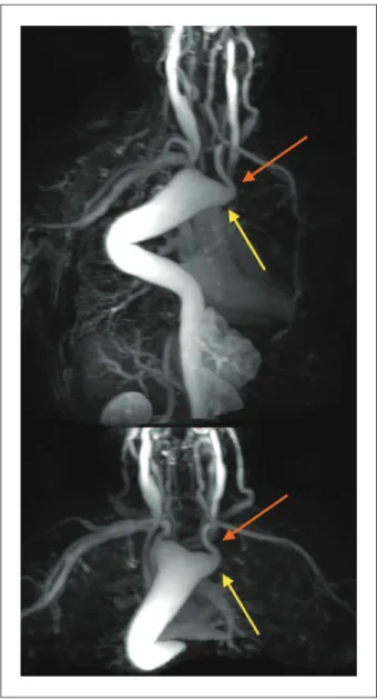
After the exam, that showed mediastinal enlargement of apparent vascular origin, the patient was referred to the image diagnostic service of Hospital das Clínicas de Niterói, Rio de Janeiro, Brazil, for magnetic resonance angiography procedure (Figures 1 and 2).
The 3D-GE TOF magnetic resonance angiography with gadolinium can identify the aortic arch at the right side with the diverticular colon and the origin of left subclavian artery. The trachea and the esophagus were in anterior position in relation to the vascular structures, as easily identified by the exam.
The study with 3D reconstruction of the thoracic aorta showed a better aortic arch at the right side, as well as aberrant left subclavian artery with origin in the Kommerell diverticulum.
The patient presented clinical improvements after taking prokinetics medication and proton pump inhibitors, as clinical follow-up and a new image exam was indicated for three months later or in case of clinical worsening.
Discussion
There are basically three types of aortic arch diverticulum: the diverticulum at left aortic arch with aberrant right subclavian artery; the diverticulum at right aortic arch with aberrant left subclavian artery; and, finally, the aortic diverticulum at aortic-ductal junction4,7-9.
The first type of diverticulum was described by Kommerell5,
in 1936, in a publication on a case of aberrant right subclavian artery, the most common anomaly of the subclavian artery, which originates in the thoracic aorta with arch at the left associated with right remnant dorsal aorta. The latter is similar to the diverticulum from which the correspondent subclavian artery originates. The Kommerell diverticulum, which corresponds to a conical dilatation in the origin of the aberrant vase, is also known as “lusoria diverticulum”, “remnant diverticulum” or “lusoria root”5.
Atypical presentation of Kommerell’s diverticulum
Adriana Dias Barranhas
1,3, João Mauricio Canavezi Indiani
1,3, Edson Marchiori
2, Alair Augusto S. M. D. dos Santos
1,3,
Carlos Eduardo Rochitte
3, Marcelo Souto Nacif
1,2,3RM Cardíaca do Hospital de Clínicas de Niterói (HCN)1, Niterói, RJ; Universidade Federal do Rio de Janeiro (UFRJ)2, Rio de Janeiro; InCor –
Figure 1 – Aortic arch at the right side (blue arrow) with rectal diverticulum (yellow line) from where the left subclavian artery originates (orange arrow). Observe the trachea (green arrow) and the esophagus (red arrow) anterior to vascular structures.
Figure 2 – Magnetic resonance angiography with 3D reconstructions of the thoracic aorta, making evident an arch at the right side and aberrant left subclavian artery (red arrow) that originates in Kommerell diverticulum (yellow arrow).
The second diverticulum type is described in dextroposed aortic arch as a rare congenital alteration (present in 0.05 to 0.1% of radiologic series6,7 and 0.04 to 0.1% of necropsy series7,9,10).
It may be associated with the aberrant left subclavian artery3,5,
which may be localized behind the esophagus (80%), precisely between the trachea and the esophagus (15%) or behind the trachea (5%)5,7,8. The right aortic arch results from an anomalous
primitive organogenesis. In general, between the fourth and fifth weeks of embryonic life, the blood leaves the heart through a single vase, the arterial trunk, which is divided into two branches: right and left ventral aorta. They communicate with the dorsal artery, respectively, through six brachial vases, known as aortic arches. The first three arches, together with their ventral and dorsal aortic connections, form the carotid system. A segment of the ventral aorta, the fourth right arch and a portion of the right dorsal aorta originate the right subclavian artery and the brachiocephalic trunk (innominate artery). The fourth left arch persists and becomes the adult aortic arch that unites with the seventh dorsal intersegmental artery to form the left subclavian artery. The fifth arches are reabsorbed, while the sixth arches form the pulmonary and arterial trunks. If the fourth left arch disappears and the right arch persists, a right aortic arch will develop. Generally, it is associated with the left dorsal aorta involution and with the right dorsal aorta persistence, resulting in a descending thoracic aorta placed in the right hemithorax. If the right dorsal aorta disappears, the dextraposed aortic arch places behind the esophagus to unite to the left dorsal aorta, while the descending thoracic aorta will unite to the left one3,8. If both arches persist, they will form a
double aortic arch or a vascular ring that encircles the trachea and the esophagus9,10.
Among the proposed classifications based on anatomical distribution of the involved structures, the most widely used is Edwards classification4,6.Three types of right aortic arches have
been described1: type I, when the big arteries branches form a
mirror-image; type II, with arterysegment enclosing area; and type III, when the isolated left subclavian artery communicates with the pulmonary area through the artery duct.
In the present case report, the aortic arch dextropositon was associated with the aberrant left subclavian artery (Edwards’ type II), which in 5 to 10% of the cases may be related to congenital cardiopathies, including Tetralogy of Fallot, pulmonary stenosis with ventricular septal defect, tricuspid atresia and ductus arteriosus2. The right aortic arch
is frequently asymptomatic. In adults, symptoms may originate from atherosclerotic disease of anomalous vases, such as aneurysm dissection or dilatation with compression of adjacent structures, causing dysfagia (dysfagia lusoria), dyspnea, stridor, noisy breathing, cough, repetition pneumonia, obstructive emphysema or thoracic pain2,3.
The third type is the aortic diverticulum in the aortic-ductal junction7,9,10. This diverticulum shows a steepening
throughout the internal portion that comprises the distal aortic
Arq Bras Cardiol 2009; 93(6) : e88-e90
Barranhas et al Atypical presentation of Kommerell’s diverticulum
e89
isthmus till the left subclavian artery. It is also known as “ductal diverticulum”, “ductal steepening” or “aortic diverticulum”9.
The term “ductal diverticulum” may also be applied to a steepening in the left pulmonary artery, which represents a remnant of the arterial duct pulmonary orifice6,10.
Final considerations
Magnetic resonance angiography may be useful as a preoperative image exam in cases of surgical planning or when images are needed for ambulatory follow-up care.
1. Salomonowitz E, Edwards JE, Hunter DW, Castaneda-Zuniga WR, Lund G, Cragg AH, et al. The three types of aortic diverticula. Am J Roentgenol. 1984; 142: 673-9.
2. Ota T, Okada K, Takanashi S, Yamamoto S, Okita Y. Surgical treatment for Kommerell’s diverticulum. J Thorac Cardiovasc Surg. 2006; 131 (3): 574-8.
3. Faucz RA, Furlan S, Barros AS, Bof AM, Lomonte ES, Leiro LC, et al. Arco aórtico direito com artéria subclávia esquerda aberrante e divertículo de Kommerell. Radiol bras. 2005; 38 (5): 381-4.
4. Edwards JE. Anomalies of the derivatives of the aortic arch system. Med Clin North Am. 1948; 32: 925-48.
5. Kommerell B. Verlagerung des oesophagus durcheine abnorm verlaufende arteria subclavia dextra (Arteria Lusoria). Fortschr Geb Roentgenstr. 1936; 54: 590-5.
6. Hastreiter AR, D’Cruz IA, Cantez T, Namin EP, Licata R. Right-sided aorta. Occurrence of right aortic arch in various types of congenital heart disease. Br Heart J. 1966; 28: 722-5.
7. Haughton VM, Fellows KE, Rosenbaum AE. The cervical aortic arches. Radiology. 1975; 114: 675-81.
8. Cinà CS, Althani H, Pasenau J, Abouzahr L. Kommerell’s diverticulum and right-sided aortic arch: a cohort study and review of the literature. J Vasc Surg. 2004; 39: 131-9.
9. Goodman PC, Jeffrey RB, Minagi H, Federle MP, Thomas AN. Angiographic evaluation of the ductus diverticulum. Cardiovasc Intervent Radiol. 1982: 5: 1-4.
10. Shuford WH, Sybers RG, Gordon IJ, Baron MG, Carson GC. Circumflex retroesophageal right aortic arch simulating mediastinal tumor or dissecting aneurysm. Am J Roentgenol. 1986; 146: 491-6.
References
Arq Bras Cardiol 2009; 93(6) : e88-e90
Barranhas et al
Atypical presentation of Kommerell’s diverticulum