Martinna de Mendonça e Bertolini Yuri Wanderley Cavalcanti
Dimorvan Bordin Wander José da Silva Altair Antoninha Del Bel Cury
Departmentof Prosthodontics and Periodontology, Piracicaba Dental School, Universidade Estadual de Campinas - Unicamp, Piracicaba, SP, Brazil.
Corresponding Author: Martinna de Mendonça e Bertolini E-mail: [email protected]
Candida albicans
biofilms and MMA
surface treatment influence the
adhesion of soft denture liners to
PMMA resin
Abstract: The effect of Candida albicans bioilms and methyl methac-rylate (MMA) pretreatment on the bond strength between soft denture liners and polymethyl methacrylate (PMMA) resin was analyzed. Speci-mens were prepared and randomly divided with respect to PMMA pre-treatment, soft liner type (silicone-based or PMMA-based), and presence or absence of a C. albicans bioilm. Samples were composed of a soft denture liner bonded between two PMMA bars. Specimens (n = 10) were incubated to produce a C. albicans bioilm or stored in sterile PBS for 12 days. The tensile bond strength test was performed and failure type was determined using a stereomicroscope. Surface roughness (SR) and scanning electron microscopy (SEM) analysis were performed on denture liners (n = 8). Highest bond strength was observed in samples containing a silicone-based soft liner and stored in PBS, regardless of pretreatment (p < 0.01). Silicone-based specimens mostly underwent adhesive failures, while samples containing PMMA-based liners predominantly underwent cohesive failures. The silicone-based specimens SR decreased after 12 days of bioilm accumulation or PBS storage, while the SR of PMMA-based soft liners increased (p < 0.01). The PMMA-based soft liners sur-faces presented sharp valleys and depressions, while silicone-based speci-mens surfaces exhibited more gentle features. In vitro exposure to C. albicans bioilms reduced the adhesion of denture liners to PMMA resin, and MMA pretreatment is recommended during relining procedures.
Descriptors:Candida albicans; Denture Liners; Tensile Strength; Polymethyl Methacrylate.
Introduction
Soft denture liners are used to form a comfortable interface between denture and soft oral tissues, reducing traumatic transmission of occlu-sal forces to severely resorbed alveolar ridges and areas recovering from surgical procedures.1 However, failure often occurs in adhesive bond
be-tween soft liner and denture base resin, resulting in tearing and mate-rial loss during clinical use.2 This damage can increase surface roughness
and create irregularities that act as sheltered sites where oral bioilms may accumulate over time.3 These bioilms are mainly composed of C.
albicans and may cause denture-induced stomatitis4 or accelerate wear
Declaration of Interests: The authors certify that they have no commercial or associative interest that represents a conflict of interest in connection with the manuscript.
Submitted: Apr 02, 2013
Accepted for publication: Sep 05, 2013 Last revision: Sep 20, 2013
and aging of soft liner and denture base.5 In order to
prevent these problems, several denture base surface treatments have been proposed to increase the bond strength between these materials.6
When compared to other denture resin pretreat-ments,6-8 methyl methacrylate (MMA) surface
pre-treatment increased tensile bond strength8 and
re-duced microleakage between the denture base and silicone-based soft liners.7 In order to evaluate the
durability of bonds between soft liners and pretreat-ed denture resins, previous studies have subjectpretreat-ed specimens to distilled water storage,9 accelerated
ag-ing in hot water,10 or thermocycling procedures8,11
prior to bond strength testing.
More recently, it was hypothesized that in vi-tro exposure of composites to oral bioilms results in clinically relevant surface degradation.5
Al-though there have been reports concerning the bond strength of soft liners to denture base resins and the effects of various pretreatment methods, until now there have been no studies considering the potential damaging effect of oral bioilms on the interface between soft liners and pretreated denture resins. Therefore, the inluence of C. albicans bioilms on the tensile bond strength between soft liners and denture resin, with or without MMA pretreatment, was analyzed. The principal hypothesis was that bioilms can cause degradation of the denture liner– PMMA interface, decreasing the bond strength.
Methodology
Experimental design
An in vitro study with blind analysis was per-formed, in which specimens were prepared and ran-domly divided according to PMMA surface treat-ment (MMA pretreattreat-ment or no treattreat-ment), denture liner type (silicone-based or PMMA-based), and presence or absence of a C. albicans bioilm. Den-ture liners were applied between two treated PMMA bars, and specimens (n = 10) were subjected to bio-ilm accumulation, or phosphate buffered saline (PBS) storage, in order to simulate conditions expe-rienced by dentures in clinical applications. Tensile bond strength was measured and the nature of fail-ure (adhesive, cohesive, or mixed) was determined using a stereomicroscope under 10× magniication.
Surface roughness (SR) and scanning electron mi-croscopy (SEM) analyses were performed on denture liner discs (n = 8) for surface characterization.
Specimen preparation
Microwave-polymerized PMMA (Vipi Wave, VIPI, Pirassununga, Brazil) resin bars (25.0 × 5.0 × 5.0 mm; n = 160), silicone-based (Ui Gel SC, VOCO, Cux-haven, Germany) and PMMA-based soft liner discs (Coe Soft, GC, Coe Laboratories Inc., Chicago, USA; 10.0 mm diameter × 2.0 mm thick; n = 32) were prepared according to manufacturers’ recom-mendations using metal master patterns.
The PMMA bars were trimmed and inished in a polishing machine (APL-4 Model; Arotec, Cotia, Brazil), using abrasive paper (320, 400, and 600 grit, Carbimet; Buehler, Lake Bluff, USA). Speci-mens were ultrasonically cleaned (Thornton T740, Thornton-Inpec Eletrônica Ltda., Vinhedo, Brazil) and immersed in distilled water at 35°C for 48 h for residual monomer release.12
The square faces of PMMA bars were either treated with MMA (180 s) or left without surface treatment prior to adhesion of the 2 mm-thick den-ture liner.8,13,14 Specimens were stored in 100%
rela-tive humidity before testing.
Soft liner specimens were ultrasonically cleaned and maintained in 100% humidity at 35°C for 24 h prior to bioilm accumulation or PBS storage. The cleaning procedure consisted of sonication for 10 minutes in 0.5% sodium hypochlorite and 10 min-utes in sterile water.12 Discs were used for surface
roughness evaluation and SEM analysis before and after PBS storage or bioilm accumulation.
Biofilm formation and PBS storage conditions
Candida albicans (OMZ 110) was reactivated in yeast nitrogen base (YNB) medium containing 50 mM glucose, and the bioilm inoculum was stan-dardized at an optical density of 0.25 in YNB con-taining 100 mM glucose. After allowing 90 minutes for initial adhesion, specimens were transferred to new tubes containing 7.0 mL of sterile YNB with 100 mM glucose for bioilm development.15 Control
Soft liner surface roughness results were evaluated using one-way ANOVA and the Bonferroni test.
Results
The highest tensile bond strength (Table 1) was observed in the groups with silicone-based soft lin-ers stored in PBS (p < 0.01), regardless of pretreat-ment. C. albicans bioilm accumulation resulted in a bond strength decrease for silicone-based specimens (p < 0.01).
Silicone-based specimens generally underwent adhesive failures while PMMA-based groups expe-rienced predominantly cohesive failures (Table 2). Cohesive failures were mainly observed in the group receiving MMA pretreatment and stored in PBS (Ta-ble 2).
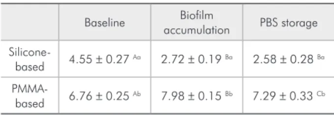
The silicone-based liners surface roughness de-creased after bioilm accumulation or PBS storage (p < 0.001, Table 3), while PMMA-based liners sur-face roughness increased after PBS storage, and even more after bioilm accumulation (p < 0.001).
SEM observations of PMMA-based samples re-vealed a smooth porous surface (Figure 1A). After PBS storage (Figure 1B) or bioilm accumulation (Figure 1C), surfaces exhibited smaller pores as well as the formation of crests and valleys. Surface modi-ications may be due to material swelling, which causes pore constriction and creates wrinkles on the surface. At baseline, silicone-based samples present-ed smooth surfaces without visible pores, but with sharp crests and depressions (Figure 1D). After PBS storage or bioilm accumulation, material swelling sets of samples were stored at 35°C with agitation.
PBS solution and bioilm culture medium were changed daily. After 12 days, the specimens were ul-trasonically cleaned and prepared for testing.
Tensile bond strength evaluation
The tensile strength test was performed in a uni-versal testing machine (4411, Instron Corp., Canton, USA) using a crosshead speed of 5.0 mm/min. Sam-ples were tested until failure. The tensile strength, in MPa, was determined by multiplying the stress (Kgf) at the time of failure by a constant (9.8) and divid-ing this result by the surface area of adhesion (mm²). Failures were examined using a stereomicroscope at 10× magniication and classiied as adhesive (total separation at the interface between the liner and res-in), cohesive (tearing within the soft liner), or mixed failures (both adhesive and cohesive).8,16
Surface roughness evaluation
Surface roughness was used to identify changes in the soft liner surface occurring during bioilm ac-cumulation or PBS storage. Measurements were ob-tained using a proilometer (Surfcorder SE1700; Ko-saka Laboratory Ltd., Tokyo, Japan) with 0.01 mm resolution and adjusted for a 0.8 mm sample length, 3.2 mm percussion of measurement, and 0.5 mm/s stylus speed. Reported roughness values are the aver-age of three measurements performed on each speci-men (n = 8).12
Scanning electron microscopy evaluation To evaluate the effect of bioilm accumulation, or PBS storage, on soft liners surface, specimens were ultrasonically cleaned and prepared for SEM. Sam-ples were examined using an acceleration voltage of 15 kV at 2000× magniication.17,18 The surfaces were
evaluated at baseline (n = 3) and after bioilm accu-mulation (n = 3) or PBS storage (n = 3).
Statistical analysis
Statistical analysis was performed using the Sig-maPlot 12 software (SigSig-maPlot v. 12.3, Systat Soft-ware Inc., San Jose, USA) at 5% signiicance. Tensile strength results were evaluated using three-way anal-ysis of variance (ANOVA) and Holm-Sidak’s test.
Table 1 - Tensile bond strength (mean ± SD, MPa) of soft liners to untreated (NT) or surface treated (MMA) PMMA following biofilm accumulation or PBS storage.
Biofilm
accumulation PBS
Silicone-based liner
MMA 3.60 ± 0.47 Aa* 5.92 ± 0.70 Ab§
NT 3.21 ± 0.78 Aa* 4.03 ± 0.70 Ab+
PMMA-based liner
MMA 1.24 ± 0.19 Bc¥ 1.11 ± 0.15 Bc¥
NT 1.31 ± 0.39 Bc¥ 1.14 ± 0.18 Bc¥
made the depressions less pronounced, resulting in a smother surface than that observed at baseline (Fig-ure 1E and 1F).
Discussion
The observation that soft liner surfaces are gen-erally rough and covered with a bioilm2 motivated
our evaluation of the effect of C. albicans bioilms on tensile bond strength between soft liners and untreated or MMA-pretreated PMMA resin. How-ever, we found that the effect of bioilms on adhe-sion was important mainly in samples employing silicone-based liners.
Our principal hypothesis was accepted in the case of silicone-based liners, in which the presence of C. albicans bioilms resulted in signiicantly low-er bond strength. This result is in accordance with a previous study5 demonstrating that in vitro
expo-sure to oral bioilms leads to clinically relevant ag-ing of dental materials.
However, no statistical difference was observed for bond strengths of specimens employing PMMA-based liners, even with MMA pretreatment. There was a relationship between failure type and MMA pretreatment, with better adhesion being associated with more mixed failures in both silicone and PMMA
liner groups, as previously reported in the literature.19
In spite of the fact that other surface pretreat-ments had previously been reported ineffective in im-proving bond strength during a hard chairside reline using PMMA acrylic resin,6 we found MMA
pretreat-ment to be effective in increasing the bond strength between silicone-based soft liners and PMMA resin stored in PBS. Considering that there is no chemical interaction between silicone-based liner and PMMA acrylic resin,8 the increase in bond strength may be
due to the ability of MMA to dissolve PMMA sur-face layer and increase the bonding sursur-face area.18
For PMMA-based groups there were no signii-cant differences in bond strength due to the presence of bioilm or following resin pretreatment. However, this result should be interpreted with caution, since the high number of cohesive failures in PMMA-based soft liners may be due to the fact that the bond to PMMA resin is stronger than the denture liner tensile strength itself,16,17,19 inducing failure in soft liner
be-fore debonding from PMMA resin occurs. However, all of the soft liners demonstrated bond strengths to denture base resin above the minimum acceptable bond strength for clinical use (0.45 MPa).11,14
Besides to the degradation between soft lin-ers and PMMA resin interface, C. albicans bioilm accumulation led to a greater overall degradation. This is probably related to the ability of C. albicans
hyphae to adhere and penetrate into soft liners,20,21
as well as the production of proteases and phos-pholipases.22,23 Thus, it is important to consider the
degradation of the soft liner itself, which makes the material more susceptible to tearing.
Storage in aqueous solutions such as PBS or growth medium promotes the release of soluble compounds and plasticizers as well as water inil-tration,11,24 both of which may contribute to
deg-Failure type
Silicone-based liner PMMA-based liner
MMA NT MMA NT
Biofilm PBS Biofilm PBS Biofilm PBS Biofilm PBS
Adhesive 90 70 100 80 10 0 40 30
Mixed 10 30 0 20 20 20 30 30
Cohesive 0 0 0 0 70 80 30 40
Table 3 - Surface roughness (mean ± SD, µm) of soft liners at baseline and after biofilm accumulation or PBS storage.
Baseline Biofilm
accumulation PBS storage
Silicone-based 4.55 ± 0.27
Aa 2.72 ± 0.19 Ba 2.58 ± 0.28 Ba
PMMA-based 6.76 ± 0.25
Ab 7.98 ± 0.15 Bb 7.29 ± 0.33 Cb
Different uppercase letters indicate statistical differences between baseline, biofilm accumulation, and PBS storage groups. Different lowercase letters indicate statistical differences between liner types.
radation and surface modiication of soft liner ma-terials.11 A large number of crests and valleys in
PMMA-based soft liners and surface modiications in silicone-based liners were evident in SEM images of the liners obtained after storage. However, more studies are necessary to conirm the effects of wa-ter uptake, and evaluations of other soft liner types should be undertaken, including other PMMA and silicone-based materials.
When immersed in MMA solution, a decrease in lexural strength is sometimes observed in PMMA resins;8 however, this would not be expected in
cli-nical practice since the MMA pretreatment involves only surface application, as was performed in the present study. The use of MMA pretreatment may re-sult in better clinical performance and greater pros-thetic survival. Bioilm accumulation seems to play an important role in degradation of the adhesive in-terface and should be avoided.5 Although the results
are based on an in vitro study, clinical application of these recommendations may contribute to a higher-strength interface with a smoother surface and less bioilm accumulation.23 However, in patients with
candidiasis, material selection alone may not
inlu-ence the C. albicans bioilms growth, particularly when oral hygiene measures are correctly applied.25
Several factors are expected to affect the bond strength between soft liners and denture base resins, including aging in water9,10 and thermocycling.8,11
This list may now also include bioilm accumulation, the use of a bonding agent, and the composition of the soft liner. Additional studies incorporating longer periods of bioilm accumulation must be conducted with the purpose of assessing material degradation and structural failures under these conditions.
It is also important to consider that aging of soft liners in the oral cavity involves more than expo-sure to bioilms: temperature variations and immer-sion in water or acidic luids from foods may also contribute to clinical aging.5 Future in vitro studies
should attempt to simulate as many of these condi-tions as possible.
Conclusion
In vitro exposure to C. albicans bioilms re-duced the adhesion of soft liners to PMMA resin, and MMA pretreatment of denture bases is recom-mended during relining procedures.
References
1. Pisani MX, Malheiros-Segundo AL, Balbino KL, Souza RF, Paranhos HF, Silva CH. Oral health related quality of life of edentulous patients after denture relining with a silicone-based soft liner. Gerodontology. 2012 Jun;29(2):474-80.
2. Mutluay MM, Oguz S, Fl∅ystrand F, Saxegaard E, Dogan A, Bek B, et al. A prospective study on the clinical performance of polysiloxane soft liners: one-year results. Dent Mater J. 2008 May;27(3):440-7.
3. Monsenego P. Presence of microorganisms on the fitting den-ture complete surface: study ‘in vivo’. J Oral Rehabil. 2000 Aug;27(8):708-13.
4. Uludamar A, Özyeşil AG, Ozkan YK. Clinical and microbiologi-cal efficacy of three different treatment methods in the manage-ment of denture stomatitis. Gerodontology. 2011 Jun;28(2):104-10.
5. Rinastiti M, Özcan M, Siswomihardjo W, Busscher HJ, van der Mei HC. Effect of biofilm on the repair bond strengths of composites. J Dent Res. 2010 Dec;89(12):1476-81.
6. Leles CR, Machado AL, Vergani CE, Giampaolo ET, Pavarina AC. Bonding strength between a hard chairside reline resin and a denture base material as influenced by surface treatment. J Oral Rehabil. 2001 Dec;28(12):1153-7.
7. Saraç YS, Başoğlu T, Ceylan GK, Saraç D, Yapici O. Effect of denture base surface pretreatment on microleakage of a silicone-based resilient liner. J Prosthet Dent. 2004 Sep;92(3):283-7. 8. Sarac D, Sarac YS, Basoglu T, Yapici O, Yuzbasioglu E. The
evaluation of microleakage and bond strength of a silicone-based resilient liner following denture base surface pretreat-ment. J Prosthet Dent. 2006 Feb;95(2):143-51.
9. Tugut F, Akin H, Mutaf B, Akin GE, Ozdemir AK. Strength of the bond between a silicone lining material and denture resin after Er:YAG laser treatments with different pulse durations and levels of energy. Lasers Med Sci. 2012 Mar;27(2):281-5. 10. Al-Athel M, Jagger R, Jagger D. Effect of ageing on the bond
strength of a permanent denture soft lining material. J Oral Rehabil. 2002 Oct;29(10):992-6.
11. Takahashi JM, Consani RL, Henriques GE, Nóbilo MA, Mes-quita MF. Effect of accelerated aging on permanent deformation and tensile bond strength of autopolymerizing soft denture liners. J Prosthodont. 2011 Apr;20(3):200-4.
12. Senna PM, Silva WJ, Faot F, Del Bel Cury AA. Microwave disinfection: cumulative effect of different power levels on physical properties of denture base resins. J Prosthodont. 2011 Dec;20(8):606-12.
13. Minami H, Suzuki S, Ohashi H, Kurashige H, Tanaka T. Effect of surface treatment on the bonding of an autopolymerizing
soft denture liner to a denture base resin. Int J Prosthodont. 2004 May-Jun;17(3):297-301.
14. Maeda T, Hong G, Sadamori S, Hamada T, Akagawa Y. Dura-bility of peel bond of resilient denture liners to acrylic denture base resin. J Prosthodont Res. 2012 Apr;56(2):136-41. 15. Gonçalves LM, Del Bel Cury AA, Sartoratto A, Garcia Rehder
VL, Silva WJ. Effects of undecylenic acid released from denture liner on Candida biofilms. J Dent Res. 2012 Oct;91(10):985-9. 16. Pinto JR, Mesquita MF, Henriques GE, Nóbilo MAA. Effect of thermocycling on bond strength and elasticity of 4 long-term soft denture liners. J Prosthet Dent. 2002 Nov;88(5):516-21. 17. Mutluay MM, Ruyter IE. Evaluation of bond strength of soft
relining materials to denture base polymers. Dent Mater. 2007 Nov;23(11):1373-81.
18. Rached RN, Del Bel Cury AA. Heat-cured acrylic resin repaired with microwave-cured one: bond strength and surface texture. J Oral Rehabil. 2001 Apr;28(4):370-5.
19. Akin H, Tugut F, Guney U, Kirmali O, Akar T. Tensile bond strength of silicone-based soft denture liner to two chemically different denture base resins after various surface treatments. Lasers Med Sci. 2013 Jan;28(1):119-23.
20. Bulad K, Taylor RL, Verran J, McCord JF. Colonization and penetration of denture soft lining materials by Candida albicans. Dent Mater. 2004 Feb;20(2):167-75.
21. Rodger G, Taylor RL, Pearson GJ, Verran J. In vitro coloniza-tion of an experimental silicone by Candida albicans. J Biomed Mater Res B Appl Biomater. 2010 Jan;92(1):226-35.
22. Nikawa H, Jin C, Hamada T, Makihira S, Kumagai H, Murata H. Interactions between thermal cycled resilient denture lining materials, salivary and serum pellicles and Candida albicans in vitro. Part II. Effects on fungal colonization. J Oral Rehabil. 2000 Feb;27(2):124-30.
23. Nikawa H, Jin C, Hamada T, Murata H. Interactions between thermal cycled resilient denture lining materials, salivary and serum pellicles and Candida albicans in vitro. Part I. Effects on fungal growth. J Oral Rehabil. 2000 Jan;27(1):41-51. 24. Garcia RM, Léon BT, Oliveira VB, Del Bel Cury AA. Effect of a
denture cleanser on weight, surface roughness, and tensile bond strength of two resilient denture liners. J Prosthet Dent. 2003 May;89(5):489-94.