Ana Paula Coelho Vieira(a) Plínio Mendes Senna(b) Wander José da Silva(c)
Altair Antoninha Del Bel Cury(c)
(a) MSc student; (b)PhD student; (c)PhD – Department of Prosthodontics and Periodontology, Piracicaba Dental School, State University of Campinas, Brazil.
Corresponding author:
Altair Antoninha Del Bel Cury Av. Limeira, 901
Piracicaba - SP - Brazil CEP: 13414-903
E-mail: [email protected]
Received for publication on May 18, 2010 Accepted for publication on Jul 01, 2010
Long-term efficacy of denture cleansers
in preventing
Candida
spp. biofilm
recolonization on liner surface
Abstract: This study evaluated the long-term eficacy of denture cleans-ers against Candida spp. bioilm recolonization on liner surface. Speci-mens were fabricated of a poly(methyl methacrylate)-based denture liner and had their surface roughness evaluated at baseline and after cleansing treatments. C. albicans or C. glabrata bioilms were formed on liner sur-face for 48 h, and then the specimens were randomly assigned to one of cleaning treatments: two alkaline peroxides (soaking for 3 or 15 min), 0.5% sodium hypochlorite (10 min) or distilled water (control; 15 min). After the treatments, the specimens were sonicated to disrupt the bio-ilm, and residual cells were counted (cell/mL). Long-term effectiveness of the cleaning processes was determined by submitting a set of cleaned specimens to bioilm growth conditions for 48 h followed by estimation of cell counts. The topography of specimens after cleaning treatments was analyzed by SEM. Data were analyzed by ANOVA and Tukey’s test (α = 0.05). Results of cell count estimation showed signiicant dif-ferences in cleanliness among the treatments (p < 0.001), and it could be observed by SEM. However, no signiicant difference (p > 0.05) was observed among the Candida species regarding the recolonization con-dition. Alkaline denture cleansers showed similar cleaning performance and both differed from the control (p < 0.001). Sodium hypochlorite was the only treatment that removed bioilm eficiently, since no viable cells were found after its use. In conclusion, alkaline peroxide denture cleans-ers were not effective in removing Candida spp. bioilm from denture liner surfaces and preventing bioilm recolonization.
Descriptors: Denture, complete; Bioilms; Candidiasis.
Introduction
Denture liners are important in clinical practice, considering that their use provide relief for sharp bony undercuts or extreme sensitivity due to submucosal exposure of the inferior alveolar nerve.1 Although
denture liners are commonly used, their physical characteristics make them susceptible to sorption, which results in dimensional changes that favor bioilm formation on their surfaces, leading to easy colonization and infection by Candida spp.2
Although C. albicans is commonly associated with CEC, other non-albicans species have been isolated from removable denture surfaces and palatal mu-cosa, in particular C. glabrata, an emerging fungal pathogen.5
Denture cleansers are increasingly used by the large consumer base in this specialized healthcare market,6 mainly due to the increase number of
elder-ly people and the use of liners. Usualelder-ly indicated as an auxiliary denture care method, denture cleansers can also be indicated as the main method for elderly patients in long-term care hospitals, who are unable to brush their dentures adequately because of dis-ease, dementia, poor dexterity and visual acuity.7
Classiied into different groups according to their main components,8,9 effervescent tablets are
classi-ied as chemical soak-type products. When dissolved in water, the sodium perborate readily decomposes to form an alkaline peroxide solution that subse-quently releases oxygen, thus enabling a mechani-cal cleaning by oxygen bubbles as well as chemimechani-cal cleaning.8,10 Although microorganism elimination
by denture cleansers has been evaluated,7,11 it is
sug-gested that denture cleansers are not effective in preventing their initial adherence to the denture lin-ers.12 Another aspect of denture cleaners that is not
fully understood is related to recolonization of the host surface by microbial bioilm after using these products. Considering that denture cleansers are ap-plied in an attempt to remove bioilm, it is impor-tant for these products to be capable of preventing, or at least, delaying surface recolonization.
This study evaluated the eficacy of denture cleansers against Candida spp. bioilm developed on liner surface and their long-term effect on bioilm surface recolonization.
Materials and Methods
Experimental designThis in vitro study was approved by the local Research Ethics Committee, and the volunteer who donated the saliva used in the study signed a written inform consent.
The present study had a randomized and blinded design regarding cell counts. Treatments with chem-ical cleansers (enzymatic cleanser solution, cleanser
solution or 0.5% sodium hypochlorite) or distilled water (as control), surface roughness and Candida
specie (C. albicans or C. glabrata) were considered as factors under study. Surface roughness and cell counts (C. albicans and C. glabrata) were the test variables. Analyses were performed immediately af-ter treatments and in a long-af-term, afaf-ter treatments and recolonization.
Acrylic resin specimens relined with a layer of a permanent denture liner were fabricated accord-ing to the manufacturer’s instructions. C. albicans
or C. glabrata bioilms were formed for 48 h, and specimens were then assigned to one of the 4 treat-ments. Remaining adherent microorganisms were removed from the treated specimens by sonication and cell counts of each microorganism were calcu-lated (immediately). The long-term eficacy of the denture cleansers was determined using another set of specimens covered with bioilm and cleaned by the same treatments. After, the specimens were inserted in new fresh culture medium and bioilm was allowed to develop for 48 h. After this time, the specimens were sonicated and the cells were quanti-ied (cell/mL). Cell count data were analyzed statis-tically. Scanning electronic microscopy (SEM) was used to evaluate the liner surface after cleansing treatment.
Specimen preparation
The specimens were prepared according to the manufacturers’ instructions at room temperature, under aseptic conditions. Cylindrical wax pattern discs (10 mm in diameter and 1.5 mm thick) were prepared using an aluminum matrix. Discs were in-vested in metallic lasks and subsequently the wax was softened and eliminated with boiling water. The heat-polymerized poly(methyl methacrylate) (PMMA) (Lucitone 550, Denstply, Rio de Janeiro, Brazil) resin was then packed and the lasks were placed in a hot water bath at 74°C during 9 h. Once processed, all lasks were allowed to bench cool for 2 h. The specimens were then removed and im-mersed in distilled water at 37°C for 12 h for residu-al monomer release.13 Next, these discs were relined
tem-perature. A uniform 1.5-mm-thick liner layer was applied by inserting each disc into a glass mould, pouring in the denture liner, placing glass slides over and both ends of the mold are irmly ixing them. The glass slides were separated after the material was polymerized and the specimens were removed from the moulds,12,14 inished and used immediately.
Surface roughness (µm) of the relined specimens was measured at baseline and after the cleansing treatments, using a proilometer (Surfcorder SE 1700; Kosaka Laboratory Ltd., Kosaka, Japan) ac-curate to the nearest 0.01-mm, calibrated at a speci-men length of 0.8 mm, 2.4-mm percussion of mea-sure, and 0.5 mm/s. Three readings were made for each specimen and a mean value was obtained.15
After, the specimens were ultrasonically cleansed (Thornton T 740; Thornton-Inpec Eletronica Ltda., Vinhedo, SP, Brazil) in sterile distilled water for 20 minutes prior to bioilm formation to remove any contaminants and artifacts from their surfaces.16
Subsequently, these specimens were randomly divid-ed into two groups according to the Candida strains (C. albicans - ATCC 90028 or C. glabrata - ATCC 2001) and were exposed to human whole saliva for acquired pellicle formation.
Inoculum and growth conditions
Prior to each experiment, the Candida strains were aerobically cultured at 37°C for 24 h on Sa-bouraud Dextrose Agar (SDA; Difco Laboratories, Detroit, MI, USA) and a loopful of yeast cultures growth was inoculated into Yeast Nitrogen Base (YNB) broth (Difco Laboratories, Detroit, MI, USA) supplemented with 50 mM glucose. After 18 to 20 h of incubation, cells were washed twice with PBS and suspended in YNB supplemented with 100 mM glu-cose and standardized to 107 cells/mLascertained
spectrophotometrically (Bausch & Lomb Spectronic 20, San Pablo, Calif, USA) at 520 nm.13
Biofilm assays
Each specimen was placed in 24-well polysty-rene tissue culture plates (TPP, Trasadingen, Swit-zerland). Subsequently, 2 ml of each cell inoculum was added to each well. Bioilms were formed on sa-liva-coated relined discs by incubation with clariied
and sterilized by 0.22 µm membrane iltration (TPP, Trasadingen, Switzerland) human whole saliva for 30 minutes at 37°C.13 All bioilm assays were
per-formed in duplicate in at least 4 independent experi-ments on different days. The organisms were grown at 37°C at 75 rpm in an orbital shaker (model NT 151; Kline Shaker; Nova Técnica Laboratório, Sao Paulo, Brazil) for 48 h to allow bioilm formation. The medium was changed every 24 h.17
Cleansing treatment
Each specimen was individually placed in a sterile beaker containing 8 ml of one of the treatment solu-tions:12 POL (alkaline peroxide containing enzyme;
Polident 3-minutes, GlaxoSmithKline; Philadelphia, PA, USA); EFF (alkali peroxide; Efferdent, Warner Lambert Co., Morris Plains, NJ, USA); HYP (0.5% sodium hypochlorite, Proderma Pharmacy, Piraci-caba, Brazil) or DW (distilled water - control). POL and EFF were prepared with distilled water follow-ing the manufacturer’s directions. The immersion periods were: 3 min for POL and 15 min for EFF, in accordance with the manufacturers’ directions; for HYP the immersion time was 10 min and for DW the immersion time was 15 min, as a reference for the longest time used for EFF.
Biofilm cell counts immediately after the treatments
After the cleansing procedure, the specimens were immersed in sterile PBS and sonicated (7 W, for 30 s) to disrupt the bioilm structure.18 The
sonicated solutions were serially diluted in PBS and 20 µL specimens were plated in triplicate onto SDA. The plates were incubated at 37°C, under aerobic conditions for 24-48 h. Yeast cells were counted us-ing a stereomicroscope (Coleman Comp. Imp., San-to André, SP, Brazil), and the results were expressed in cell counts/mL.17
Long-term efficacy of the treatments
and transferred to a new sterile 24-well plate con-taining 2 ml YNB supplemented with 100 mM glu-cose. The plates were incubated for 48 h at 37°C at 75 rpm in an orbital shaker. At each 24 h incubation period, all specimens were washed with PBS fol-lowed by the addition of 2.0 ml of fresh medium. After 48 h, cell count estimation was performed.17
Scanning electron microscopy
After the treatments, the specimens were rinsed with sterile PBS and prepared for SEM performance. The surface features of the bioilm were visualized with a SEM (JEOL JSM5600LV; Tokyo, Japan) in high vacuum mode at 15 kV.
Statistical analysis
All analyses were performed using the SAS soft-ware (SAS Institute Inc., version 9.0, Cary, USA) with the level of signiicance set at 5%. The normal-ity of error distribution and degree of non-constant variance were checked for the response variables and data were transformed as suggested by the software.
The cell count data were transformed by logarithm [log10 (χ)]. All data were analyzed using two-way ANOVA and Tukey’s test.
Results
After the treatments, the surface of relined speci-mens was rougher when compared with the baseline values (P = 0.013; Table 1).
After the treatments, denture cleansers showed similar cleaning performance and both presented lower counts compared to the control (P < 0.001) for C. albicans. However, no differences (P > 0.05) were found for C. glabrata after the treatments us-ing DW or alkaline peroxides (Table 2).
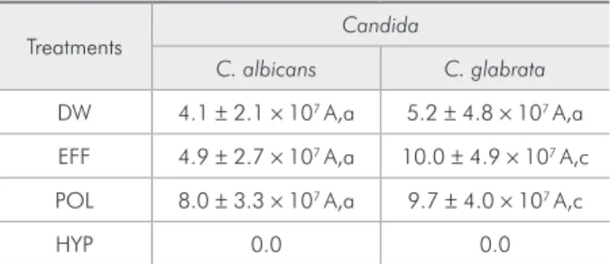
Regarding surface recolonization, alkaline per-oxides and DW treatments showed statistically simi-lar results for C. albicans (P > 0.05), while for C. glabrata, both alkaline peroxides showed higher counts when compared to the control (P < 0.001) (Table 3).
The only effective treatment to clean the liner surfaces was the use of HYP, since no Candida cell growth was observed under both conditions and for both strains (Tables 2 and 3).
C. glabrata showed signiicantly higher cell counts in comparison to C. albicans when treated with both alkaline denture cleansers (P < 0.001). However, regarding recolonization, no differences were found between the Candida strains after treat-ment with DW or two alkaline peroxides (P > 0.05) (Table 3).
SEM micrographs of specimen surface after the cleansing treatments are presented in Figure 1.
Table 1 - Surface roughness (µm) of relined specimens at baseline and after the treatments (Mean ± SD; n = 8).
Treatments Baseline After treatments
DW (Control) 3.8 ± 0.5 a 4.0 ± 0.3 b
HYP 3.5 ± 0.9 a 4.0 ± 0.9 b
EFF 3.2 ± 0.4 a 3.8 ± 0.6 b
POL 3.2 ± 0.5 a 3.4 ± 0.4 b
Different lowercase letters indicate significant differences between baseline and after treatments (P = 0.013).
Table 2 - Cell counts/mL for Candida spp. immediately af-ter cleansing treatments (Mean ± SD; n = 8).
Treatments Candida
C. albicans C. glabrata
DW 5.8 ± 5.4 × 10⁶ A,a 5.0 ± 2.3 × 10⁶ A,a
EFF 0.17 ± 0.23 × 10⁶ A,b 4.8 ± 3.2 × 10⁶ B,a
POL 0.07 ± 0.1 × 10⁶ A,b 5.7 ± 7.0 × 10⁶ B,a
HYP 0.0 0.0
Different uppercase letters in rows and different lowercase letters in columns indicate statistically significant differences between Candida spp., and among denture cleansers, respectively.
Table 3 - Cell counts/mL for the Candida spp. after the treatments and surface recolonization (Mean ± SD; n = 8).
Treatments Candida
C. albicans C. glabrata
DW 4.1 ± 2.1 × 10⁷ A,a 5.2 ± 4.8 × 10⁷ A,a
EFF 4.9 ± 2.7 × 10⁷ A,a 10.0 ± 4.9 × 10⁷ A,c
POL 8.0 ± 3.3 × 10⁷ A,a 9.7 ± 4.0 × 10⁷ A,c
HYP 0.0 0.0
HYP-cleaned specimens showed a cell-free surface (b), while POL (c), EFF (d) and DW (a) showed sur-faces with adhered cells.
Discussion
This study evaluated for the irst time the eficacy of denture cleansers on denture liner bioilms imme-diately after the cleansing treatments and after the treatments followed by surface recolonization. Sur-face roughness before and after treatments was also evaluated. The bioilm growth model used simulated
in vivo conditions of static bioilm growth found on the tissue-contacting surface of a denture.10
A rougher surface was found after all treatments, which indicated that changes in denture surface probably occur in the mouth, considering that it is
immersed in saliva while it is worn. The increased roughness associated with surface irregularities, such as cracks and pits, found in denture liners, pro-vide a larger surface area and a more sheltered envi-ronment for bioilm to develop and protect micro-organisms from being removed by cleaning.19 The
results of the present study showed that the speci-mens treated with denture cleansers POL and EFF presented lower cell counts compared to DW treat-ment. Nevertheless, these cleansing solutions were not able to remove bioilm completely. When the
Candida species were compared, the performance of cleansers POL and EFF was the same as that of the control for C. glabrata. These results corroborate those found by Ferreira et al.,12 who used the same
denture cleansers and Candida species, and Sousa et
Figure 1 - Representatives SEM micrographs of biofilm developed on denture liner surface after treatment with the different denture cleansers: (A) surface after DW treatment; (B) surface after HYP treatment; (C) surface after POL treatment; (D) surface after EFF treatment.
A B
al.,20 who found ineficiency on reduction of C.
al-bicans cells after using a similar peroxide cleanser. Another clinical study21 also found that POL and
EFF had similar performances, showing that bioilm growth in the present study model mimicked the in vivo environment.
Furthermore, irregularities can serve as reser-voirs for fungal species, ready for recolonization of the surface.8 In the present study, residual bioilm
cells are clearly seen on such irregularities in the SEM images. POL and EFF are likely to promote a greater disturbance on bioilm structure, consider-ing the effervescent action (Figures 1C and D), com-pared to DW, in which bioilm was poorly disturbed (Figure 1A). Results for POL and EFF were not dif-ferent from each other, even taking in consideration the time elapsed between both treatments.
After 48 h, liner surfaces treated with EFF and POL showed similar counts of viable cells, and it was also observed after the treatments and recolo-nization. Therefore, cells disturbed by the POL and EFF treatments could develop freely, while DW-treated cells remained on a steady state, which could be a reasonable explanation for the increase in cell counts observed after POL and EFF treatments and recolonization (Table 3).
The results of this study also showed that soak-ing specimens in 0.5% HYP, which is considered a fungicidal agent, was the only effective treatment against Candida species under both conditions, since no viable cells were found after its use for both
Candida species, which is in accordance with the
indings of a previous study.19 In addition to its
fun-gicidal effects, sodium hypochlorite acts dissolving mucin and other organic substances, such as extra-cellular polymeric matrix.2 Although satisfactory
results were found for HYP, the dental literature has shown that this product has the potential to bleach denture-base and may cause surface corrosion,8
es-pecially of the metal content in partial removable dentures. HYP is thus not indicated for daily use. Nevertheless, these problems seem to have been ex-aggerated and further studies are required to evalu-ate different concentrations and immersion times.
The objective of immersing a denture in a disin-fectant is to remove bioilm and to decontaminate the surface by destroying the microorganisms, since dentures may function as a reservoir of pathogens.22
Thus, one of the most important purposes of a den-ture cleansing protocol is to avoid recolonization of the oral cavity.12 In the present study, the fungal
levels returned to the initial levels within 48 h with-out signiicant difference between the Candida spe-cies. Although this study does not fully reproduce the oral environment, these results may suggest the need for stipulating a routine protocol for denture cleaning.
Conclusion
Within the limitations of this study, it may be concluded that alkaline peroxide denture cleansers were not effective in removing Candida spp. bioilm from denture liner surfaces and bioilm recoloniza-tion was not prevented.
References
1. Benting DG, Pesun IJ, Hodges J. Compliance of resilient denture liners immersed in effervescent denture cleansers. J Prosthodont. 2005 Sep;14(3):175-83.
2. Nikawa H, Yamamoto T, Hamada T, Sadamori S, Agrawal S. Cleansing efficacy of commercial denture cleansers: ability to reduce Candida albicans biofilm activity. Int J Prosthodont. 1995 Nov-Dec;8(6):527-34.
3. Dar-Odeh NS, Shehabi AA. Oral candidosis in patients with removable dentures. Mycoses. 2003 Jun;46(5-6):187-91. 4. Radford DR, Challacombe SJ, Walter JD. Denture plaque and
adherence of Candida albicans to denture-base materials in
vivo and in vitro. Crit Rev Oral Biol Med. 1999;10(1):99-116.
5. Li L, Redding S, Dongari-Bagtzoglou A. Candida glabrata: an emerging oral opportunistic pathogen. J Dent Res. 2007 Mar;86(3):204-15.
6. Coulthwaite L, Verran J. Potential pathogenic aspects of den-ture plaque. Review. Br J Biomed Sci. 2007;64(4):180-9. 7. Gornitsky M, Paradis II, Landaverde G, Malo AM, Velly AM.
8. Jin C, Nikawa H, Makihira S, Hamada T, Furukawa M, Murata H. Changes in surface roughness and colour stability of soft denture lining materials caused by denture cleansers. J Oral Rehabil. 2003 Feb;30(2):125-30.
9. Nikawa H, Hamada T, Yamashiro H, Kumagai H. A review of in vitro and in vivo methods to evaluate the efficacy of denture cleansers. Int J Prosthodont. 1999 Mar-Apr;12(2):153-9. 10. Pusateri CR, Monaco EA, Edgerton M. Sensitivity of
Candida albicans biofilm cells grown on denture acrylic to antifungal proteins and chlorhexidine. Arch Oral Biol. 2009 Jun;54(6):588-94.
11. Nakamoto K, Tamamoto M, Hamada T. Evaluation of denture cleansers with and without enzymes against Candida albicans. J Prosthet Dent. 1991 Dec;66(6):792-5.
12. Ferreira MA, Pereira-Cenci T, Rodrigues de Vasconcelos LM, Rodrigues Garcia RC, Del Bel Cury AA. Efficacy of denture cleansers on denture liners contaminated with Candida spe-cies. Clin Oral Investig. 2009 Jun;13(2):237-42.
13. Moura JS, Silva WJ, Pereira T, Del Bel Cury AA, Rodrigues Garcia RC. Influence of acrylic resin polymerization methods and saliva on the adherence of four Candida species. J Prosthet Dent. 2006 Sep;96(3):205-11.
14. Nikawa H, Yamamoto T, Hamada T, Rahardjo MB, Murata H. Commercial denture cleansers--cleansing efficacy against Candida albicans biofilm and compatibility with soft denture-lining materials. Int J Prosthodont. 1995 Sep-Oct;8(5):434-44.
15. Lima EM, Moura JS, Del Bel Cury AA, Garcia RC, Cury JA. Effect of enzymatic and NaOCl treatments on acrylic
roughness and on biofilm accumulation. J Oral Rehabil. 2006 May;33(5):356-62.
16. Luo G, Samaranayake LP. Candida glabrata, an emerging fungal pathogen, exhibits superior relative cell surface hydro-phobicity and adhesion to denture acrylic surfaces compared with Candida albicans. APMIS 2002 Sept; 110(9):601-610. 17. da Silva WJ, Seneviratne J,Samaranayake LP, Del Bel Cury
AA. Bioactivity and architecture of Candida albicans bio-films developed on poly(methyl methacrylate) resin surface. J Biomed Mater Res B Appl Biomater 2010 Jul;94(1):149-56. 18. Aires CP, Del Bel Cury AA, Tenuta LM, Klein MI, Koo H,
Duarte S, et al. Effect of starch and sucrose on dental biofilm formation and on root dentine demineralization. Caries Res 2008 Sept;42(5):380-6.
19. Pereira-Cenci T, Cury AA, Cenci MS, Rodrigues-Garcia RC. In vitro Candida colonization on acrylic resins and denture liners: influence of surface free energy, roughness, saliva, and adhering bacteria. Int J Prosthodont. 2007 May-Jun;20(3):308-10.
20. Sousa FA, Paradella TC, Koga-Ito CY, Jorge AO. Effect of sodium bicarbonate on Candida albicans adherence to thermally activated acrylic resin. Braz Oral Res. 2009 Oct-Dec;23(4):381-5.
21. Moore TC, Smith DE, Kenny GE. Sanitization of dentures by several denture hygiene methods. J Prosthet Dent. 1984 Aug;52(2):158-63.