Braz. J. Cardiovasc. Surg. vol.17 número4
Texto
Imagem
Documentos relacionados
[7], studied 48 patients randomly divided in two groups, 1 using epsilon-aminocaproic acid and the other tranexamic acid, reported that there was no statistical difference in the
Also in the right lower limbs, 16 cases of iliac regurgitation at an intensity of less than 26 cm/s (Classes 1 to 3) occurred, which is similar to the great saphenous ostial in
The FC IV, the serous creatinine level > 1.5 mg/dL, the LVEF < 65%, the AP < 60%, the cardiopulmonary bypass time > 120 minutes, the time of aortic cross-clamping >
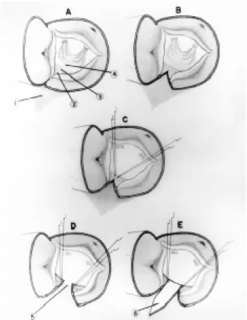
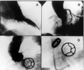
Surgical techniques for the treatment of ostial lesions of the coronary arteries, with the exception of endarterectomy [1], utilize patches of autologous tissue (bovine
The right internal mammary artery was anastomosed in the anterior descending artery and a great saphenous vein graft was utilized for the first marginal branch of the circumflex
Surgical Technique – The interventricular communication was corrected with a Teflon patch fixed at seven points and the mitral valve was evaluated which presented with severe
regurgitation disease in both left and right iliac veins sustain this hypothesis for the study of treatment by endovascular (valved stents) or direct surgical implantation of
changes of P wave in the electrocardiogram performed after the mitral valvuloplasty in patients with mitral stenosis and left atrial enlargement, and there is even less
