Brazilian
Journal
of
OTORHINOLARYNGOLOGY
www.bjorl.org
ORIGINAL
ARTICLE
Histologic
correlation
of
expression
of
Ki-67
in
squamous
cell
carcinoma
of
the
glottis
according
to
the
degree
of
cell
differentiation
夽
Rodrigo
Gonzalez
Bonhin
a,∗,
Guilherme
Machado
de
Carvalho
a,
Alexandre
Caixeta
Guimarães
a,
Carlos
Takahiro
Chone
a,
Agrício
Nubiato
Crespo
a,
Albina
Messias
de
Almeida
Milani
Altemani
b,
Eliane
M.I.
Amstalden
baDisciplineofOtorhinolaryngology,HeadandNeck,UniversidadeEstadualdeCampinas(UNICAMP),Campinas,SP,Brazil bDepartmentofPathology,UniversidadeEstadualdeCampinas(UNICAMP),Campinas,SP,Brazil
Received9February2013;accepted11March2014 Availableonline11June2014
KEYWORDS Carcinoma, squamouscell; Glottis;
Biologicalmarkers; Cellproliferation; Headandneck neoplasms
Abstract
Introduction:Squamouscellcarcinomaisthemostcommonneoplasmofthelarynxandglottis, anditsprognosisdependsonthesizeofthelesion,leveloflocalinvasion,cervicallymphatic spread, andpresenceofdistantmetastases.Ki-67(MKI67) isaprotein presentinthecore, whosefunctionisrelatedtocellproliferation.
Aim:ToevaluatetheexpressionofmarkerKi-67insquamouscellcarcinomaofthelarynxand glottisanditscorrelationtopathologicalfindings.
Methods:ExperimentalstudywithimmunohistochemistryanalysisofKi-67,calculatingthe per-centageofthecellproliferationindexinglotticsquamouscellcarcinomas.
Results:Sixteencaseswereanalyzed,withsixwell-differentiatedand10poorly/moderately differentiatedtumors.Therewasacorrelationbetweencellproliferationindexanddegreeof celldifferentiation,withhigherproliferationinpoorly/moderatelydifferentiatedtumors. Conclusion:Thecellproliferationindex,asmeasuredbyKi-67,maybeusefulinthe character-izationofhistologicaldegreeinglotticsquamouscelltumors.
© 2014Associac¸ãoBrasileira de Otorrinolaringologiae CirurgiaCérvico-Facial. Publishedby ElsevierEditoraLtda.Allrightsreserved.
夽 Pleasecitethisarticleas:BonhinRG,deCarvalhoGM,GuimarãesAC,ChoneCT,CrespoAN,AltemaniAM,etal.Histologiccorrelation
ofexpressionofKi-67insquamouscellcarcinomaoftheglottisaccordingtothedegreeofcelldifferentiation.BrazJOtorhinolaryngol. 2014;80:290---5.
∗Correspondingauthor.
E-mail:[email protected](R.G.Bonhin). http://dx.doi.org/10.1016/j.bjorl.2014.05.016
PALAVRAS-CHAVE Carcinomadecélulas escamosas;
Glote; Marcadores biológicos; Proliferac¸ãode células;
Neoplasiasdecabec¸a epescoc¸o
Correlac¸ãohistológicadaexpressãodoKi-67noCarcinomaEpidermoideGlóticode
acordocomgraudediferenciac¸ãocelular
Resumo
Introduc¸ão: O carcinoma de células escamosas é a neoplasia mais frequente da laringe e daregião glótica,seuprognósticodependedotamanhodalesão,doníveldeinvasãolocal, disseminac¸ãocervicallinfáticaedaexistênciademetástasesàdistância.Ki-67(MKI67)éuma proteínapresentenonúcleo,cujafunc¸ãoestárelacionadacomaproliferac¸ãocelular. Objetivos: AvaliaraexpressãodomarcadorKi-67emcarcinomadecélulasescamosasdalaringe glóticosecorrelacionarsuaexpressãocomosachadosanatomopatológicos.
Método: TrabalhoexperimentaldeanáliseimunohistoquímicadoKi67atravésdocálculo per-centualdoíndicedeproliferac¸ãocelularemprodutosdecarcinomasepidermóidesglóticos. Resultados: Dezesseis casos foram analisados, sendo seis bem diferenciados e dez pouco/moderadamentediferenciados.Houvecorrelac¸ãoentreoíndicedeproliferac¸ão celu-lareograudediferenciac¸ãocelular,sendoaproliferac¸ãomaiornospouco/moderadamente diferenciados.
Conclusão:Oíndicedeproliferac¸ãocelular,medidopeloKi-67podeserútilnacaracterizac¸ão dograuhistológicoemtumoresglóticosdecélulasescamosas.
©2014Associac¸ãoBrasileiradeOtorrinolaringologiaeCirurgiaCérvico-Facial.Publicado por ElsevierEditoraLtda.Todososdireitosreservados.
Introduction
Squamouscellcarcinomaisthemostcommonneoplasmof thelarynxandglotticregion,anditsprognosisdependson the size of the lesion, level of local invasion, lymphatic spread,andpresenceofdistantcervicalmetastases.1---5The
behaviorofthesquamouscellcarcinomaismarkedbythe degree of cell proliferation and differentiation, and this indexcanbederivedbymeasuringKi-67.6---10
Histologically, the epidermoid or squamous cell carci-nomaintheseregionsissimilartotheothers.Itisdefined bytheneoplasticproliferationofsquamouscellsin differ-entdegreesofdifferentiationandtheirinfiltrativenature. Depending on the power of keratinization and cellular atypia,theyareclassifiedasfollows:well,moderately,or poorlydifferentiated.1
Thewell-differentiatedtypeischaracterizedbyits sim-ilarity with normal squamous epithelium, high power of keratinization,andverymildcellatypia;themoderately dif-ferentiatedtypecontainsdistinctnuclearpleomorphismand mitoticactivityispresent,includingatypicalfigures.Inthis type,keratinizationismuchlower.1,2Inthepoorly
differen-tiatedtype,thereisapredominanceofimmaturecells,with numerousmitoticfiguresandvirtuallyabsentkeratinization. The levelofinvasionmaybelimitedtotheepithelium, respectingthebasallamina,whichiscalledinsitu.Whenit affectsonlythelaminapropria,itisconsidered microinva-siveorsuperficiallyinvasive.2
Asforthefranklyinvasivecarcinoma,itmanifestsasthe destruction of the basal lamina, clearly extending to the underlying tissues,possiblyaccompanied by stromal reac-tion. Perineural invasion and angiolymphatic invasion are signs of increased aggressiveness and change in staging, respectively.3
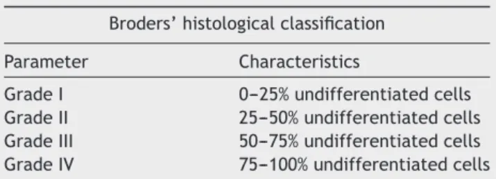
Broders proposed a grading system for squamous cell carcinomas.11,12 The classification established in 1920
and subsequently reviewed in 1925 was based on the
Table1 Broders’classification.
Broders’histologicalclassification
Parameter Characteristics
GradeI 0---25%undifferentiatedcells GradeII 25---50%undifferentiatedcells GradeIII 50---75%undifferentiatedcells GradeIV 75---100%undifferentiatedcells
fundamental principleof cellulardifferentiation, and was completely dissociated from clinical history. Carcinomas were divided into four grades, ranging from I to IV. Car-cinomas of grade I had up to 25% undifferentiated cells. GradeIItumorshad25---50%undifferentiatedcells.GradeIII tumorsconsistedof50---75%undifferentiatedcellsandGrade IVtumorshad75---100%undifferentiatedcells(Table1).11,12
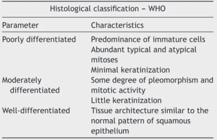
The histopathological classification of malignancy pro-posedby theWorldHealthOrganization (WHO, 2005) was basedonthedegreeofcelldifferentiationandallowedthe classificationof this malignancy into threecategories, as well-,moderatelyandpoorlydifferentiated(Table2).2,11---13
The well-differentiated tumor is characterized by sim-ilarity with normal squamous epithelium, high power of keratinization,and very mild cell atypia; the moderately differentiatedcontainsdistinctnuclearpleomorphism, pres-enceof mitotic activity,includingatypical figures.In this type, keratinization is much lower.2 In the poorly
differ-entiatedtype,immaturecellspredominatewithnumerous mitotic figures and keratinization is virtually absent. Regardingthe power ofinvasion, itcan belimited tothe epithelium,respectingthebasal lamina,when itis called insitu.Whenitonlyaffectsthelaminapropria,itis consid-eredmicroinvasiveorsuperficiallyinvasive.2
Table2 ClassificationrecommendedbytheWorldHealth Organization(WHO).
Histologicalclassification---WHO
Parameter Characteristics
Poorlydifferentiated Predominanceofimmaturecells Abundanttypicalandatypical mitoses
Minimalkeratinization Moderately
differentiated
Somedegreeofpleomorphismand mitoticactivity
Littlekeratinization
Well-differentiated Tissuearchitecturesimilartothe normalpatternofsquamous epithelium
underlyingtissues, possiblyaccompanied bystromal reac-tion. Perineuralinvasion and angiolymphatic invasion are signs of increased aggressiveness and change in staging, respectively.3
Themeasurementofmitoticfigures,acommonpractice intheevaluationoftumorsregardingtheirgrowthrateand degreeof aggressiveness,is farfromitsdesired accuracy, sinceitisnotpossibletoknow,throughhistologicalsections routinelystainedwithH&E,whichareexactlythecellsthat enteredthecelldivisioncycle.Thisisduetothefactthat thephasesG1,S,andG2ofthecellcyclearenotidentified byopticalmicroscopy(Fig.1).12
Ki-67(MKI67)isaproteinpresentinthenucleus,whose function is related to cell proliferation. This protein is onlyexpressedinthecelldivisioncycle:interphase(G1,S, G2),prophase,metaphase,anaphase,andtelophase,andis absentintheG0phase,whenthecellisquiescent; there-fore,itisanexcellentmarkerofcelldivision.11---13
The monoclonal antibody Ki-67 is used todemonstrate theexpression ofthisnuclearprotein.Thisantibody binds toitsspecific-antigen(MKi67)presentinthenuclei of pro-liferatingcells,stainingthemandhighlightingallcellsthat haveenteredthecelldivisioncycle.12,13
Through optical microscopy it is possible tocount the numberof cells per fieldor ‘‘hot spot’’ areas, which are stained, and to calculate the proliferation index (Ki-67
DNA synthesis
S
G1
G M
G0 Cell growth and protein synthesis
Quiescence and differentiation
Division
Figure1 Phasesofcellcycle.G1,S,G---presenceofMKI67.
index). The analysis of this index is what allows for the characterization of thedegree of tumor cell proliferation (growth rate) and consequently, itsdegree of aggressive-ness,increased risk of newtumor clones, and mutations, whichhindertumorrestrictionbytheimmunesystem.12,13
ThisstudyaimedtoevaluatetheexpressionoftheKi-67 markeringlotticsquamouscellcarcinomaofthelarynxand correlateitregardingpathologicalfindingsandcell prolifer-ationcharacteristics.
Materials
and
methods
This wasan experimental clinical study performed witha cross-section in a historicalcohort, using resection speci-mensfromglotticsquamouscellcarcinomaofpatientsfrom atertiarycareuniversityhospital.Thepatientswere submit-tedtohemi-laryngectomy,totallaryngectomy,andremoval ofthevocalcordswiththeirrespectivelymphadenectomy.
To facilitate the statistical analysis of glottic tumors, they were divided into well-differentiated and moder-ately/poorlydifferentiated.
Cases of glottic squamouscell carcinoma tumors were obtained from the database of the Pathological Anatomy Departmentofatertiaryuniversityhospitalintheperiodof 1994---2004.Thissampleincluded99casesintotal:85cases weremoderately/poorlydifferentiatedtumors,ofwhich41 cases were products of surgical specimens and 44 cases werebiopsiesthatwerenottreatedsurgically (chemother-apy/radiotherapy).Fourteencaseswerewell-differentiated tumors,witheightcases representingproductsof surgical specimensandsixbiopsycasesthatwerenottreated surgi-cally(chemo/radiotherapy).
The study only included cases that had surgical speci-mens,excludingbiopsycases,astheywouldbringbiastothe studybyincludingrandomtumorfragments.Ofeight well-differentiatedproductsofsurgicalspecimens,twocasesdid nothaveparaffinblocksavailablefornewsections.
Therefore, the study was conducted with six cases of well-differentiatedglotticlaryngealtumors,and10casesof moderately/poorly differentiated glottic laryngeal tumors wererandomlyselectedtocomprisethecontrolgroup.The groupsdidnothavethesamenumberofcases,astherewere notenoughcasesofwell-differentiatedtumorstocomplete thegroup.
Theinclusion criteriawereglottic laryngealtumorsfor which paraffin blocks were identified and that contained enoughmaterialfornewsections.Clinicaldataandtumor stagingwereobtainedfrommedicalrecords(Table3).
Thestudywasapprovedbytheethicscommitteeofthe institution(no.485/11).
Immunohistochemical analysiswas performed with the avidin---biotin---peroxidase method and used the following antibody:Ki-67(CloneMIB-1,IgG1,1:150,Dakobrand, pre-treatedTris---EDTA).
Positiveandnegativecontrolsofthemarkerwereused, inordertocomparethemwiththestudiedcases.Lymphoid tissuewasusedaspositivecontrol.
Table3 Clinicaldataofstudypatients.
Clinicaldata Group1 Group2 Obs
TNM T1N0M0---fourcases T1N0M0---threecases
T2NOMO---onecase T2N0M0---fivecases T3N0M0---onecase T2N1M0---onecase
T3N0M0---onecase Alcoholconsumption 50%(3cases)
Smoking 67%(4cases)
Gender 4M:2F 10M:0F
Age 53(37---73) 58(39---70)
Preop.clinicalneck Negative 1positivecase(N1)
Margins 50%positive 100%negative
Vascularinvasion 100%negative 80%negative 2cases+
Neuralinvolvement 100%negative 90%negative 1case+
Extracapsularextension 100%negative 100%negative
Preop.radiotherapy 0cases 1case
Postop.radiotherapy 50%(3cases) 20%(2cases)
Preop.chemotherapy 0cases 0cases
Postop.chemotherapy 0cases 0cases
Total 6patients 10patients
Group1,well-differentiatedglotticSCC;Group2,CECmoderately/poorlydifferentiatedglotticSCC;Preop.,preoperative;Postop., postoperative.
controls. Pictures scanned at high magnification (400×) weretakenfromhighlyreactive areas(hotspots),usinga Nikoncamera(Coolpix995).Subsequently,theimageswere uploaded to the computer for histological analysis, using the software Imagelab 2000. All parameters were blindly evaluated.
The evaluation of the cell proliferation rate was per-formed using commonpercentage ratio,asit already has been used in literature.8,9 Five areas of greater intensity
ofimmunomarkerexpression(hotspots)wereselected.At least 500 cells were counted, and the ratiobetween the numberofcellsthathadapositivelystainednucleusandthe totalnumberofcellswascalculated.Thepercentageratio wasstratifiedasfollows:<5%---lowcellproliferationrate; between5%and10%---mildcellproliferationrate;between 10%and30%---moderatecellproliferationrate;and>30% ---highcellproliferationrate.
To simplify and better analyze the data, they were groupedasfollows:
Cellproliferationindex:
--- Low/mildcellproliferation. --- Moderate/highcellproliferation.
Histologicaltype:
--- Glottic,well-differentiated.
--- Glottic,moderately/poorlydifferentiated.
Statisticalanalysis
Afterpoolingofthedata,thestatisticalanalysiswascarried outwithIBMSPSSStatistics®software,usingtheChi-squared
testandFisher’sexacttest.
Results
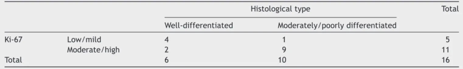
A total of 16 cases were evaluated, with six well-differentiated and 10 moderately/poorly differentiated cases.
Among the well-differentiated glottic tumors (n=6 cases),fourcaseshadlow/mildcellproliferation andtwo caseshadmoderate/highcellproliferation.Amongthe mod-erately/poorlydifferentiatedglottictumors(n=10cases), onecasehadlow/mildcellproliferationandninecaseshad moderate/highcellproliferation.
Inthecorrelationanalysisofcelldifferentiationdegree versuscellproliferation index,statistical significance was demonstrated(p<0.05)whencomparingthegroupsthrough two-tailedFisher’sexacttest,withp=0.036.
Table4 Distributionofcasesaccordingtohistologicaltypeandcellproliferation.
Histologicaltype Total
Well-differentiated Moderately/poorlydifferentiated
Ki-67 Low/mild 4 1 5
Moderate/high 2 9 11
Consideringthehistologicaltypeasthegoldstandardto assesscelldifferentiationinlaryngealcarcinomas,thecell proliferation index had a sensitivity of 0.9, specificity of 0.67,positive predictive value of 0.82,and negative pre-dictivevalueof0.8(Table4).
Discussion
Thecellproliferationindexwaspositivelycorrelatedwith thedegreeofcelldifferentiation,andwashigherin moder-ately/poorlydifferentiatedcases.Thisfindingcorroborates theideathatthemoreundifferentiatedtheneoplasm,the poorer the control of the cell division process and the greater the proliferation.8 This evaluation showed
statis-ticalsignificancebetweentumor typecorrelationandcell proliferationindex(p<0.05).
The studies by Rodrigues et al. found no correlation betweentheimmunohistochemicalexpressionofKi-67and prognosticvalue;7,14,15however,itappearsthatKi-67
over-expression associated with the idea of mitotic disarray, uncontrolledcellgrowth,andproliferationwouldbe associ-atedwithprognosticfactorsandapproachprioritizationof thesepatients.
An interesting fact was pointed out in a Canadian retrospective study on the clinical use of Ki-67, which demonstratedthatitsoverexpressioniscorrelatedwithhigh risk of cervical recurrenceafter radiotherapy and, there-fore,suchcases shouldundergomoretargeted treatment atearlierstages.16
Otherhead andneck tumorsites,suchastheoral cav-ity,alsoprovideevidencethatover expression ofmarkers suchasp53andKi-67 constituteagreaterrisk andpoorer prognosisforthistypeofsquamouscellcarcinoma.17
In a study with animals exposed to tobacco, only the developmentofbenignlesionswasobserved,withno pro-gression to malignant tumors. Histological evaluation of these animals showed absenceof p53marking, but Ki-67 positivityassociated with the tobacco-induced lesion was observed.18
Whenassessingtheclinicalprognosisofpatientswith epi-dermoidcarcinomasofthelarynx,itwasobservedthatthe Ki-67markerassociatedwithKi-S11isanimportanttoolto aidinclinicalmanagementandprognosticanalysis.Patients withlow cellproliferationrateassessed bythesemarkers have a lower recurrence rate with statistical significance (p<0.05)and improved five-year survival when compared tothegroupwithhighcellproliferationrate.19
Regarding the clinical application of Ki-67, it was also shownthatthegroupofpatientswithexpressionconsidered tobepositivehadhigherrateoflocalrecurrence,evenafter radiotherapy.Itissuggestedthatthesepatientshavea per-sonalizedandmoredetailedfollow-upprotocol.16However,
inanothergroup,itwasidentifiedthatpatientswithglottic tumorsinvolvingtheanteriorcommissureandshowingover expressionof Ki-67had abetteroutcome andhad tumors consideredtoberadio-sensitive,withsignificantrelevance ofresults.20
The literature also indicates the methodology used in the anatomopathological evaluation of advanced epider-moidcarcinomaofthelarynx.Intheaforementionedstudy, theheterogeneityinKi-67expressionwasdemonstratedin
areasoftumor cellactivityand,therefore,anassessment methodologyshouldbe well-established,andthe variabil-ity of previous studies that correlated the expression of immunomarkers withprognostic factors can be explained bysuchcharacteristic.21
Theevaluationofcarcinomaprecursorlesions (hyperpla-sia, carcinomain situ)wasalsoperformed instudieswith immunomarkerssuchasKi-67andp-16.Oftheselesions, car-cinomainsitutendstohaveahigherpositivitythanisolated hyperplasias.22
Although the present study found a significant corre-lation between cell proliferation degree and histological tumordifferentiation,thereweretwocases(18%)inwhich the rate of cell proliferation was moderate/high even in well-differentiated tumors. Considering that even some well-differentiated tumors showed high cell proliferation index, these tumors possibly exhibit a more aggressive behavior,aswiththeincreasedpopulation oftumor cells, thechanceofmitoticdisarrayisgreater.
Newclonesthatmaybemoreaggressiveduetomutations andthathinderrestrictionbytheimmunesystemmayhave betterresponsetochemotherapyandradiotherapy.Future studies that assess the behavior of well-differentiated tumorswithhighrateofcellproliferationmayassistinthe managementofthesepatients.
It is noteworthy that although this study had a sam-ple of 16 patients, limited by the difficulty in obtaining surgical materialfromwell-differentiated glotticcases, it emphasized theanalysis ofthis specific regionof the lar-ynx,differentfromotherstudieswithlargersamplesthat included patients with carcinomas in any region of the larynx.Therefore,thepresentstudy corroboratesthe cor-relation between increased expression of Ki-67 in more undifferentiatedtumors,8 anddemonstratesthatthis
cor-relationalsoappliesspecificallytolaryngealglottictumors. The authors believe that the association between the expression of Ki-67 with clinical criteria and prognosis remainsunclear.Thisassociationisstillquitecontroversial in themedical literature,in both clinical and experimen-talstudies.Therelevanceofusingimmunomarkersisclear; however,furtherstudiesarenecessarytobetterdefinethe patientsinwhichimmunomarkersshouldbeusedinclinical practice.
Conclusion
Thisstudydemonstratedthattherateofcellproliferation measuredbyKi-67maybeusefulincharacterizingthe histo-logicalgradeinglotticsquamouscelltumorsofthelarynx. Furtherstudiesareneededtoevaluatethebehaviorof well-differentiated tumors with high rate of cell proliferation todefinethebesttherapeuticapproachinthesepatients. Otherfactorsmustbeinvolvedregardingthepathogenesis andtumorprogressionofthisdiseaseintheglottis.
Funding
Conflict
of
interest
Theauthorsdeclarenoconflictsofinterest.
References
1.AltemaniAMAM, Amstalden EMI.Bogliolo. Patologia.8thed. GuanabaraKoogan;2009.
2.Diagnostichistopathologyoftumors.3rded.Elsevier;2007.p. 158---65.
3.Sternberg’s diagnostic surgical pathology. 5th ed. Wolters KluwerHealth/LippincottWillian&Wilkins;2009.p.777---92. 4.Carde-saA,GaleN,NadalA,ZidarN.Pathologyandgeneticsof
headandnecktumors(WHO/OMS).IARCPress;2005.p.118---21. 5.CarvalhoGM,ChoneCT, GuimarãesAG,KohlerHF,GrippFM, CrespoAN,etal.Plannedneckdissection:evaluationofviable metastases.RevBrasCirCabec¸aPescoc¸o.2011;40:133---7. 6.MohanS,EpsteinJB.Carcinogenesisandcyclooxygenase:the
potentialroleofCOX-2inhibitioninupperaerodigestivetract cancer.OralOncol.2003;39:537---46.
7.RodriguesRB,MottaRR,MachadoSMS,CambruzziE,ZettlerEW, ZettlerCG,etal.Prognosticvalueoftheimmunohistochemistry correlationofKi-67andp53insquamouscellcarcinomasofthe larynx.BrazJOtorhinolaryngol.2008;74:855---9.
8.AshrafMJ,MaghbulM,AzarpiraN, KhademiB.Expression of Ki67andP53inprimarysquamouscellcarcinomaofthelarynx. IndianJPatholMicrobiol.2010;53:661---5.
9.HaroonS,HashmiAA,KhurshidA,KanpurwalaMA,MujubaS, MalikB,etal.Ki67indexbreastcancer:correlationwithother prognosticmarkersand potentialinPakistanipatients.Asian PacJCancerPrev.2014;14:4353---8.
10.BoonkitticharoenV,KulapaditharomB,LeopairutJ,Kraiphibul P, Larbcharoensub N, Cheewaruangroj W, et al. Vascular endothelialgrowthfactor a and proliferationmarkerin pre-diction of lymph node metastasis in oral and pharyngeal squamouscell carcinoma.Arch Otolaryngol Head NeckSurg. 2008;134:1305---11.
11.BrodersAC.Themicroscopicgradingofcancer.SurgClinNorth Am.1941;21:947---62.
12.Lourenc¸o SQC, Schueler AF, Camisasca DR, Lindenblatt RC, BernardoVG.Histologicalclassificationsoforalsquamouscell carcinoma:areviewoftheproposedsystems.RevBras Can-cerol.2007;53:325---33.
13.WardLS.Entendendo oprocessomoleculardatumorigênese. ArqBrasEndocrinolMetab.2002;46:351---60.
14.BubánT,TóthL,TanyiM,KappelmayerJ,Antal-SzalmásP.Ki-67 ---newfacesofanoldplayer.OrvHetil.2009;150:1059---70. 15.CalliC,CalliA,PinarE,OncelS,TatarB.Prognosticsignificance
ofp63,p53andki67expressioninlaryngealbasaloidsquamous cellcarcinomas.B-ENT.2011;7:37---42.
16.Nichols AC,WhelanF,Basmaji J, DhaliwalS, DowthwaiteS, ChapeskieC,etal.Ki-67expressionpredictsradiotherapy fail-ure in early glottic cancer. J Otolaryngol Head Neck Surg. 2012;41:124---30.
17.MottaRdaR,ZettlerCG,CambruzziE,JotzGP,BerniRB.Ki-67 and p53 correlationprognosticvalueinsquamous cell carci-nomasoftheoralcavityandtongue.BrazJOtorhinolaryngol. 2009;75:544---9.
18.deOliveiraSemenzatiG,deSouzaSalgadoB,RochaNS,Michelin MatheusSM,deCarvalhoLR,GarciaMartinsRH.Histologicaland immunohistochemicalstudyoftheexpressionofp53andki-67 proteinsinthemucosaofthetongue,pharynxandlarynxofrats exposedtocigarettesmoke.InhalToxicol.2012;24:723---31. 19.Cordes C, Münzel AK, Rudolph P, Hoffmann M, Leuschner I,
Gottschlich S. Immunohistochemical staining of Ki-67 using the monoclonal antibody Ki-s11 is a prognostic indica-tor for laryngeal squamous cell carcinoma. Anticancer Res. 2009;29:1459---65.
20.AhmedWA,SuzukiK,ImaedaY,HoribeY.Ki-67,p53and epider-malgrowthfactorreceptorexpression inearlyglotticcancer involvingtheanteriorcommissuretreatedwithradiotherapy. AurisNasusLarynx.2008;35:213---9.
21.WittekindtC,SittelC,GreissJ,DrebberU, EckelHE,Preuss SF.MappingofKi-67proteindistributiononwholeorganserial sectionsofthelarynx.ActaOtolaryngol.2008;128:207---12. 22.PavlovicB,DjukicV,MilovanovicJ,TomanovicN,Milovanovic