*Correspondence: S. L. Fialho. Departamento de Desenvolvimento Far-macotécnico e Biotecnológico, Fundação Ezequiel Dias. Rua Conde Pereira Carneiro, 80 - Gameleira - 30510-010 - Belo Horizonte - MG, Brazil. Email: [email protected]; [email protected]
A
vol. 48, n. 1, jan./mar., 2012
Poly-
ε
-caprolactone microspheres containing interferon
alpha as alternative formulations for the treatment
of chronic hepatitis C
Cristiane da Silva Melo
1, Bruno Gonçalves Pereira
2, Armando Silva-Cunha
1, Sílvia Ligório Fialho
2,*1Faculty of Pharmacy, Federal University of Minas Gerais, 2 Department of Pharmaceutical and Biotechnological Development, Fundação Ezequiel Dias
Interferon-alpha (alpha) is one of the main drugs used in the treatment of hepatitis C. Use of IFN-alpha has some limitations that result in poor treatment eficacy and low patient compliance. Therefore, the aim of this study was to develop poly-ε-caprolactone (PCL) microspheres containing IFN-alpha as an alternative for the treatment of chronic hepatitis C. Microspheres were prepared using the multiple emulsion followed by solvent evaporation technique. Particle size, surface morphology, drug content and encapsulation eficiency of the microspheres produced were evaluated. The stability of the formulation was assessed after 90 days at -20ºC. An in vitro release study was performed in PBS. In vitro cytotoxicity
of the formulation was studied using hepatic cell line. The freeze-dried microspheres had mean particle size, IFN-alpha content, and encapsulation eficiency of 38.52 ± 4.64 µm, 15.52 ± 3.28% and 83.93 ±
5.76%, respectively. There were no signiicant changes during storage and the structural integrity of the protein was not compromised by the preparation technique. A total of 82% of the IFN-alpha was released after 28 days and the developed microspheres did not present cytotoxicity to the hepatic cell
line. In vivo studies are currently underway to evaluate the biological activity of IFN-alpha encapsulated
into microspheres.
Uniterms: Interferon alfa. Hepatitis C/treatment. Poly- ε-caprolactone/microspheres/development.
O interferon alfa (IFN-alfa) é um dos principais fármacos utilizados no tratamento de hepatite C, mas o seu uso apresenta limitações que resultam em baixa eicácia do tratamento e não adesão do paciente. Diante disso, este estudo objetiva o desenvolvimento de microesferas de poli-ε-caprolactona (PCL) contendo IFN-alfa como alternativa ao tratamento de hepatite C crônica. As microesferas foram preparadas pelo
método de emulsão múltipla seguido de evaporação do solvente e caracterizadas quanto ao diâmetro
médio das partículas, morfologia da superfície, taxa e eiciência de encapsulamento. A estabilidade da formulação foi acompanhada durante 90 dias a -20 ºC. O estudo de liberação in vitro foi realizado em PBS.
A citotoxicidade da formulação foi avaliada utilizando linhagem de células hepáticas. As microesferas lioilizadas apresentaram diâmetro médio, taxa de encapsulamento e eiciência de encapsulamento de 38,52 ± 4,64 µm, 15,52 ± 3,28% e 83,93 ± 5,76%, respectivamente. Não foram observadas alterações
signiicativas durante o armazenamento e a integridade estrutural da proteína foi mantida após o preparo. Oitenta e dois por cento de IFN-alfa foram liberados em 28 dias e a formulação desenvolvida não apresentou toxicidade para as células testadas. Estudos in vivo estão em andamento para avaliar a
atividade biológica do IFN-alfa encapsulado nas microesferas.
INTRODUCTION
Hepatitis is deined as an inlammatory liver dis -ease which is caused mainly by virus, bacteria, fungus or drugs. Although several virus types may cause hepatitis, only types B, C and D are responsible for chronic hepa -titis (Ferreira, Carvalho 1998). The virus responsible for hepatitis C disease belongs to the Flaviviridae family and
is recognized as the leading cause of chronic hepatitis and liver diseases for which liver graft is indicated. An esti -mated 170 to 220 million people in the world are infected by this virus worldwide and 3 to 4 million new cases of hepatitis C infection emerge annually. Some 55 to 85 % of patients with the acute infection have an elevated risk of developing hepatocellular injury, cirrhosis, carcinoma or liver failure (Parolin et al., 2006; Siciliano, Boulos, 2004;
Weigand et al., 2007).
The treatment of hepatitis C consists, initially, of suppressing viral replication. For this purpose, interferon, ribavirin and PEG-interferon are the main drugs used in clinics (Almeida, Souto, 2007).
Interferon-alpha (IFN-alpha) was approved for clinical use in 1986, being one of the irst biotherapeutic products approved by the FDA. It belongs to a class of macromolecules that naturally occurs in the body and is produced by cells of the immune system (Sanchez et al., 2003). During the last 20 years, IFN-alpha has had
increasingly broad clinical application, being used in the treatment of conditions such as hepatitis B and C, multiple sclerosis, viral myocarditis, Kaposi’s sarcoma, hairy cell leukaemia, condyloma acuminatum and cutaneous T cell lymphomas with considerable success (Beilharz 2000). However, the clinical use of IFN-alpha has some limita -tions, including a short half-life and rapid loss of biologi -cal activity, long-term frequent injections, luctuation of plasma concentration and serious side effects. These dif -iculties may result in poor eficacy of the treatment and low compliance among patients (Beilharz, 2000; Sanchez
et al., 2003; Hua et al., 2011).
The increasing number of peptides and proteins approved by the regulatory authorities for human use represents an important ield of research. However, their use poses major challenges due to a number of obstacles to therapeutic application that hamper effective delivery and release in the body (Almeida, Souto, 2007). To guarantee eficacy of the proteins and peptides, they must target the site of action without being degraded and remain at the site for a long period to promote the desired effect (Segers, Lee, 2007).
Several delivery systems of IFN-alpha have been proposed to improve its efficacy and half-life. Among
these systems, pegylated IFN-alpha has proved highly successful as it can promote the sustained release of this protein over a period of one week. However, according to the literature, although pegylation can improve bio -distribution, evidence suggests that polyethylene glycol (PEG), due to its characteristic of non-biodegradability, may accumulate in the body. Also, it has been suggested that PEG conjugates may suppress protein immunogenic -ity and antigenic-ity, which may in turn reduce the conju -gate residence time in the blood stream (Hua et al., 2011;
Segers, Lee, 2007). Furthermore, it has been described that pegylated IFN-alpha is more prone to induce side effects than IFN-alpha (Li et al., 2011).
Recently, several attempts have been made to develop IFN-alpha delivery systems composed of biode -gradable polymers, such as micro and nanoparticles, that can effectively delivery the active protein at a sustained rate, and at a concentration within its therapeutic window (Mohl, Winter, 2004)
These systems are able to maintain adequate serum levels of the drug for long periods of time and protect the macromolecules from degradation before they reach the target site, thus reducing side effects and increasing pharmacological activity (Teixeira et al., 2005). The main
objective of the development of delivery systems contain -ing IFN-alpha is to improve patient compliance to the treatment by lowering the number of injections, reducing side effects, and promoting prolonged release of the drug at the site of action (Jorgensen et al., 2006). Among these
systems, microparticles may represent a promising alterna -tive as they can facilitate controlled delivery of IFN-alpha by non-invasive routes, such as oral and nasal.
Among the biodegradable polymers, poly-ε-caprolactone (PCL) is of particular interest as it allows for a long, sustained and possibly modulated drug release rate (Chawla, Amiji, 2002). PCL is a biodegradable and biocompatible polymer with a very slow degradation rate, making it suitable for long-term delivery. Furthermore, it is biocompatible and extensively used in the pharma -ceutical and biomedical ields as a biomaterial (suture, osteosynthesis material, artiicial skin, support for cellular regeneration) and for prolonged drug delivery systems targeting speciic tissues within the body (Sinha et al., 2004). PCL micro or nanoparticles have indeed been widely explored in recent years for the administration of drugs by different routes and for the treatment of differ -ent diseases (Barbault-Foucher et al., 2002; Blanco et al.,
2003; Lemmouchi et al., 1998; Sinha et al., 2004; Sun et al., 2006; Verger et al., 1998).
-ity and physical/chemical interactions. Consequently, as described in previous studies, its co-encapsulation with stabilizing substances can be necessary to assure stability of the peptides and proteins (Li et al., 2011). Substances
evaluated to date include human serum albumin (HSA), which has proven effective for protecting interferon against potential chemical and physical degradation mech -anisms (Ruiz et al., 2006).
Therefore, the objective of this study was to develop PCL microspheres containing IFN-alpha co-encapsulated with HSA as an alternative to pegylated-interferon for the treatment of chronic hepatitis C. Characterization and in
vitro cytotoxicity of the formulation and its components
in hepatic cell line were studied.
MATERIALS AND METHODS
Materials
Human recombinant IFN-alpha was purchased from Pestka Biomedical Laboratories Interferon Source (USA). Polymer poly-ε-caprolactone [(PCL) molecular weight (Mw) 14kDa] and human serum albumin [(HSA), Frac -tion V, Mw 66kDa] were obtained from Sigma-Aldrich (Brazil). Poloxamer 188 (Pluronic® F68) was kindly do -nated by Basf Chemicals (USA). Ultrailtrated water was obtained from Milli Q plus, Millipore (USA). All other chemicals were of analytical grade.
Preparation of IFN-alpha-loaded microspheres
The multiple emulsion followed by solvent evapo -ration was the technique used for the prepa-ration of the microspheres. Firstly, a solution of IFN-alpha was diluted in phosphate-buffered saline (PBS, pH 7.4) containing HSA to form the inner aqueous phase. This solution was added to the organic phase, containing PCL dissolved in dichloromethane, with vigorous stirring using an UltraTur -rax device (IKA Labortechnik, Germany) to form a single emulsion. This emulsion was added to a water solution containing the surfactant (poloxamer 188) under stirring again using an Ultra Turrax device (IKA Labortechnik, Germany) until multiple emulsion formation. The solvent (dichloromethane) was eliminated by evaporation under magnetic stirring and the microspheres thus obtained were collected by centrifugation (25700xg for 15 minutes, three times). The supernatant obtained was collected for IFN-alpha analysis and the pellets were immediately stored at -20 ºC (E-C Apparatus, USA). Prior to lyophilization, the samples stored at -20 ºC were placed in liquid nitro -gen for 2 minutes. The samples were then placed in the
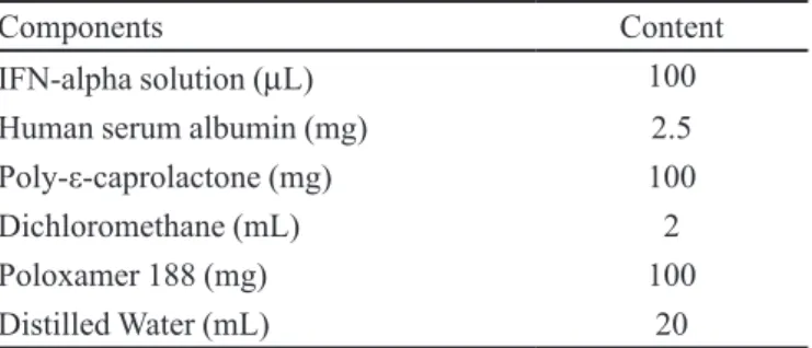
TABLE I - Composition of the developed microsphere
suspensions
Components Content
IFN-alpha solution (µL) 100
Human serum albumin (mg) 2.5
Poly-ε-caprolactone (mg) 100
Dichloromethane (mL) 2
Poloxamer 188 (mg) 100
Distilled Water (mL) 20
lyophilization apparatus, which was set at a temperature of -50 ºC. After 4 hours, the samples were removed from the equipment and immediately stored at -20 ºC. Formula -tions without IFN-alpha were also prepared by the same technique and served as controls. The composition of the developed formulation is described in Table I.
Analysis of IFN-alpha
For determination of drug content and encapsulation eficiency, and also of the amount of IFN-alpha released during the in vitro study, the ELISA technique with the
VerikineTM Human IFN Alpha Elisa Kit (Pestka Biomedi -cal Laboratories Interferon Source, USA) was used. The method adapted from Yang et al. (2010) was followed, as
described below.
A known weight of freeze-dried microspheres was added to 10 mL of 6 N HCL solution, which was then placed in a water bath set at 40 ºC for 36 hours. A 8 N NaOH solution was added to the resultant solution and the amount of IFN-alpha was then determined in supernatant liquid us -ing the ELISA kit. The amount of IFN alpha was calculated using the equation obtained from a standard curve.
The validation of the method showed the absence of interference of the components used in the microsphere formulations, ruling out the risks of over or under estima -tion of the concentra-tion of IFN-alpha.
Characterization of IFN-alpha-loaded microspheres developed
Particle size
-struments, England). For the analysis, water was used as a dispersing agent and the obscurity rate was set at 12%. All analyses were performed at room temperature. The results were expressed as mean particle size ± standard deviation of values from six different samples.
Zeta potential
The Zeta potential, that measures the surface charge of the particles, was evaluated by dynamic light scatter -ing set at a wavelength of 633 nm at an angle of 90º us-ing a Zetasizer apparatus (Zetasizer HSA 3000, Malvern, Instruments Ltd., UK). All analyses were realized at the temperature of 25 ºC and the samples were diluted 40 times to analysis. The results were expressed as zeta potential ± standard deviation of values from six different samples.
Morphological studies
The surface morphology of the developed micro -spheres after freeze-drying was observed by scanning electron microscopy (SEM) using a FEI microscope (FEI, USA) operating at 15 kV. Before visualization, the freeze-dried samples were placed into coverslips, which were dried for 72 hours in a vacuum dessicator at room temperature. After drying, the coverslips containing the samples were mounted on aluminum stubs. Prior to mi -croscopic examination, the samples were sputter coated with a gold layer under an argon atmosphere. Microsphere surfaces were viewed at 300 x to 1000 x magniication, and the images transferred to a computer by means of a digital image transfer interface.
Drug content and encapsulation efficiency
The drug content was calculated based on the ratio of amount of IFN-alpha found in the microspheres to total amount of microspheres used. The encapsulation eficiency was calculated based on the ratio of amount of IFN-alpha found in the microspheres to total amount of drug added to the formulation.
The amount of IFN-alpha was determined using the technique described in the section on Analysis of
IFN-alpha. All values were expressed as percentage ± standard
deviation from six different samples.
Stability assessment for microspheres developed
Between preparation and beginning of the study, the developed freeze-dried microspheres were stored in impermeable containers that provided a permanent barrier to entry of moisture or solvent.
The particle size, drug content and encapsulation efficiency were monitored over 7, 30, 60 and 90 days after preparation of the freeze-dried microspheres stored
at a temperature of -20° C. Before the analysis of particle size, the freeze-dried microspheres were resuspended in water containing 0.03% polysorbate 80.
The drug content was measured using the method described in the section on Analysis of IFN-alpha.
In vitro release study
A 50 mg amount of IFN-alpha-loaded freeze-dried microspheres was placed in sterile low-binding microtubes and suspended in 2.0ml of phosphate buffered saline (PBS, pH 7.4) containing 0.02% of polysorbate 80. The micro -tubes were incubated at 37 ºC and centrifuged at 90 rpm. At pre-determined intervals, samples were centrifuged at 2000xg for 15 min and 1.0 mL of the supernatant was collected and replaced with fresh PBS to maintain the sink conditions. The amount of IFN-alpha released was determined using the method described in the section on
Analysis of IFN-alpha.
Evaluation of IFN-alpha structural integrity
For the evaluation of IFN-alpha structural integrity after microsphere preparation, during the storage time, and also during the in vitro release study, sodium dodecyl
sulfate polyacrylamide gel electrophoresis (SDS-PAGE) analysis was performed using a Bio-Rad apparatus under non-reduced conditions. The samples were prepared under reduced conditions for application onto a gel consisting of 5 and 15% stacking and resolving gel, respectively.
In vitro cytotoxicity study
Microspheres and components
For this study, the following samples were used: microspheres alone, IFN-alpha-loaded microspheres co-encapsulated with HSA and the components of the formulation (PCL, poloxamer, IFN-alpha, HSA) at the concentration used in the preparation. The cytotoxicity of these samples was evaluated using a hepatic cell line (HepaRGTM, Invitrogen, USA).
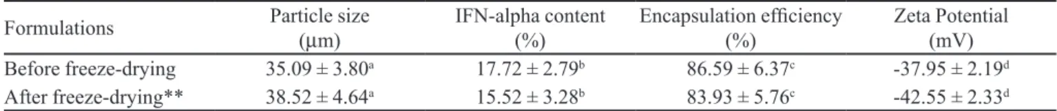
TABLE II - Characteristics of developed microspheres before and after freeze-drying*
Formulations Particle size ( µm)
IFN-alpha content
(%) Encapsulation eficiency (%) Zeta Potential (mV)
Before freeze-drying 35.09 ± 3.80a 17.72 ± 2.79b 86.59 ± 6.37c -37.95 ± 2.19d
After freeze-drying** 38.52 ± 4.64a 15.52 ± 3.28b 83.93 ± 5.76c -42.55 ± 2.33d
Different letters in the same column means values are signiicantly different (p< 0.05). * All results expressed as mean ± standard
deviation, n = 6. ** Results related to freeze-dried microspheres after resuspension in water containing 0.03% polysorbate 80.
USA). Briely, culture medium was removed, wells were washed once with PBS, and MTT diluted in culture medium containing 1% FBS was added to each well. After 3 h of incubation at 37 ºC and 5% CO2 atmosphere, the plate was centrifuged at 1.000 rpm for 10 minutes in order to precipi -tate the formazan crystals. The supernatant was removed and 50 µL of dimethylsulphoxide (DMSO) was added to
each well to dissolve the formazan crystals. The absorbance values were measured at 595 nm using a microplate reader (Spectramax M5e, Molecular Devices, USA).
As controls for cellular viability, the same cells used in this study received no treatment (positive control) or hydrogen peroxide (30 mM) (negative control). A positive control was considered as 100% of cellular viability. The microspheres or its components were considered cytotoxic if less than 50% of cellular viability was found. The results are expressed as percentage cellular viability ± standard deviation from six different samples.
Microsphere degradation products
Samples of microspheres alone and IFN-alpha-loaded microspheres co-encapsulated with HSA were used after freeze-drying.
For this experiment, hydrolysis and oxidation degradation methods were used to simulate microsphere degradation in vivo. A suficient amount of freeze-dried
microspheres was weighed to a final concentration of 0.1 mg/mL in 30 % hydrogen peroxide solution. The samples were incubated for 24 hours at the temperature of 85 ºC until complete visual degradation. Subsequently, the solutions containing the biodegradation products obtained were evaluated using the hepatic cell line, fol -lowing the same procedure as described for Microspheres
and components.
As controls for cellular viability, the same cells used received no treatment (positive control) or hydrogen peroxide (30 mM) (negative control). A positive control was considered as 100% of cellular viability. The micro -sphere degradation products were considered cytotoxic if less than 50% of cellular viability was found. The results are expressed as percentage cellular viability ± standard deviation from six different samples.
Statistical analysis
The unpaired t-test was used to analyze differences in particle size, IFN-alpha content and encapsulation ef -iciency of the developed microspheres. Values of p< 0.05
were considered statistically signiicant.
The One-way ANOVA Kruskal-Wallis test was used to evaluate the differences for each parameter throughout the timeframe of the stability assessment test. Values of
p< 0.05 were considered statistically signiicant.
RESULTS
Preparation and characterization of IFN-alpha-loaded microspheres
The freeze-dried microspheres co-encapsulated with HSA were successfully prepared by the multiple emulsion followed by solvent evaporation technique and the results of their characterization are shown in Table II. It was observed that the freeze-drying technique did not signiicantly change microsphere characteristics.
Scanning electron photomicrographs of the micro -spheres without any protein and those containing IFN-alpha co-encapsulated with HSA are depicted in Figure 1. It was observed that independent of composition, all microspheres were spherical with a smooth surface and homogeneous size. For both samples evaluated, no signiicant difference in the surface of the microspheres was detected.
FIGURE 1 - Scanning electron photomicrographs of freeze-dried
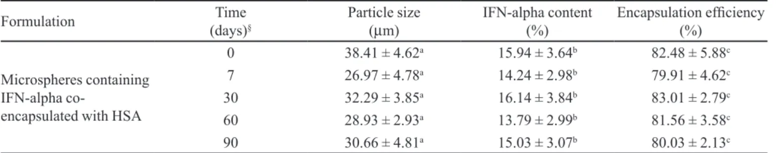
The stability assessment study showed that the mi -crospheres could be easily resuspended in 0.03% polysor -bate 80. There was no signiicant change in particle size, suggesting they do not agglomerate during storage. (Table III). This was expected because the packaging selected for this product is impermeable and should not allow entry of moisture or any other solvent that could cause microsphere agglomeration or degradation. The structural integrity of the protein does not appear to be compromised by the preparation technique or during storage as evidenced by gel electrophoresis analysis.
In vitro release study
The cumulative release proile of IFN-alpha from the microspheres prepared with PCL co-encapsulated with HSA under in vitro sink conditions is plotted in Figure 2.
The results showed a fast initial release and continu -ous release, during which the drug was released from mi -crospheres in a sustained manner (Figure 2). The structural integrity of the protein does not appear to be compromised by this study as evidenced by gel electrophoresis analysis.
In vitro cytotoxicity study
To evaluate the cytotoxicity of the microspheres and their components, as well as the microspheres’ degradation products, the MTT assay on a hepatic cell line was per -formed. MTT, a tetrazolium salt, is cleaved by mitochon -drial dehydrogenase in living cells forming the product formazan, which is dark blue and easily measurable. If the cells sustain damage or are dead, dehydrogenase activity is reduced and lower levels of formazan will be produced compared to live cells (Thomas et al., 2009).
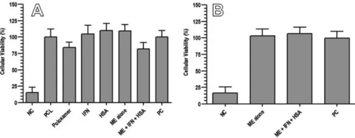
As seen in Figure 3, cell viability after application of the positive control was considered 100% and results
obtained for the samples were calculated related to the positive control. The cellular viability after hydrogen per -oxide 30 mM (negative control) treatment was near 15%, showing that it is highly cytotoxic to the evaluated cells.
The results obtained in this study showed that the developed microspheres and all their components did not present cytotoxicity to the evaluated cells as all showed a percentage of cellular viability higher than 50% (Figure 3A). Comparing microspheres alone and those containing IFN-alpha co-encapsulated with HSA, it was observed that the cellular viability was slightly higher for the mi -crospheres alone than for those containing the proteins. Nevertheless, cellular viability was still over 50% for both formulations, showing that it is not toxic.
Regarding microsphere degradation products, the cytotoxicity study showed that in the samples submitted to degradation, the products obtained were not toxic to TABLE III - Stability assessment for developed microspheres*#
Formulation Time
(days)§
Particle size (µm)
IFN-alpha content
(%) Encapsulation eficiency (%)
Microspheres containing IFN-alpha
co-encapsulated with HSA
0 38.41 ± 4.62a 15.94 ± 3.64b 82.48 ± 5.88c
7 26.97 ± 4.78a 14.24 ± 2.98b 79.91 ± 4.62c
30 32.29 ± 3.85a 16.14 ± 3.84b 83.01 ± 2.79c
60 28.93 ± 2.93a 13.79 ± 2.99b 81.56 ± 3.58c
90 30.66 ± 4.81a 15.03 ± 3.07b 80.03 ± 2.13c
IFN-alpha – interferon alpha. HSA - human serum albumin. Different letters in same column means values are signiicantly different (p< 0.05). * All results expressed as mean ± standard deviation, n = 6. # Results related to freeze-dried microspheres
after resuspension in water containing 0.03% polysorbate 80. § Time was determined immediately after preparation, considering
freeze-dried microspheres stored at -20 ºC.
FIGURE 2 - Cumulative interferon-alpha release from
FIGURE 3 – Percentage of hepatic cell line viability after: A – application of developed microspheres and all their components;
and B – application of microsphere degradation products. NC: negative control; PCL: poly-ε-caprolactone; IFN: interferon-alpha; HSA: human serum albumin; ME alone: microspheres without protein; ME + IFN + HSA: microspheres containing interferon-alpha and human serum albumin; PC: positive control (Values are shown as mean ± standard deviation, n = 6).
the evaluated cells as the percentage cellular viability was higher than 50% for all the groups (Figure 3B).
DISCUSSION
The multiple emulsion followed by solvent evapora -tion is the technique most used to encapsulate hydrophilic substances, including proteins, into microspheres, as they can be entrapped in the inner aqueous phase, thus leading to higher drug content results. According to the literature, it can sometimes be necessary to use stabilizing agents in order to maintain the physical stability of proteins (San -chez et al. 2003). Human serum albumin (HSA) at a con -centration of 0.1% can protect IFN-alpha against chemical and physical degradation when it is under solution (Ruiz
et al., 2006). For this reason, and also because of its avail -ability and low cost, it was decided to co-encapsulate IFN-alpha with HSA.
In this study, it was observed that microsphere particle size slightly increased after freeze-drying but as this was not signiicant, it can be assumed that the freeze-drying process does not influence particle size in this formulation, even when it is resuspended (Table II).
When evaluating the content and encapsulation eficiency, the encapsulation eficiency for freeze-dried microspheres was slightly different compared with micro -spheres that were not freeze-dried (Table II).
In polymeric matrix systems, such as microspheres, the drug is dispersed, homogeneously inside the matrix material. Slow diffusion of the drug through the polymeric system provides its sustained release (Dash and Cudworth
II 1998). Drug release from biodegradable monolithic de -vices can occur by diffusion, degradation of the polymer, or a combination of these two mechanisms (Jain, 2000).
PCL is a biodegradable polymer that degrades by hydrolysis producing ε-hydroxycaproic acid by cleavage of the polymeric chains at the ester linkage (Merkli et al., 1998). Its small polymeric fragments from the matrix
undergo phagocytosis and are easily eliminated from the body. The degradation of PCL matrices occurs at very low rates, making it suitable for the sustained and prolonged release of interferons because it can both reduce the number of injections for treatment and increase patient compliance. According to the literature, their degradation products are not toxic, corroborated by the in vitro cyto -toxicity study performed here showing that the developed system and degradation products were not toxic to the evaluated cells. Further studies involving other cell types and performed in vivo will be conducted to conirm the
absence of toxicity of the developed formulations.
The in vitro release proiles obtained from the
Considering that IFN-alpha is highly soluble, it could freely move from the inside of the microspheres to the aqueous medium where drug release was not predomi -nantly dependent on polymer degradation.
As it has been reported by Nam et al. (2000), the
mechanism of protein release from biodegradable micro -spheres is mainly governed by diffusion rather than ero -sion for up to 6 weeks. In this experiment, the IFN-alpha was released from the PCL microspheres for 28 days.
The in vitro release proiles obtained showed that
the microspheres developed are suitable for the prolonged release of IFN-alpha. The microspheres co-encapsulated with HSA showed a maximum percent release of 82% in 28 days.
The determination of IFN-alpha by the ELISA technique is possible to indicate its stability because of the specificity and high affinity of antibodies. Clearly, this antigen reactivity may not be related to the biological activity of the protein as, according to the authors, the pro -tein may lose biological activity without losing its epitope reactivity (Sanchez et al., 2003). Hence, in vivo studies are being carried out to evaluate IFN-alpha release from the developed microspheres as well as biological activity.
As a irst step, the developed microspheres will be studied for parenteral administration as an alternative to pegylated-interferon. Further experiments will then be conducted to evaluate the potential of these systems for protecting the protein against enzymatic degradation in order to use PCL microspheres for oral administration of IFN-alpha.
CONCLUSION
This work demonstrated that PCL microspheres are able to release IFN-alpha for a prolonged period of time. The microspheres, their components, and degradation products did not present cytotoxicity to the hepatic cell line as observed in the test performed. Further studies are being conducted in vivo to conirm if the structural
integrity of the protein was maintained and also to evalu -ate the biological activity of IFN-alpha encapsul-ated into PCL microspheres.
Based on this preliminary study, it can be concluded that the developed microspheres represent a feasible al -ternative for the treatment of chronic hepatitis C that will be able to improve the quality of life of this patient group.
ACKNOWLEDGEMENTS
CNPq and Fapemig for inancial support.
REFERENCES
ALMEIDA, A.J.; SOUTO, E. Solid lipid nanoparticles as a drug delivery system for peptides and proteins. Adv. Drug Deliv. Rev., v.59, p.478-490, 2007.
BARBAULT-FOUCHER, S.; GREF, R.; RUSSO, P.; GUECHOT, J.; BOCHOT, A. Design of poly-e-caprolactone nanospheres coated with bioadhesive hyaluronic acid for ocular delivery. J. Control. Release, v.83, p.365-375, 2002.
BEILHARZ, M.W. Therapeutic potential for orally administered type 1 interferons. PSTT, v.3, 193-197, 2000.
BLANCO, M.D.; BERNARDO, M.V.; SASTRE, R.L.; OLMO, R.; MUNIZ, E.; TEIJO, J.M. Preparation of bupivacaine-loaded poly(e-caprolactone) microspheres by spray drying: drug release studies and biocompatibility. Eur. J. Pharm. Biopharm., v.55, p.229-236, 2003.
CHAWLA, J.S.; AMIJI, M.M. Biodegradable poly(e-caprolactone) nanoparticles for tumor-targeted delivery of tamoxifen. Int. J. Pharm., v.249, p.127-138, 2002.
DASH, A.K.; CUDWORTH II, G.C. Therapeutic applications of implantable drug delivery systems. J. Pharmacol. Toxicol. Methods, v.40, p.1-12, 1998.
FERREIRA, M.S.; CARVALHO, A.M. Hepatites. In: ROCHA, M.O.C.; PEDROSO, E.R.P.; FONSECA, J.G.M.; SILVA, A.O. (Eds). Terapêutica clínica. Rio de Janeiro: Guanabara
Koogan, 1998. p.1043-1051.
HUA, H.; JO, R.J.; YEON, J.H.; HONG, J.T.; JUNG, K.H.; YOO, S.K.; JANG, B.C. Preparation of branched dextran microspheres of soluble interferon-alpha and its activity
in vitro and in vivo. J. Microbiol. Biotechnol,. v.21,
p.176-182, 2011.
JAIN, R.A. The manufacturing techniques of various drug loaded biodegradable poly (lactide-co-glycolide) (PLGA) devices. Biomaterials, v.21, p.2475-2490, 2000.
JORGENSEN, L.; MOELLER, E.H.; VAN DE WEERT, M.; NIELSEN, H.M.; FROKJAER, S. Preparing and evaluating delivery systems for proteins. Eur. J. Pharm. Sci., v.29,
LEMMOUCHI, Y.; SCHACHT, E.; LOOTENS, C. In vitro
release of trypanocidal drugs from biodegradable implants
based on poly(ecaprolactone) and poly(D,L-lactide). J.
Control. Release, v.55, p.79-85, 1998.
LI, Z.; LI, L.; LIU, Y.; ZHANG, H.; LI, X.; LUO, F.; MEI, X. Development of interferon alpha-2b microspheres with constant release. Int. J. Pharm., v.410, p.48-53, 2011.
MERKLI, A.; TABATABAY, C.; GURNY, R.; HELLER, J. Biodegradable polymers for the controlled release of ocular drugs. Prog. Polym. Sci, v.23, p.563-580, 1998.
MOHL, S.; WINTER, G. Continuous release of rh-interferon α-2a from triglyceride matrices. J. Control. Release, v.97,
p.67-78, 2004.
NAM, Y.S.; SONG, S.H.; CHOI, J.Y.; PARK, T.G. Lysozyme microencapsulation within biodegradable PLGA microspheres: urea effect on protein release and stability.
Biotechnol. Bioeng., v.70, p.270-277, 2000.
PAROLIN, M.B.; RÉA, R.; VARGAS, R.M.; ALMEIDA, A.C.; BALDANZI, G.R.; LOPES, R.W. Prevalence of hepatitis C infection in patients with type 2 Diabetes mellitus. Arq. Gastroenterol., v.43, p.77-80, 2006.
RUIZ, L.; REYES, N.; AROCHE, K.; BÁEZ, R.; ALDANA, R.; HARDY, E. Some factors affecting the stability of interferon alpha 2b in solution. Biologicals, v.34, p.15-19, 2006.
SÁNCHEZ, A.; TOBÍO, M.; GONZÁLEZ, L.; FABRA, A.; ALONSO, M.J. Biodegradable micro- and nanoparticles as long-term delivery vehicles for interferon-alpha. Eur. J. Pharm. Sci., v.18, p.221-229, 2003.
SEGERS, V.F.M.; LEE, R.T. Local delivery of proteins and the use of self-assembling peptides. Drug Discov. Today, v.12,
p.561-568, 2007.
SICILIANO, R.F.; BOULOS, M. A revision of hepatitis C
treatment. Arq. Gastroenterol., v.41, p.1-2, 2004.
SINHA, V.R.; BANSAL, K.; KAUSHIK, R.; KUMRIA, R.;
TREHAN, V. Poly-ε-caprolactone microspheres and
nanospheres: an overview. Int. J. Pharm., v.278, p.1-23,
2004.
SUN, H.; MEI, L.; SONG, C.; CUI, X.; WANG, P. The in
vivo degradation, absorption and excretion of PCL-based
implant. Biomaterials, v.27, p.1735-1740, 2006.
TEIXEIRA, M.; ALONSO, M.J.; PINTO, M.M.M.; BARBOSA, C.M. Development and characterization of PLGA nanospheres and nanocapsules containing xanthone and
3-methoxyxanthone. Eur. J. Pharm. Biopharm., v.59,
p.491-500, 2005.
THOMAS, C.; GUPTA, V.; AHSAN, F. Inluence of surface charge of PLGA particles of recombinant hepatitis B surface antigen in enhancing systemic and mucosal immune responses. Int. J. Pharm., v.379, p.41-50, 2009.
VERGER, M.L.; FLUCKIGER, L.; KIM, Y.; HOFFMAN, M.; MAINCENT, P. Preparation and characterization of nanoparticles containing an antihypertensive agent. Eur. J. Pharm. Biopharm., v.46, p.137-143, 1998.
WEIGAND, K.; WOLFGANG, S.; ENCKE, J. Treatment of hepatitis C virus infection. World J. Gastroenterol., v.13, p.1897-1905, 2007.
YANG, F.; SONG, F.L.; PAN, Y.F.; WANG, Z.Y.; YANG, Y.Q.; ZHAO, Y.M.; LIANG, S.Z.; ZHANG, Y.M. Preparation and characteristics of interferon-alpha poly (lactic-co-glycolic acid) microspheres. J. Microencapsul., v.27, p.133-141,
2010.
Received for publication on 13th September 2011