*Correspondence: Dian-Ming Jiang. Department of Orthopedics, The First
Afiliated Hospital of Chongqing Medical University, Chongqing 400016, People’s Republic of China. E-mail: [email protected]
A
vol. 51, n. 1, jan./mar., 2015 http://dx.doi.org/10.1590/S1984-82502015000100014
Tetramethylpyrazine protects Schwann cells from ischemia-like
injury and increases cell survival in cold ischemic rat nerves
Ming-Ming Yang, Wei Huang, Dian-Ming Jiang
*Department of Orthopedics, The First Afiliated Hospital of Chongqing Medical University, Chongqing, People’s Republic of China
Tetramethylpyrazine (TMP), a major active ingredient of Ligusticum wallichi Franchat extract (a Chinese herb), exhibits neuroprotective properties in ischemia. In this study, we assessed its protective effects on Schwann cells (SCs) by culturing them in the presence of oxygen glucose deprivation (OGD) conditions and measuring cell survival in cold ischemic rat nerves. In the OGD-induced ischemic injury model of SCs, we demonstrated that TMP treatment not only reduced OGD-induced cell viability losses, cell death, and apoptosis of SCs in a dose-dependent manner, and inhibited LDH release, but also suppressed OGD-induced downregulation of Bcl-2 and upregulation of Bax and caspase-3, as well as inhibited the consequent activation of caspase-3. In the cold ischemic nerve model, we found that prolonged cold ischemic exposure for four weeks was markedly associated with the absence of SCs, a decrease in cell viability, and apoptosis in preserved nerve segments incubated in University of Wisconsin solution (UWS) alone. However, TMP attenuated nerve segment damage by preserving SCs and antagonizing the decrease in nerve iber viability and increase in TUNEL-positive cells in a dose-dependent manner. Collectively, our results indicate that TMP not only provides protective effects in an ischemia-like injury model of cultured rat SCs by regulating Bcl-2, Bax, and caspase-3, but also increases cell survival and suppresses apoptosis in the cold ischemic nerve model after prolonged ischemic exposure for four weeks. Therefore, TMP may be a novel and effective therapeutic strategy for preventing peripheral nervous system ischemic diseases and improving peripheral nerve storage.
Uniterms: Tetramethylpyrazine. Schwann cell. Oxygen glucose/deprivation. Peripheral nerve. Ischemia/
experimental study. University of Wisconsin solution.
Tetrametilpirazina (TMP), o principal componente do extrato de Ligusticum wallichi Franchat (erva chinesa), apresenta propriedades neuroprotetoras na isquemia. Nesse estudo, avaliamos seus efeitos protetores nas células de Schwann (SC), cultivando-as na presença de condições de depleção de oxigênio da glicose (OGD) e medindo a sobrevivência dos nervos de ratos isquêmicos pelo resfriamento. No modelo de lesão isquêmica em SC induzida por OGD, demonstramos que o tratamento com TMP não somente reduziu as perdas de viabilidade celular induzida por OGD, a morte celular, a apoptose de SC dose-dependente e inibiu a liberação de LDH, mas, também, suprimiu a infra-regulação do Vcl-2 e a supra-regulação de Bax e caspase-3, e inibiu a consequente ativação da caspase-3. No modelo de nervo isquêmico por resfriamento, observamos que a exposição prolongada ao resfriamento por quatro semanas estava, marcadamente, associada com a ausência de SC, com o decréscimo da viabilidade celular e a apoptose em segmentos de nervo incubados na solução da Universidade de Wisconsin apenas. Entretanto, a TMP atenuou o dano no segmento do nervo preservando SC e antagonizando a diminuição da viabilidade da ibra nervosa e o aumento das células TUNEL-positiva de modo dose-dependente. De forma conjunta, nossos resultados indicam que o TMP não só fornece efeitos protetores em um modelo de dano semelhante à isquemia de SC de ratos cultivados pela regulação de BCl-2, Bax e caspase 3, mas, também, aumenta a sobrevivência celular e suprime a apoptose no modelo de isquemia por resfriamento por exposição prolongada por quatro semanas. Então, TMP pode ser uma estratégia terapêutica eicaz para prevenir doenças isquêmicas do sistema nervoso periférico e melhora a armazenagem do nervo periférico.
Unitermos: Tetrametilpirazina. Célula de Schwann. Oxigênio da glicose/depleção. Nervo periférico.
INTRODUCTION
Nerve autografts have remained the gold standard for peripheral nerve gap repair for over 50 years. However, peripheral nerve injuries with extensive nerve deficits are still a challenge to reconstructive surgeons, which is compounded by limited amount of expendable nerves available for autografts and the inevitable and concomitant donor site morbidity that is associated with harvesting autografts. Tubulization techniques, including biological conduits (artery (Itoh et al., 1996)or vein (Chiu et al., 1982)) and artiicial conduits (silicone or polyglycolic acid polymer), are only feasible for repairing short nerve gaps (Mackinnon, 1989; Siemionow, Brzezicki, 2009). Advancements in our understanding of alloimmunity have resulted in a renewed interest in nerve allografts, which are now considered to be an available option for massive nerve tissue loss and a useful alternative to nerve autografts. The irst description of the use of human peripheral nerve allografts provided encouraging results (Mackinnon, Hudson, 1992). However, peripheral nerve allograft transplantation requires tissue typing of both the donor and the recipient, and often transportation of graft materials over long distances. These preoperative requirements can result in variable storage times and conditions between the time of harvesting of the graft from the organ donor and implantation into the host. If an optimal storage method can be developed whereby peripheral nerves can be preserved for a suficient amount of time for transport, then nerve allografts would be the irst choice in peripheral nerve injury reconstruction. Ideally, the preservation of peripheral nerve grafts would result in the establishment of a nerve tissue bank.
The maintenance of Schwann cell (SC) viability in peripheral nerves during cold storage appears to be an important determinant in the successful repair of massive nerve deficits. SCs are the major cellular component of peripheral nerves and are vital in the axonal regeneration process. It has been reported that acellular nerve grafts generally fail to support axonal
regeneration beyond 3cm (Hess et al., 2007). Ideally,
the ability to successfully preserve nerve grafts prior to transplantation would ensure that the time needed to meet preoperative requirements would not jeopardize the viability of the grafts.
University of Wisconsin solution (UWS) is commonly used for flushing and cold storage of solid organs. This well-established cold storage medium has also been used for preserving segments of peripheral nerves, but it was shown that peripheral nerve segments stored in UWS over a long period of time (≥3 weeks)
resulted in nerves with few viable SCs that were essentially
acellular conduits (Levi et al., 1994). Ischemic injury
may be partly responsible for the decrease in viable SCs, because it can initiate a cascade of events that results in cell death by apoptosis and necrosis (Faubel, Edelstein, 2005).
Tetramethylpyrazine (TMP), one of the major and most important active ingredients of traditional Chinese medicine Ligusticum wallichii Franchat (Chuan Xiong), has been used to treat ischemic disorders for many years
(Chun-Sheng et al., 1978; Ho, Wen, Lee, 1989). However,
limited data exist on the protective effect of TMP on ischemia-like injury of SCs, and no reports have described the use of a TMP solution for nerve tissue preservation. In this study, we examined the protective effects of TMP in an oxygen glucose deprivation (OGD)-induced SCs injury model, and explored the possible mechanism for the observations (Study 1). In addition, we hypothesized that TMP may increase cell survival in a cold ischemic rat nerve model after prolonged ischemia exposure of up to four weeks (Study 2), which would improve peripheral nerve storage, allow longer periods of preservation, and aid in peripheral nerve allotransplantation clinically available.
MATERIAL AND METHODS
All animal care procedures complied with the guidelines of the Chongqing Medical University Animal Care and Use Committee and the study was approved by the ethics committee of Chongqing Medical University.
Chemicals and reagents
serum (FBS) were purchased from Gibco (Grand Island, NY, USA). University of Wisconsin solution (UWS) was obtained from Bristol-Myers Squibb Company (Princeton, New Jersey, USA).
Study 1
Schwann cell cultures
SC cultures were obtained using the method described in Brockes et al., 1980, with minor modiications. Briely, sciatic nerves from 2-3-day-old Sprague-Dawley rats were harvested and digested with 0.1% collagenase
and 0.25% trypsin at 37 °C for 15 min. Twenty-four
hours after planting, the cells were treated with cytosine arabinoside for 48 h to eliminate proliferating ibroblasts. Following this treatment, the culture medium was replaced with fresh medium supplemented with 20 ng/mL bFGF to allow for SC proliferation. After eight days, the SCs were detached with 0.1% EDTA-trypsin and passaged. The culture medium was changed three times per week. Cultured cells were fixed with 4% paraformaldehyde and the purity of SCs was assessed by immunochemical staining of S-100 protein.
Oxygen glucose deprivation (OGD)
OGD was induced according to the method
described by Keihoff et al., 2008. To initiate OGD, the
original cell culture medium was removed and cells were gently washed with a glucose-free phosphate-buffered saline (PBS). Cells were then incubated in a fresh glucose-free salt buffer solution containing 109.5 mM NaCl, 5.4
mM KCl, 1.8 mM CaCl2, 0.8 mM MgSO4, 25 mM HEPES
25, and 0.91 mM NaH2PO4. The cells were maintained in
a hypoxic environment in a tri-gas cell culture incubator (Thermo Fisher Scientific, Basingstoke, UK) flushed
with 5% CO2 and 95% N2 for 6 h followed by 1 h of
reoxygenation in a standard incubator with 5% CO2. The
concentration of oxygen, which was lower than 1%, was determined by an oxygen analyzer (Vascular Technology, NH, USA). TMP was freshly prepared as a stock solution with dimethylsulfoxide (DMSO) and diluted with PBS (pH 7.3). The inal concentration of DMSO added to the cells never exceeded 0.1%. A 0.1% solution of DMSO, which was the highest concentration in the medium, was used as a vehicle control. TMP (0, 25, 50, 100 or 200 μg/ mL) was added to the culture at the start of OGD exposure and continued until the SC cultures were collected and analyzed. Normal control cultures were always maintained in normal medium under normal incubation conditions. The second passage of SCs were exposed to OGD and treated with TMP, and cells at 80% conluence were used
for all experiments. Three replicates were used for each experiment.
Cell viability analysis
Cell viability was evaluated by two methods: an MTT assay and calcein-AM/ethidium homodimer I (EthD-I) staining. For the MTT assay, the MTT solution (20μl) in PBS was added to each well at a inal concentration of 5 mg/mL and then incubated for an
additional 4 h at 37 °C. The medium containing MTT was
then removed and the blue MTT-formazan complex was dissolved in DMSO (100 μL). Optical density (OD) values were measured on a microplate reader at a wavelength of 570 nm. Cell viability rates were expressed as the mean percentage of values of normal control cells.
A LIVE/DEAD Viability/Cytotoxicity Kit was also used to measure cell viability, which takes advantage of the phenomenon that living cells are able to hydrolyze calcein-AM (2 µM) by intracellular esterases resulting in green fluorescence. In parallel, dead cells were labeled with ethidium homodimer I (EthD-I, 4 µM), which interacts with nucleic acids to yield a red luorescence in cell nuclei. Each reagent was incubated with the cells for 30 min and then assessed on a luorescence microscope.
LDH release assay
Cell cytotoxicity was quantitatively assessed by measuring the amount of LDH released from the damaged cells into the culture medium. LDH release into the culture medium was detected using a cytotoxicity detection kit according to the manufacturer’s instructions. Cultures under normal conditions (normal control group) represented basal LDH release, and the data were expressed as the mean percent of the maximal LDH released in the OGD alone group.
Analysis of apoptosis by flow cytometry
Apoptosis was measured by annexin V-FITC/PI staining and low cytometry. Briely, cells were harvested after the treatment and resuspended in PBS at a density of 106 cells/mL). Aliquots of 105 cells were stained with 5 μL
of annexin V-FITC and 10 μL of PI for 15 min at room temperature in the dark. The apoptosis rates were then quantiied with a FACS Vantage SE low cytometer (BD Biosciences, San Jose, Calif., USA). The cell populations of annexin V+/PI− and annexin V+/PI+ were calculated as
a representation of apoptotic cells.
Real-Time PCR analysis
instructions, and the RNA concentration was measured by ultraviolet spectroscopy. Primers were designed and synthesized by Shanghai Sangon (Shanghai, China), with β-actin used as an endogenous control for gene expression analysis. The primer sequences were as follows: Bax forward, 5’-ATG GCT GGG GAG ACA CCT GA-3’; Bax reverse, 5’-GCA AAG TAG AAG AGG GCA ACC-3’; Bcl-2 forward, 5’-CCT TCT TTG AGT TCG GTG-3’; Bcl-2 reverse, 5’-GAG ACA GCC AGG AGA AAT-3’; Caspase-3 forward, 5’-ATG CTT ACT CTA CCG CAC CCG-3’; Caspase-3 reverse, 5’-GGT TAA CAC GAG TGA GGA TGT GC-3’; β-actin forward, 5’-CGT TGA CAT CCG TAA AGA CCT C-3’; and β-actin reverse, 5’-TAG GAG CCA GGG CAG TAA TCT-3’. Quantitative
real-time PCR was performed using the SYBRTM Green
real-time PCR Master Mix kit (TaKaRa, Japan) according
to the manufacturer’s instructions, on a SLAN® Real-Time
PCR Detection System (Hongshi Medical Technology Co., Ltd, Shanghai, China). The PCR cycling conditions were as follows: 95 °C for 1 min, followed by 40 cycles of denaturation at 95 °C for 15 s, annealing at 58 °C for 20 s, and extension at 72 °C for 20 s. The relative transcript amount of target gene was normalized to that of β-actin
using the 2-ΔΔct method, as previously described (Livak,
Schmittgen, 2001).
Western blot analysis
SCs (1.0×10⁶) from the different treatment groups
were lysed in 200 μL lysis buffer (Beyotime Institute of Biotechnology, JiangSu, China) containing 100 mM phenylmethylsulfonyl fluoride (PMSF; Sigma) on ice for 30 min and then centrifuged at 14,000 x g for 10 min at 4 °C. Protein quantiication was performed with the Bradford reagent (Beyotime Institute of Biotechnology,
JiangSu, China). Protein samples were boiledfor 10 min
with loading buffer and then separated on 12% SDS-PAGE gels. Each lane was loaded with 40 μg protein. After separation, the proteins were transferred to polyvinylidene luoride (PVDF) membranes (Millipore, USA), and the membranes were blocked in 0.05% Tween-20 in TBS (TBS-T) containing 5% nonfat milk for 1 h. The membranes were then incubated overnight with antibodies to Bax (1:200 dilution), Bcl-2 (1:200 dilution), and caspase-3 (1:1000 dilution), followed by incubation with the respective HRP-conjugated secondary antibodies (1:2000 dilution). In addition, β-actin (1:1000 dilution) was used as a loading control to normalize the samples. Immunoblots were developed by enhanced chemiluminescence (ECL) (Pierce, USA). Quantitative analysis of the visualized protein bands was performed using an AlphaEaseFC imaging system (Alpha Innotech).
Study 2
Harvesting and preserving nerve grafts
Forty-two adult male Sprague-Dawley rats weighing 200-250g were deeply anesthetized by an intraperitoneal injection of chloral hydrate (300mg/kg). Both sciatic nerves were removed under sterile technique using a muscle splitting incision. The nerve was transected proximally at the sciatic notch and distally at the point of trifurcation to harvest a 15 mm nerve graft segment. After the bilateral sciatic nerves were excised, the animals were sacriiced by cervical dislocation while still anesthetized. The nerve segments (n=84) were divided randomly into six equal groups:
• Fresh-nerve group (n=14): Nerve segments were
removed without any storage.
• 100 mg/mL TMP-nerve group (n=14): Nerve seg
-ments were immersed in UWS containing TMP
(100 mg/mL) for 4 weeks at 4 °C.
• 200 mg/mL TMP-nerve group (n=14): Nerve seg
-ments were immersed in UWS containing TMP
(200 mg/mL) for 4 weeks at 4 °C.
• 300 mg/mL TMP-nerve group (n=14): Nerve seg
-ments were immersed in UWS containing TMP
(300 mg/mL) for 4 weeks at 4 °C.
• 400 mg/mL TMP-nerve group (n=14): Nerve seg
-ments were immersed in UWS containing TMP
(400 mg/mL) for 4 weeks at 4 °C.
• UW-nerve group (n=14): Nerve segments were im
-mersed in UWScontaining 0.1% DMSOfor 4 weeks
at 4 °C.
TMP was freshly prepared as a stock solution with DMSO. Importantly, the inal concentration of DMSO added to UWS never exceeded 0.1%. UWS was prepared immediately before nerve harvesting with the following additives: 200000 U/L penicillin G, 40 U/L humulin, and 16 mg/L dexamethasone.
Electron microscopy
Three nerve segments from each study group were fixed in 3% (v/v) phosphate-buffered glutaraldehyde solution, post-ixed with 1% (w/v) phosphate-buffered osmic acid, and inally embedded in epoxy resin. Ultrathin sections of the nerve segments were stained with 5% (w/v) uranyl acetate in 70% methanol and lead citrate, dried, and then examined using transmission electron microscopy (TEM; Hitachi H-7500, Japan).
Assessment of nerve viability
Nerve viability was assessed by the method
of each study group were directly stained with a calcein-AM/EthD-I stain (Live/Dead Viability/Cytotoxity Kit) to investigate the viability of the cells of the nerve segments as described above for the SCs.
TUNEL staining
For the TUNEL assay, five nerve segments from each study group were fixed in freshly prepared 4% paraformaldehyde-PBS solution, embedded in parafin, and then cut in 5 µm thick sections. The nerve tissue section was stained using an in situ apoptosis detection kit according to the manufacturer’s instructions (Roche, SUI). The total number of TUNEL-positive cells was counted in ive random microscopic ields (original magniication 400x) from each section. Data were expressed as the number of TUNEL-positive cells per ield.
Statistical analysis
Experimental data were analyzed using the statistical software package SPSS 16.0 (IBM, USA). All data were presented as mean±standard deviations (SD). Statistical differences among multiple groups were evaluated by one-way analysis of variance (ANOVA) followed by the least signiicant difference multiple-comparisons test as appropriate. A P-value < 0.05 was considered statistically
signiicant.
RESULTS
Study 1
Primary cultured SCs with high purity
To assess the purity of the culture, we used a rabbit polyclonal antibody raised against S-100 to immunostain primary cultured SCs. S-100, which is a specific SC marker, is a calcium-binding protein. Cultures were stained with an anti-S-100 antibody to mark the SCs (green), and all cells showed Hoechst 33342 nuclear labeling (blue). The merged images from confocal luorescence imaging, and the blue nuclei that overlapped with the green cell bodies were identiied as SCs, while other cells present in the cultures were ibroblasts. Approximately 90% of the cells in the primary culture were veriied as SCs (Figure 1).
Protective effects of TMP on SCs against OGD induced injury
We found that OGD induced substantial cell injury, as indicated by the MTT assay and calcein-AM/ EthD-I staining. As shown in Figure 2, SC viability was markedly decreased after the cells were exposed to OGD.
FIGURE 1 - Immunohistochemistry of primary cultured SCs. The S-100 positive cells (green) represent SCs, and all cells showed Hoechst 33342 nuclear labeling (blue). Scale bar: 200 μm.
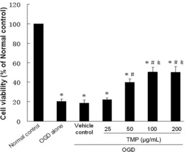
FIGURE 2 - MTT assay. The effects of TMP on SC viability subjected to OGD were assessed with the MTT reduction test. Data are expressed as the mean percentage of values of the normal control group. Values are expressed as mean ± standard
deviation (SD). *P < 0.05 vs. normal control group. #P < 0.05 vs.
cells exposed to OGD alone. &P < 0.05 vs. 50 μg/mL TMP group.
The viability of the OGD alone group and vehicle control group (20.22±2.34 and 18.62±3.22%, respectively) was signiicantly lower than the normal control group (both
P<0.01). In addition, the difference betweenthe OGD
alone group and vehicle control group was not statistically significant (P>0.05), which indicated that the vehicle
signiicantly attenuated in a TMP dose-dependent manner, and cell viabilities increased to 39.70±3.70, 50.41±4.80, and 50.09±5.97%, respectively, compared to the OGD alone
group (all P<0.05). However, treatment with 25 μg/mL
TMP provided no signiicant protection of OGD-induced cell toxicity compared to the OGD alone group
(22.14±1.68 vs. 20.22±2.34%, respectively, P > 0.05).
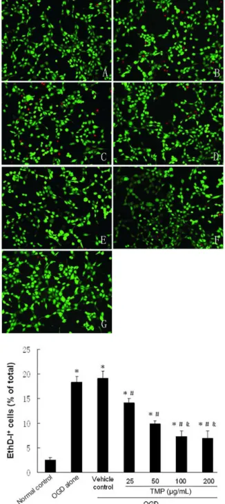
Inhibition of SC death with TMP was also conirmed by double-labeling with calcein-AM/EthD-I (Figure 3).
A LDH colorimetric assay was used to quantify cell death. As shown in Figure 4, LDH leakage signiicantly increased after OGD exposure compared to the normal
control group (14.26±0.48%, P<0.05). Importantly, TMP
(50, 100, and 200 μg/mL) signiicantly attenuated OGD-induced cell death, which reduced the LDH release to 51.50±5.17, 44.43±5.72, and 46.42±3.50%, respectively, compared to the OGD alone group (all P<0.05). However,
treatment with 25μg/ml TMP (95.52±3.34%) provided no significant protection of OGD-induced cell death compared to the OGD alone group (P>0.05). In addition,
no statistically signiicant difference was found between the OGD alone and vehicle control groups (98.70±6.33%,
P>0.05).
Protective effects of TMP on SCs against OGD-induced apoptosis
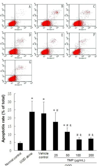
In order to verify whether TMP could protect cells from OGD-induced apoptotic death, further analyses using annexin V-FITC/PI staining were performed.
The cell populations of annexin V+/PI− and annexin
V+/PI+ were calculated as a representation of apoptotic
cells, respectively, and the data were presented as the percentage of apoptotic cells. As shown in Figure 5, treatment of SCs with OGD significantly induced cell apoptosis compared to the normal control (23.83±6.59 vs. 4.44±0.86%, respectively, P<0.05), and no statistically
signiicant difference was found between the OGD alone and vehicle control groups (23.83±6.59 vs. 22.83±7.03%, respectively, P>0.05). Treatment with 25, 50, 100, and
200 µg/mLTMP signiicantly attenuated OGD-induced apoptosis compared to the OGD alone group (17.60±5.73, 11.43±2.48, 6.78±1.10, and 7.18±0.86 vs. 23.83±6.59%, respectively, all P<0.05).
Since the highest cell viability and lowest level of LDH release and percentage of apoptotic cells was detected in the 100 or 200 μg/mL TMP treatment groups, the 100μg/ml concentration was chosen for real-time PCR and Western blot analyses.
Real-time PCR and Western blot analyses
Real-time PCR analysis revealed a modulation of
FIGURE 3 - Calcein-AM/EthD-I staining and quantitative evaluation. A: Normal control group; B: OGD alone group; C: Vehicle control group; D: 25 μg/mL TMP group; E: 50 μg/mL TMP group; F: 100 μg/mL TMP group; and G: 20.0 μg/mL TMP group. The magniication is 400x. The viability of Schwann cells was determined with the double-labeling fluorescence technique, whereby living cells, hydrolyzing calcein-AM by intracellular esterases, generate a green luorescence. Dead cells are labeled with EthD-I, which interacts with DNA resulting in a red fluorescence of cell nuclei. The percentage of dead cells (EthD-I positive) within the total number of Schwann cell proiles in the respective culture was counted and expressed as
mean ± SD. *P < 0.05 vs. normal control group. #P < 0.05 vs.
FIGURE 4 - LDH release assay. SC death was evaluated by LDH release into the culture medium. Data are expressed as the mean percentage of the maximal LDH released in the OGD alone group. Values are expressed as mean ± standard deviation (SD).
*P<0.05 vs. normal control group. #P < 0.05 vs. cells exposed to
OGD alone. &P < 0.05 vs. 50 μg/mL TMP group.
FIGURE 5 - TMP reduces OGD-induced apoptosis of SCs. A:
Normal control group; B: OGD alone group; C: Vehicle control group; D: 25 μg/mL TMP group; E: 50 μg/mL TMP group; F: 100 μg/mL TMP group; and G: 200 μg/mL TMP group. The percentage of apoptotic cells detected by annexin V-FITC/ PI staining and low cytometry. The apoptotic cells included
annexin V+/PI− and annexin V+/PI+ cells. Results are expressed
as the mean ± SD of ive independent experiments. *P < 0.05
vs. normal control group. #P < 0.05 vs. cells exposed to OGD
alone. &P < 0.05 vs. 25 μg/mL TMP group.
apoptosis-relevant genes in SCs exposed to OGD. As shown in Figure 6, the mRNA expression of Bax and caspase-3 genes was significantly increased 1.5-fold
compared to the normal control (both P<0.05), whereas
mRNA expression of Bcl-2 was downregulated by
approximately 80% (P<0.05). No signiicant differences
in the expression of Bax, Bcl-2, and caspase-3 genes were observed compared to the OGD alone and vehicle control groups (all P>0.05). Importantly, treatment with
TMP inhibited the induction of caspase-3 and Bax genes expression, whereas expression of Bcl-2 was elevated
compared to the OGD alone group (all P<0.05).
The results from Western blot analyses also proved that the expression of Bax increased, whereas Bcl-2 expression decreased after exposure to OGD. Moreover, the 32 kDa pro-enzyme form of caspase-3 and its cleavage into the active 17 kDa isoform were both induced by OGD. However, treatment with 100 μg/ mL TMP signiicantly inhibited caspase-3 expression and activation, elevated Bcl-2 levels, and reduced Bax expression relative to the OGD alone group (Figure 7). Taken together, these results clearly demonstrate that TMP can prevent OGD-induced apoptosis by upregulating anti-apoptotic Bcl-2 and downregulating the apoptotic markers Bax and caspase-3.
Study 2
Electron microscopy
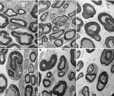
Histological examination revealed a remarkable
difference in the state of nerve segments from each group (Figure 8). In the group treated with 200-400 mg/mL TMP, there was some evidence of degenerative changes, but the basal lamina as well as the cytoplasmic membrane remained intact and SCs were present. In contrast, the group receiving 100mg/ml TMP, as well as the UWS control, had no detectable SCs.
Assessment of nerve graft viability
calcein-AM/EthD-I staining were performed. The fresh-nerve ibers displayed an intense green luorescence along their entire length with little red luorescence. There was no clear difference in the confocal microscopic images between the 200, 300, and 400 mg/mL TMP treatment groups, and intense green luorescence with dispersed red luorescence was observed in most nerve ibers. In marked contrast, the 100 mg/mL TMP-nerve and UW-nerve groups displayed intense red luorescence with little green staining, indicating that the ibers were dead (Figure 9).
Assessment of nerve graft apoptosis
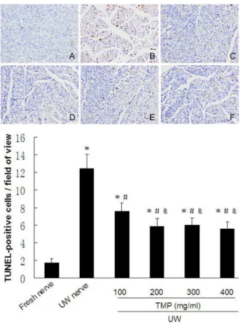
Prolonged cold ischemic exposure for four weeks was highly associated with the presence of cell apoptosis in nerve segments preserved with UWS alone. The number of TUNEL-positive cells was dramatically higher in the UW-nerve group compared to the fresh-nerve group
(12.43±1.62 vs. 1.71±0.49%, respectively, P<0.05). In
contrast, TUNEL-positive cells were signiicantly lower in the TMP-treated nerve groups treated with 100, 200, 300, and 400 mg/mL compared to the UW-nerve group control (7.57±0.98, 5.86±0.90, 6.00±0.82, and 5.57±0.79 vs. 12.43±1.62%, respectively, all P<0.05;Figure 10).
Therefore, these results indicate that adding TMP into UWS can reduce cell apoptosis in a
concentration-FIGURE 6 - Real-Time PCR analysis of the ratio of Bax, Bcl-2,
and caspase-3 mRNA levels in SCs. The results were expressed
as the mean ± SD. *P < 0.05 vs. normal control group. #P < 0.05
vs. cells exposed to OGD alone.
FIGURE 7 - Representative immunoblot of Bax, Bcl-2, caspase-3, and cleaved caspase-3 proteins. A: Normal control group; B: OGD alone group; C: Vehicle control group; and D: 100 μg/ml TMP group. The results were expressed as the mean
± SD. *P < 0.05 vs. normal control group. #P < 0.05 vs. cells
exposed to OGD alone.
FIGURE 8 - Electron microscopy of Fresh-, TMP-, and
UW-nerve cross-sections after four weeks of preservation. A: Fresh-nerve group; B: UW-nerve group; C-F: 100-400 mg/mL TMP-nerve group. SCs are preserved in the nerve segments from 200, 300, and 400 mg/mL TMP-nerve group, while the nerve segments stored for four weeks in 100 mg/ml TMP or UWS alone show myelin breakdown and an absence of SCs.
FIGURE 9 - Confocal microscopy of Fresh-, TMP-, and
UW-nerve ibers stained with calcein-AM/EthD-I. A: Fresh-UW-nerve group; B: UW-nerve group; C-F: 100-400 mg/mLTMP-nerve group. Fresh-nerve fibers present a typical uniform green calcein luorescence with almost no detectable labeling of the SC nucleus by red EthD- I fluorescence. Nerve fibers from 200, 300, and 400 mg/mL TMP-nerve groups display intense green luorescence with dispersed red luorescence, while the 100 mg/mL TMP-nerve and UW-nerve groups have obvious red EthD-I luorescence, demonstrating that SCs within these ibers are dead. Scale bars: 75 μm.
dependent manner in a cold ischemic nerve segment model after prolonged ischemia exposure for four weeks.
DISCUSSION
Ischemic injury can initiate a cascade of events that results in inlammation and cell death by apoptosis and necrosis (Faubel, Edelstein, 2005). Apoptosis is a type of cell death that occurs as a cascade of cellular machinery. It is believed that the ultimate vulnerability of cells to multiple apoptotic stimuli is influenced or determined by the Bcl-2 family (Yang, Korsmeyer, 1996), among
FIGURE 10 - TUNEL-stained nerve sections obtained from each
group. A: Fresh-nerve group; B: UW-nerve group; C-F: 100-400 mg/mL TMP-nerve group. The magniication is 100-400x. The number of TUNEL-positive cells was counted in ive random microscopic ields from each section. Data are expressed as the number of TUNEL-positive cells per ield. Values are expressed
as mean ± SD. *P < 0.05 vs. Fresh-nerve group. #P < 0.05 vs.
UW-nerve group. &P < 0.05 vs. 100 mg/mL TMP-nerve group.
which the pro-apoptotic protein Bax plays a dominant role in initiating cell death. The translocation of Bax to the mitochondrial membrane may cause a loss of mitochondrial membrane potential and an increase of mitochondrial permeability, which consequently disrupts the integrity of the mitochondrial membrane, hastens the release of cytochrome c from the mitochondria, and leads to caspase-3 activation and ultimately apoptosis
(Chinnaiyan et al., 1996; Condorelli et al., 1999). In
contrast, the anti-apoptotic protein 2 of the Bcl-2 family, which are potential inhibitors of apoptosis (Henshall et al., 2002; Creson et al., 2009), prevent the release of cytochrome c from mitochondria and caspase
activation (Akhtar, Ness, Roth, 2004; Kuhn et al., 2005;
apoptotic pathway, mediates the cleavage of itself and other downstream caspases, and is centrally important in apoptotic cell death (Liu et al., 1997; Nunez et al., 1998). Therefore, Bax, Bcl-2, and caspase-3, which are all closely involved in the apoptotic pathway, were used as markers to determine the role of TMP in this study.
In Study 1, we demonstrated that TMP could partially alleviate OGD-induced injury of SCs in vitro. Our results from the MTT assay, calcein-AM/EthD-I staining, LDH release assay, and low cytometry provide evidence that OGD induces a signiicant decrease in SC survival, whereas TMP preserves cell viability and inhibits OGD-mediated apoptosis and LDH release in a dose-dependent manner. Moreover, we found that the maximal protective effect was observed at TMP concentrations of 100 or 200 μg/mL. In addition, real-time PCR and Western blot analysis showed that TMP (100 μg/mL) attenuated the OGD-mediated upregulation of the pro-apoptotic protein Bax and downregulation of the anti-apoptotic protein Bcl-2, as well as inhibited the activation of caspase-3. These data suggest that TMP may provide a protective effect
against OGD-induced death of SCs in vitro, which may
occur through the regulation of Bcl-2 and Bax expression and suppression of caspase-3 activation.
Previous studies have indicated that TMP has a remarkable neuroprotective effect during ischemic injury. Intraperitoneal administration of TMP was found to signiicantly protect the brain from ischemic insult, as shown by the reduction in infarction volume, neuronal loss, behavioral disturbance, and brain edema (Liao et al.,
2004; Kao et al., 2006). TMP may provide neuroprotection
against ischemia brain injury by inducing thioredoxin
transcription (Zhu et al., 2009) or by reducing nNOS
expression (Xiao et al., 2010). In addition, some studies have reported that TMP has protective effects against cardiovascular diseases and ischemia-reperfusion damage
in the kidney (Dai, Bache, 1985; Zhou et al., 2004; Chen
et al., 2006; Feng et al., 2011). Moreover, several studies
have shown that TMP has potent and effective anti-apoptotic activity. TMP was found to attenuate anti-apoptotic injury in rat cerebellar granule cells and the renal tubular cell line NRK-52E after iron-induced or adriamycin-induced damage (Zhang et al., 2003; Cheng et al., 2006).
The inhibition of H2O2-induced PC12 cell apoptosis
by TMP may be related to the inhibition of caspase-3 activation, suppression of cytochrome c release, and increase in the ratio of Bcl-2/Bax (Cheng et al., 2007). The anti-apoptosis activity of Ligusticum chuanxiong, which is the Chinese medicine where TMP originates, may also be mediated through a Protein Kinase A/cAMP Response Element-Binding (PKA/CREB)-dependent pathway in
serum deprivation-induced PC12 cell apoptosis (Lin et
al., 2007). During rat subarachnoid hemorrhage, TMP
may ameliorate cerebral vasospasm by inhibiting the caspase-3-dependent apoptosis pathway (Gao et al., 2008). In spinal cord ischemia-reperfusion injury, alleviation of the damage and inhibition of apoptosis by TMP was linked to Bcl-2 and Bax expression regulation (Fan et al.,
2006). Chang et al., demonstrated that TMP may provide
neuroprotective effects by inhibiting the activation of Hypoxia-Inducible Factor-1 alpha (HIF-1α), Tumor Necrosis Factor alpha (TNF-α), and caspase-3, resulting in a reduction in the infarct volume in ischemia-reperfusion
brain injury (Chang et al., 2007). Taken together, these
studies and our current findings indicate that TMP is an effective anti-apoptotic and ischemic factor that can attenuate ischemia-induced SC death. Based on these
in vitro indings, we further investigated if TMP could
attenuate ischemia injury and increase graft survival in a cold ischemic rat nerve model after prolonged ischemia exposure, which could be beneicial for peripheral nerve preservation.
Preservation techniques are essential for maintaining graft viability outside of the body prior to transplantation, and optimization of preservation solution components will ultimately help improving transplantation outcomes. A major focus of nerve allograft transplant research has been on preserving nerve tissues without compromising the viability and function of the transplanted nerve tissues. Ultimately, the goal would be to establish a nerve bank where an abundance of nerve graft materials of various sizes and from different HLA-type donors would be available immediately to recipients. To date, there is still no consensus on the optimal nerve graft preservation method that would allow for longer ischemic exposure during preservation without jeopardizing the viability of the graft.
UWS is an intracellular solution characterized by a relatively low sodium and high potassium concentration that is capable of maintaining organ quality over a long period of oxygen deprivation. Moreover, UWS can minimize hypothermic-induced cell swelling, prevent interstitial edema and intracellular acidosis, scavenge oxygen-free radicals, and provide substrates for regenerating high-energy phosphate compounds (Belzer, Southard, 1988). The solution is the gold standard for graft preservation and has been successfully used to preserve explanted liver, heart, and kidney prior to transplantation
(Lopez-Andujar et al., 2009; George et al., 2011;
Hoeger et al., 2011). Thus, we chose UWS as nerve graft
preservation solution in this experiment. In addition, the
method for preserving peripheral nerves in UWS at 4 °C
Previous studies have tried using UWS for nerve graft preservation in peripheral nerve allotransplantation (Levi et al., 1994; Evans et al., 1995). However, it has been found that while short storage intervals of less than one week in UWS alone resulted in peripheral nerve segments containing functioning SCs, the long-term preservation of peripheral nerve segments for more than
three weeks caused a substantial loss of SCs (Levi et
al., 1994). SCs outnumber other non-neuronal cells in
the peripheral nerve and play an indispensable role in promoting axonal regeneration by providing surface cell
adhesion molecules (such as N-CAM and Ng-CAM/L1),
synthesizing basement membrane components containing extracellular matrix proteins (such as laminin and ibronectin), and producing several neurotrophic factors and receptors (Fu, Gordon, 1997; Siemionow, Brzezicki, 2009). Most pretreatment methods, such as chemical treatment, irradiation, freezing, lyophilization, and freeze-thawing could effectively reduce nerve graft antigenicity; however, nerve regeneration remains inferior to autografts. One problem has been acellularizing the graft (Song, Yang, Russell, 2009), and although short nerve deicits (less than 3 cm) can be bridged by acellular grafts, longer deficits may be more dependent upon the presence of
donor-derived viable SCs (Hems, Glasby, 1992; Evans et
al., 1995; Strauch et al., 1996; Song, Yang, Russell, 2009), because viable SCs are essential for successful axonal regeneration through longer deicits (Hall, 1986; Feneley, Fawcett, Keynes, 1991; Bunge, 1993). Furthermore,
Blais, Grenier, Berthod and Sun et al. found that
tissue-engineered skin or acellular nerve allografts seeded with SCs could improve nerve regeneration (Blais, Grenier,
Berthod, 2009; Sun et al., 2009). Therefore, sustaining
donor-derived viable SCs of preserved nerve graft until the graft is repopulated with host SCs is a critical feature in determining the outcome of nerve regeneration for peripheral nerve allografts.
Ischemia, even at low temperatures, has been found to be associated with apoptosis (Turkmen et al., 2011) and may be partly responsible for the decrease in viable SCs during nerve graft preservation. Ischemia time has been shown to correlate significantly with apoptosis rate in human cadaveric renal transplants (Castaneda et al., 2003).
Moreover, Jani et al. demonstrated that cold ischemia
results in pro-apoptotic caspase-3, -2, -8, and -9 activation, as well as increased apoptosis and tubular brush-border injury in a model of non-donation after cardiac death
(non-DCD) kidney preservation with UWS (Jani et al.,
2004). Recently, it was also found that cold ischemia activated caspase-3 and caused widespread tubular cell apoptosis in DCD kidneys (Jani et al., 2011). The addition
of a caspase inhibitor to the preservation solution was found to effectively reduce apoptosis (Natori et al., 1999; Natori et al., 2003; Jani et al., 2004). It was also found that adding green tea polyphenols, which can protect tissues from ischemia, to UWS, resulted in sustaining never graft viability (Ikeguchi et al., 2003). Thus, these studies together with our inding that TMP can protect SCs from ischemic insult, led us to hypothesize that the addition of TMP to UWS may attenuate ischemia injury and increase graft survival after prolonged cold ischemic exposure.
In Study 2, we demonstrated that prolonged cold ischemic exposure for four weeks was highly associated with the absence of viable SCs and an increase in apoptosis in nerves preserved in UWS alone. However, the addition of TMP to the UWS increased graft survival and reduced cell apoptosis in a concentration-dependent manner, with the maximum protective effect observed at a TMP concentration equal to or greater than 200 mg/mL. Therefore, our results have shown that the addition of TMP (≥200 mg/mL) in UWS can improve the cell survival of nerve grafts even after prolonged cold ischemia exposure for four weeks, which indicated that peripheral nerves may be preserved in a TMP solution. However, the exact mechanism by which TMP maintains graft viability and blocks apoptosis in a cold ischemic nerve model will require further studies. In addition, several key questions require further exploration before TMP can be used for routine peripheral nerve storage. For example, additional studies are need to conirm that nerve grafts preserved in a TMP solution after prolonged cold ischemia exposure can indeed be beneicial in the repair of massive nerve deicits and functional regeneration of in vivo models. In addition, we will need to determine the maximum period of time that nerve grafts can be preserved in TMP without any loss in viability.
Taken together, the findings presented here demonstrate that TMP exerts anti-ischemic properties on OGD-induced primary cultured SC injury and apoptosis by abrogating the activation of caspase-3 and altering the expression of Bcl-2 and Bax. Moreover, the addition of TMP to UWS can improve the preservation properties of the solution for use in nerve grafts by maintaining graft survival and reducing apoptosis in a concentration-dependant manner. Therefore, TMP may be useful in the preservation of nerve grafts for repairing deficits and improving regeneration.
ACKNOWLEDGMENTS
project of Chongqing Municipal Health Bureau (2009-1-10).
REFERENCES
AKHTAR, R. S.; NESS, J. M.; ROTH, K. A. Bcl-2 family regulation of neuronal development and neurodegeneration.
Biochim. Biophys. Acta,v.1644, n.2-3, p.189-203, 2004.
BELZER, F. O.; SOUTHARD, J. H. Principles of solid-organ
preservation by cold storage. Transplantation,v.45, n.4,
p.673-676, 1988.
BLAIS, M.; GRENIER, M.; BERTHOD, F. Improvement of nerve regeneration in tissue-engineered skin enriched with
schwann cells. J. Invest. Dermatol.,v.129, n.12,
p.2895-2900, 2009.
BROCKES, J. P.; RAFF, M. C.; NISHIGUCHI, D. J.; WINTER, J. Studies on cultured rat Schwann cells. III. Assays for
peripheral myelin proteins. J. Neurocytol.,v.9, n.1,
p.67-77, 1980.
BUNGE, R. P. Expanding roles for the Schwann cell: ensheathment, myelination, trophism and regeneration.
Curr. Opin. Neurobiol.,v.3, n.5, p.805-809, 1993.
CASTANEDA, M. P.; SWIATECKA-URBAN, A.; MITSNEFES, M. M.; FEUERSTEIN, D.; KASKEL, F. J.; TELLIS, V.; DEVARAJAN, P. Activation of mitochondrial apoptotic pathways in human renal allografts after
ischemiareperfusion injury. Transplantation, v.76, n.1,
p.50-54, 2003.
CHANG, Y.; HSIAO, G.; CHEN, S. H.; CHEN, Y. C.; LIN, J. H.; LIN, K. H.; CHOU, D. S.; SHEU, J. R. Tetramethylpyrazine suppresses HIF-1alpha, TNF-alpha, and activated caspase-3 expression in middle cerebral artery occlusion-induced
brain ischemia in rats. Acta Pharmacol. Sin.,v.28, n.3,
p.327-333, 2007.
CHEN, S. Y.; HSIAO, G.; HWANG, H. R.; CHENG, P. Y.; LEE, Y. M. Tetramethylpyrazine induces heme oxygenase-1 expression and attenuates myocardial ischemia/reperfusion
injury in rats. J. Biomed. Sc.,v.13, n.5, p.731-740, 2006.
CHENG, C.Y.; SUE, Y.M.; CHEN, C.H.; HOU, C.C.; CHAN, P.; CHU, Y. L.; CHEN, T. H.; HSU, Y. H. Tetramethylpyrazine attenuates adriamycin-induced apoptotic injury in rat renal
tubular cells NRK-52E. Planta Med.,v.72, n.10,
p.888-893, 2006.
CHENG, X. R.; ZHANG, L.; HU, J. J.; SUN, L.; DU, G. H. Neuroprotective effects of tetramethylpyrazine on hydrogen
peroxide-induced apoptosis in PC12 cells. Cell Biol. Int.,
v.31, n.5, p.438-443, 2007.
CHINNAIYAN, A. M.; ORTH, K.; O’ROURKE, K.; DUAN, H.; POIRIER, G. G.; DIXIT, V. M. Molecular ordering of the cell death pathway. Bcl-2 and Bcl-xL function upstream
of the CED-3-like apoptotic proteases. J. Biol .Chem.,v.271,
n.9, p.4573-4576, 1996.
CHIU, D. T.; JANECKA, I.; KRIZEK, T. J.; WOLFF, M.; LOVELACE, R. E. Autogenous vein graft as a conduit for
nerve regeneration. Surgery, v.91, n.2, p.226-233, 1982.
CHUN-SHENG, L.; HSIAO-MENG, Y.; YUN-HSIANG, H.; CHUN, P.; CHI-FEN, S. Radix salviae miltiorrhizae and
Rhizoma ligustici wallichii in coronary heart disease. Chin.
Med. J.(Engl),v.4, n.1, p.43-46, 1978.
CONDORELLI, G.; MORISCO, C.; STASSI, G.; NOTTE, A.; FARINA, F.; SGARAMELLA, G.; DE RIENZO, A.; RONCARATI, R.; TRIMARCO, B.; LEMBO, G. Increased cardiomyocyte apoptosis and changes in proapoptotic and antiapoptotic genes bax and bcl-2 during left ventricular adaptations to chronic pressure overload in the rat.
Circulation,v.99, n.23, p.3071-3078, 1999.
CRESON, T. K.; YUAN, P.; MANJI, H. K.; CHEN, G. Evidence for involvement of ERK, PI3K, and RSK in induction of
Bcl-2 by valproate. J. Mol. Neurosci.,v.37, n.2, p.123-134,
2009.
DAI, X. Z.; BACHE, R. J. Coronary and systemic hemodynamic
effects of tetramethylpyrazine in the dog. J. Cardiovasc.
Pharmacol., v.7, n.5, p.841-849, 1985.
EVANS, P. J.; MACKINNON, S. E.; BEST, T. J.; WADE, J. A.; AWERBUCK, D. C.; MAKINO, A. P.; HUNTER, D. A.; MIDHA, R. Regeneration across preserved peripheral
nerve grafts. Muscle Nerve, v.18, n.10, p.1128-1138, 1995.
FAN, L. H.; WANG, K. Z.; CHENG, B.; WANG, C. S.; DANG, X. Q. Anti-apoptotic and neuroprotective effects of Tetramethylpyrazine following spinal cord ischemia in
rabbits. BMC Neurosci., v.7, p.48, 2006.
FAUBEL, S.; EDELSTEIN, C. L. Caspases as drug targets in
ischemic organ injury. Curr. Drug Targets Immune Endocr.
FENELEY, M. R.; FAWCETT, J. W.; KEYNES, R. J. The role of Schwann cells in the regeneration of peripheral nerve
axons through muscle basal lamina grafts. Exp. Neurol.,
v.114, n.3, p.275-285, 1991.
FENG, L.; KE, N.; CHENG, F.; GUO, Y.; LI, S.; LI, Q.; LI, Y. The protective mechanism of ligustrazine against renal
ischemia/reperfusion injury. J. Surg. Res.,v.166, n.2,
p.298-305, 2011.
FU, S. Y.; GORDON, T. The cellular and molecular basis of
peripheral nerve regeneration. Mol. Neurobiol.,v.14, n.1-2,
p.67-116, 1997.
GAO, C.; LIU, X.; LIU, W.; SHI, H.; ZHAO, Z.; CHEN, H.; ZHAO, S. Anti-apoptotic and neuroprotective effects of Tetramethylpyrazine following subarachnoid hemorrhage
in rats. Auton. Neurosci.,v.141, n.1-2, p.22-30, 2008.
GEORGE, T. J.; ARNAOUTAKIS, G. J.; BAUMGARTNER, W. A.; SHAH, A. S.; CONTE, J. V. Organ storage with University of Wisconsin solution is associated with improved outcomes after orthotopic heart transplantation.
J. Heart Lung Transplant.,v.30, n.9, p.1033-1043, 2011.
HALL, S. M. Regeneration in cellular and acellular autografts
in the peripheral nervous system. Neuropathol. Appl.
Neurobiol.,v.12, n.1, p.27-46, 1986.
HEMS, T. E.; GLASBY, M. A. Comparison of different methods of repair of long peripheral nerve defects: an experimental
study. Br. J. Plast. Surg.,v.45, n.7, p.497-502, 1992.
HENSHALL, D. C.; ARAKI, T.; SCHINDLER, C. K.; LAN, J. Q.; TIEKOTER, K. L.; TAKI, W.; SIMON, R. P. Activation of Bcl-2-associated death protein and counter-response of Akt within cell populations during seizure-induced neuronal
death. J. Neurosci.,v.22, n.19, p.8458-8465, 2002.
HESS, J. R.; BRENNER, M. J.; FOX, I. K.; NICHOLS, C. M.; MYCKATYN, T. M.; HUNTER, D. A.; RICKMAN, S. R.; MACKINNON, S. E. Use of cold-preserved allografts seeded with autologous Schwann cells in the treatment of
a long-gap peripheral nerve injury. Plast. Reconstr. Surg.,
v.119, n.1, p.246-259, 2007.
HO, W. K.; WEN, H. L.; LEE, C. M. Tetramethylpyrazine for treatment of experimentally induced stroke in Mongolian
gerbils. Stroke, v.20, n.1, p.96-99, 1989.
HOEGER, S.; LUEG, G.; TSAGOGIORGAS, C.; SCHNEIDER, M.; THEISINGER, S.; THEISINGER, B.; FONTANA, J.; WALDHERR, R.; KRAMER, B. K.; SCHNUELLE, P.; YARD, B. UW is superior compared with HTK after
prolonged preservation of renal grafts. J. Surg. Res., v.170,
n.1, p.e149-e157, 2011.
IKEGUCHI, R.; KAKINOKI, R.; OKAMOTO, T.; MATSUMOTO, T.; HYON, S. H.; NAKAMURA, T. Successful storage of peripheral nerve before transplantation using green tea polyphenol: an experimental study in rats.
Exp. Neurol., v.184, n.2, p.688-696, 2003.
ITOH, S.; SHINOMIYA, K.; SAMEJIMA, H.; OHTA, T.; ISHIZUKI, M.; ICHINOSE, S. Experimental study on nerve regeneration through the basement membrane tubes
of the nerve, muscle, and artery. Microsurgery,v.17, n.10,
p.525-534, 1996.
JANI, A.; LJUBANOVIC, D.; FAUBEL, S.; KIM, J.; MISCHAK, R.; EDELSTEIN, C. L. Caspase inhibition prevents the increase in caspase-3, -2, -8 and -9 activity
and apoptosis in the cold ischemic mouse kidney. Am. J.
Transplant.,v.4, n.8, p.1246-1254, 2004.
JANI, A.; ZIMMERMAN, M.; MARTIN, J.; LU, L.; TURKMEN, K.; RAVICHANDRAN, K.; PACIC, A.; LJUBANOVIC, D.; EDELSTEIN, C. L. Perfusion storage reduces apoptosis in a porcine kidney model of donation
after cardiac death. Transplantation,v.91, n.2, p.169-175,
2011.
KAO, T. K.; OU, Y. C.; KUO, J. S.; CHEN, W. Y.; LIAO, S. L.; WU, C. W.; CHEN, C. J.; LING, N. N.; ZHANG, Y. H.; PENG, W. H. Neuroprotection by tetramethylpyrazine
against ischemic brain injury in rats. Neurochem. Int.,v.48,
n.3, p.166-176, 2006.
KEILHOFF, G.; SCHILD, L.; FANSA, H. Minocycline protects Schwann cells from ischemia-like injury and promotes axonal outgrowth in bioartificial nerve grafts lacking
Wallerian degeneration. Exp. Neurol.,v.212, n.1,
p.189-200, 2008.
KUHN, H. G.; BIEBL, M.; WILHELM, D.; LI, M.; FRIEDLANDER, R. M.; WINKLER, J. Increased generation of granule cells in adult Bcl-2-overexpressing mice: a role for cell death during continued hippocampal
neurogenesis. Eur. J. Neurosci.,v.22, n.8, p.1907-1915,
LEVI, A. D.; EVANS, P. J.; MACKINNON, S. E.; BUNGE,
R. P. Cold storage of peripheral nerves: an in vitro assay of
cell viability and function. Glia,v.10, n.2, p.121-131, 1994.
LIAO, S. L.; KAO, T. K.; CHEN, W. Y.; LIN, Y. S.; CHEN, S.Y.; RAUNG, S. L.; WU, C. W.; LU, H. C.; CHEN, C. J. Tetramethylpyrazine reduces ischemic brain injury in rats.
Neurosci. Lett.,v.372, n.1-2, p.40-45, 2004.
LIN, Y. L.; LEE, Y. C.; HUANG, C. L.; LAI, W. L.; LIN, Y. R.; HUANG, N. K. Ligusticum chuanxiong prevents rat pheochromocytoma cells from serum deprivation-induced apoptosis through a protein kinase A-dependent pathway.
J. Ethnopharmacol.,v.109, n.3, p.428-434, 2007.
LIU, X.; ZOU, H.; SLAUGHTER, C.; WANG, X. DFF, a heterodimeric protein that functions downstream of caspase-3 to trigger DNA fragmentation during apoptosis.
Cell,v.89, n.2, p.175-184, 1997.
LIVAK, K. J.; SCHMITTGEN, T. D. Analysis of relative gene expression data using real-time quantitative PCR and the
2(-Delta Delta C(T)) Method. Methods,v.25, n.4,
p.402-408, 2001.
LOPEZ-ANDUJAR, R.; DEUSA, S.; MONTALVA, E.; SAN JUAN, F.; MOYA, A.; PAREJA, E.; DEJUAN, M.; BERENGUER, M.; PRIETO, M.; MIR, J. Comparative prospective study of two liver graft preservation solutions:
University of Wisconsin and Celsior. Liver Transpl.,v.15,
n.12, p.1709-1717, 2009.
MACKINNON, S. E. Surgical management of the peripheral
nerve gap. Clin. Plast. Surg.,v.16, n.3, p.587-603, 1989.
MACKINNON, S. E.; HUDSON, A. R. Clinical application
of peripheral nerve transplantation. Plast. Reconstr. Surg.,
v.90, n.4, p.695-699, 1992.
MATSUMOTO, T.; KAKINOKI, R.; IKEGUCHI, R.; HYON, S. H.; NAKAMURA, T. Optimal conditions for peripheral nerve storage in green tea polyphenol: an experimental
study in animals. J. Neurosci. Methods,v.145, n.1-2,
p.255-266, 2005.
NATORI, S.; HIGUCHI, H.; CONTRERAS, P.; GORES, G. J. The caspase inhibitor IDN-6556 prevents caspase activation and apoptosis in sinusoidal endothelial cells during liver
preservation injury. Liver Transpl.,v.9, n.3, p.278-284,
2003.
NATORI, S.; SELZNER, M.; VALENTINO, K. L.; FRITZ, L. C.; SRINIVASAN, A.; CLAVIEN, P. A.; GORES, G. J. Apoptosis of sinusoidal endothelial cells occurs during liver preservation injury by a caspase-dependent mechanism.
Transplantation,v.68, n.1, p.89-96, 1999.
NUNEZ, G.; BENEDICT, M. A.; HU, Y.; INOHARA, N. Caspases: the proteases of the apoptotic pathway.
Oncogene,v.17, n.25, p.3237-3245, 1998.
SHACKA, J. J.; ROTH, K. A. Regulation of neuronal cell death and neurodegeneration by members of the Bcl-2 family:
therapeutic implications. Curr. Drug Targets CNS Neurol.
Disord.v.4, n.1, p.25-39, 2005.
SIEMIONOW, M.; BRZEZICKI, G. Chapter 8: Current
techniques and concepts in peripheral nerve repair. Int. Rev.
Neurobiol.,v.87, p.141-172, 2009.
SINGH, M.; SHARMA, H.; SINGH, N. Hydrogen peroxide induces apoptosis in HeLa cells through mitochondrial
pathway. Mitochondrion,v.7, n.6, p.367-373, 2007.
SONG, J. W.; YANG, L. J.; RUSSELL, S. M. Peripheral nerve:
what’s new in basic science laboratories. Neurosurg. Clin.
N. Am., v.20, n.1, p.121-131, 2009.
STRAUCH, B.; FERDER, M.; LOVELLE-ALLEN, S.; MOORE, K.; KIM, D. J.; LLENA, J. Determining the maximal length of a vein conduit used as an interposition
graft for nerve regeneration. J. Reconstr. Microsurg.,v.12,
n.8, p.521-527, 1996.
SUN, X. H.; CHE, Y. Q.; TONG, X. J.; ZHANG, L. X.; FENG, Y.; XU, A. H.; TONG, L.; JIA, H.; ZHANG, X. Improving nerve regeneration of acellular nerve allografts seeded
with SCs bridging the sciatic nerve defects of rat. Cell Mol.
Neurobiol.,v.29, n.3, p.347-353, 2009.
TURKMEN, K.; MARTIN, J.; AKCAY, A.; NGUYEN, Q.; RAVICHANDRAN, K.; FAUBEL, S.; PACIC, A.; LJUBANOVIC, D.; EDELSTEIN, C. L.; JANI, A. Apoptosis and autophagy in cold preservation ischemia.
Transplantation,v.91, n.11, p.1192-1197, 2011.
XIAO, X.; LIU, Y.; QI, C.; QIU, F.; CHEN, X.; ZHANG, J.; YANG, P. Neuroprotection and enhanced neurogenesis by tetramethylpyrazine in adult rat brain after focal ischemia.
YANG, E.; KORSMEYER, S. J. Molecular thanatopsis: a
discourse on the BCL2 family and cell death. Blood, v.88,
n.2, p.386-401, 1996.
ZHANG, Z.; WEI, T.; HOU, J.; LI, G.; YU, S.; XIN, W. Iron-induced oxidative damage and apoptosis in cerebellar granule cells: attenuation by tetramethylpyrazine and ferulic
acid. Eur. J. Pharmacol., v.467, n.1-3, p.41-47, 2003.
ZHOU, Y.; HU, C. P.; DENG, P. Y.; DENG, H. W.; LI, Y. J. The protective effects of ligustrazine on ischemia-reperfusion and DPPH free radical-induced myocardial injury in
isolated rat hearts. Planta Med.,v.70, n.9, p.818-822, 2004.
ZHU, X. L.; XIONG, L. Z.; WANG, Q.; LIU, Z. G.; MA, X.; ZHU, Z. H.; HU, S.; GONG, G.; CHEN, S. Y. Therapeutic time window and mechanism of tetramethylpyrazine on transient focal cerebral ischemia/reperfusion injury in rats.
Neurosci. Lett.,v.449, n.1, p.24-27, 2009.
Received for publication on 07th September, 2013