ORIGINAL
ARTICLE
Enterotoxins, colonization factors, serotypes and
antimicrobial resistance of enterotoxigenic
Escherichia
coli
(ETEC) strains isolated from hospitalized children
with diarrhea in Bolivia
Authors
Claudia Rodas1 Rosalía Mamani2 Jorge Blanco3 Jesus Eulogio Blanco3 Gudrun Wiklund4 Ann-Mari Svennerholm5 Åsa Sjöling5
Volga Iniguez6 1MSc, Microbiology; PhD student, Department of Microbiology and Immunology, Institute of Biomedicine, University of Gothenburg, Sweden. 2MsC, Bioscience; PhD student, Laboratorio de Referencia de Escherichia coli, Departamento de Microbiología y Parasitología, Universidad de Santiago de Compostela, Lugo, Spain 3Dr., PhD, WHO Collaborating Centre for Reasearch on ETEC and Department of Microbiology and Immunology, Institute of Biomedicine, University of Gothenburg, Sweden 4MSc, WHO Collaborating Centre for Reasearch on ETEC and Department of Microbiology and Immunology, Institute of Biomedicine, University of Gothenburg, Sweden 5MD, Professor, WHO Collaborating Centre for Reasearch on ETEC and Department of Microbiology and Immunology, Institute of Biomedicine, University of Gothenburg, Sweden 6Dr., PhD, Intituto de Biología Molecular y Biotecnología, Facultad de Ciencias Puras y Naturales, Universidad Mayor de San Andrés, Bolivia
Submitted on: 08/11/2010 Approved on: 09/27/2010
Correspondence to:
Claudia Rodas Department of Microbiology and Immunology, Institute of Biomedicine, University of Gothenburg, Box 435, 405 30 Gothenburg, Sweden. Phone: +46-31-7866233 Fax: +46-317866205 claudia.rodas@ microbio.gu.se
We declare no conflict of interest.
ABSTRACT
Enterotoxigenic Escherichia coli (ETEC) is recognized as the main cause of bacterial diarrhoea among children in Asia, Africa and Latin America but less investigated in Bolivia. Objective: To determine the relation between enterotoxins, CFs and serotypes as well as the antimicrobial resist-ance patterns in a set of ETEC isolates collected from hospitalized children with acute diarrhea. In the present study we characterized 43 ETEC strains isolated from 2002 to 2006 from hospitalized children (0-5 years) with acute diarrhea in Bolivia. The strains were analyzed for heat-labile (LT) and heat-stable (ST) enterotoxins and colonization factor (CF) profiles, as well as for serogroups and antimicrobial resistance using phenotypic (ELISA, dot blot, slide agglutination and disc diffu-sion) and genotypic (Multiplex PCR) methods. Among the ETEC isolates tested, 30 were positive for LT, 3 for STh and 10 for LT/STh. Sixty-five percent (28/43) of the strains expressed one or more CF. The most common CFs were CS17 (n = 8) and CFA/I (n = 8). The phenotypical and geno-typical results for toxins and CFs were congruent except for CS21 that was amplified in 10 of the strains by multiplex PCR, but CS21 pili was only detected phenotypically in four of these strains. The ETEC strains had diverse O and H antigens and the most common types were O8:H9 LT CS17 (n = 6; 14%) and O78:HNM LT-ST CFA/I (n = 4; 9%). The analysis of antibiotic resistance showed that 67% (n = 29/43) of the strains were resistant to one or several of the antimicrobial agents tested. Presence of CFs was associated with antibiotic resistance. Conclusion: The most common toxin profile was LT 70%, LT/STh 23% and STh 7%. High antimicrobial resistance to ampicillin among serogroups O6, O8 and O78 were the most common.
Keywords: enterotoxigenic Escherichia coli; enterotoxins; drug resistance; Bolivia.
[Braz J Infect Dis 2011;15(2):132-137]©Elsevier Editora Ltda.
INTRODUCTION
Acute diarrheal diseases are an important health problem among children under five in developing countries and annually cause between 1.4 to 2.5 million deaths.1,2 Diar-rheal diseases cause high rates of pediatric morbidity and mortality in Bolivia, leading annually to more than 500,000 cases and be-tween 13,000 and 15,000 deaths respectively.3 Enterotoxigenic Escherichia coli (ETEC) is an important cause of diarrhea among children in developing countries and among travel-ers to ETEC-endemic areas.4 Clinical human ETEC isolates produce one or more of three enterotoxins; the heat stable toxins (STp and STh) and heat labile toxin (LT) and may pro-duce one or more of several colonization fac-tors (CFs).5 The CFs mediate adherence to the small intestinal mucosa, and to date, more
than 22 different CFs have been described in human ETEC strains (e.g CFA/I, CS1-CS8, CS12-CS15, and CS17-CS22).5-8 In developing countries, the incidence of ETEC-associated diarrhea decreases during the first five years of life9 whereas children and adults from in-dustrialized areas who travel to these coun-tries are susceptible to this type of diarrhea, indicating that natural immunity develops.4,10
resistance among ETEC strains is increasing, perhaps due to indiscriminate use of antibiotics. Although rehydration therapy is the most important part of therapy for acute di-arrhea, antimicrobials are important adjuvants for therapy and their use results in a marked decrease in overall stool volume and decreased length of illness. Most cases of bacte-rial gastroenteritis are self-limiting and in otherwise healthy patients, administration of antibiotics is not necessary. How-ever in infants, elderly people, granulocytopenic or immu-nodepressed patients with enteritis, and for patients with extra-intestinal infections, particularly when bacteremia is suspected, antibiotic therapy is fundamental for disease con-trol.12 The knowledge of antimicrobial resistance patterns locally in different countries is important when prescrib-ing antibiotics for diarrheagenic bacteria, and can serve as a guide for implementing public health interventions against diarrheal disease.
In this study, we determined the relation between en-terotoxins, CFs and serotypes, as well as the antimicrobial resistance patterns in a set of ETEC isolates collected from hospitalized children with acute diarrhea.
MATERIAL AND METHODS
Bacterial strains and determination of toxins and CFs
ETEC strains were isolated from acute watery non-bloody stool samples obtained from hospitalized children with di-arrhea under five years of age in La Paz, Bolivia, between 2002-2006. ETEC strains were grown on MacConkey agar plates, incubated at 37°C for 18 hours and five lactose posi-tive colonies were tested for the presence of ETEC toxins and CFs by GM1 ELISA, toxin multiplex PCR and dot blot.13 Confirmed ETEC strains were stored at -70°C in Luria broth (Difco) containing 25% glycerol and subsequently shipped to Sweden on deep agar stabs. All strains were re-analyzed twice by toxin GM1-ELISA, dot blot and Multiplex PCR in Sweden.13,14 For the dot blot the MAbs used were CFA/I, CS1 to CS6, CS8, CS12, CS14, CS17, CS19 and CS21. From the original set of ETEC strains (n = 79), we analyzed 43 ETEC strains that survived the transport, had identical toxin and CF profiles when tested both in Bolivia, Spain and Sweden and that were not mixed infections for serotype and antimi-crobial resistance.
Serotyping
O- and H-antigens were determined according to the meth-od described by Guinée et al.15 and employing all available O (O1-O185) and H (H1-H56) antisera in the E. coli Ref-erence laboratory (LREC) at the Universidad of Santiago de Compostela, Lugo, Spain. All antisera were obtained and ab-sorbed with the corresponding cross-reacting antigens to re-move nonspecific agglutinins. The O antisera were produced in the LREC and the H antisera were obtained from the
Stat-ens Serum Institute (Copenhagen, Denmark). Isolates that did not react with any of the O and H antisera used were classified as non-typeable (ONT and HNT), and those that were non-motile were denoted HNM.16
Antimicrobial resistance
Colonies from McConkey agar cultures were homogenized in 0.85% saline, and the turbidity was adjusted to that of a 0.5 McFarland standard. The inoculum suspension was spread on a Mueller-Hinton agar plate surface with a swab, and incubated in room temperature for 15 min before an-timicrobial disks were applied. The anan-timicrobials tested were ampicillin, nalidixic acid, chloramphenicol, ampicil-lin-sulbactam, tetracycline, ciprofloxacin, trimethoprim-sulfamethoxazole, gentamicin, and cefoxitin. The bacterial cultures were incubated for 20 hours at 37°C, and the zone of inhibition was determined. The National Committee for Clinical Laboratory Standards17 breakpoints were used to differentiate between susceptible and resistant isolates.
E. coli ATCC 25922 was used as a reference strain for quality control purposes.
Statistics
The two-tailed Fishers’ exact test was used for statistical analysis using the GraphPad InStat Software and a 2 x 2 contingency table. A p-value less than 0.05 was consid-ered significant.
RESULTS
ETEC strains were obtained from a prospective study in children under five years of age seeking care in three differ-ent hospitals in La Paz, Bolivia during 2002-2006. The chil-dren all suffered from non-bloody, acute diarrhea. In total, 853 clinical samples were analyzed and ETEC was isolated from 79 patients. Forty-tree individual ETEC strains isolat-ed from watery non-bloody diarrhea samples with only one microbial pathogen isolated and with confirmed enterotoxin and CF expression in La Paz, Santiago de Compostela and Gothenburg were selected for further studies (Table 1).
The most common toxin profile among the selected strains was LT in 30 strains (70%), followed by LT/STh in 10 strains (23%) and STh in three strains (7%). The presence of STp was not recorded in Bolivia and when analyzed for STp in Sweden and in Spain none of the strains harbored this toxin.
Table 1. Characterized ETEC strains collected from Bolivian children with diarrhea
Strain Children Characteristics of strains
Patient ID Age Sex Toxin CFsa Serotypeb Antibiotic resistancec,d,e
1 9 18 m F LTSTh CFA/I CS21 O1:H45 AMP, SAM, TET
2 18 15 m M LT CS17 O8:H9 AMP, CHL , SAM
3 19 3 m M LT CS17 O8:H9 AMP, CHL , SAM, TET
4 34 18 m F LTSTh CS1 CS3 CS21 O6:H16 AMP, SAM , SXT
5 95 47 m F LT CS2 CS3 (CS21) O21:HNT
-6 98 13 m M LT CS2 CS3 O21: HNT AMP, CHL TET, SXT, GEN
7 100 48 m F LT - O120:H10
-8 104 9 m F LT - O174:H40 TET
9 110 12 m F LT - ONT:HNM nt
10 119 11 m F LT CS17 O6:H9 AMP, CHL , SAM
11 121 11 m M LT CS17 O8:H9 AMP, CHL , SAM
12 124 9 m M LT CS12 O159: H4 AMP, TET, SXT
13 126 17 m M LT - O41:H32
-14 132 14 m M LT CS17 O8:H9 AMP, SAM , CHL
15 134 24 m F LT CS17 O8:H9 AMP, SAM , SXT
16 137 12 m F STh CS6 (CS21) O148: H28 AMP, SAM , SXT
17 162 14 m M LT CS17 O6: H16 AMP, SAM
18 172 14 m M LT - O120:H10
-19 174 4 m F LTSTh - O8:H9
-20 191 16 m M LT - ONT:H40 TET
21 193 19 m F LTSTh CFA/I (CS21) O78:HNM AMP, SAM , SXT
22 204 11 m F LT - O8:H9 AMP, SAM
23 207 5 m M STh CFA/I O153: H45 AMP, SAM , TET
24 220 16 m M LT CS7 O114:H49 TSX
25 226 28 m F LT CS7 O78: H10 AMP, SAM , SXT
26 235 14 m M STh CFA/I CS21 ONT:HNM AMP, SAM , TET
27 237 24 m F LT - ONT:H40
-28 241 7 m M LT - ONT:HNM AMP, SAM , SXT
29 244 18 m F LTSTh CFA/I (CS21) O78:HNM AMP
30 245 36 m F LT - ONT:H40
-31 389 9 m M LTSTh CS1 CS3 CS21 O6:H16 AMP, NAL, SXT
32 390 9 m F LTSTh CFA/I (CS21) O78:HNM
-33 409 9 m M LTSTh CFA/I O6:H16 AMP, NAL, SAM , SXT
34 475 5 m M LT CS1 CS3 ONT:H40
-35 520 12 m M LT - ONT: HNM AMP. SAM , TET
36 536 17 m M LT - O34:H25 AMP, SAM , TET, SXT, GEN, CEF
37 554 17 m F LT - O159:H4
-38 574 19 m M LT - ONT:HNM
-39 577 36 m F LT CS17 O8:H9 AMP, SAM , TET, SXT
40 598 14 m F LTSTh CFA/I (CS21) O78:HNM
-41 631 17 m M LT CS12 O159:H21 AMPS, TET, SXT
42 689 6 m M LTSTh CS2 CS3 O1 AMP, SAM
43 743 12 m F LT CS12 O159:H4
-aCS21 within brackets is positive in PCR but negative in dot blot.
bNT, non typeable; HNM, non-motile.
cResistant to AMP, ampicillin; SAM, ampicillin sulbactam; CIP, ciprofloxacin; CHL, chloramphenicol; GEN, gentamicin;
NAL, naldixic acid; CEF, cefoxitin; SXT, trimethoprim; sulfamethoxazole; TET, tetracycline.
dnt, not tested.
association with other CFs, especially with CFA/I and the CFA/II group (CS1, CS2 and CS3). CS21 was detected by multiplex PCR in 10 strains but was only detected by phe-notypic dot blot test in four of the strains. The pattern of phenotypically silent CS21 has previously been reported in Bolivian and Bangladeshi strains by us.14
When analyzing the serogroups of the ETEC strains, we found that they belonged to a diverse set of O groups (Table 1). Of these serogroups, O6, O8, O78, O114, O120, O148, O153 and O159 have frequently been associated with ETEC in previous studies18-20 and these 8 O-groups were detected in 26 of the 43 ETEC strains. A small num-ber of additional strains were of O serogroups that have infrequently been detected in ETEC strains, namely, O1, O41 and O21.19 The remaining serogroups O34 and O174 have to our knowledge not previously been described for any category of diarrheagenic E. coli. The strains that had O groups commonly found in ETEC were statistically more often CF positive than strains that either possessed an O serogroup not commonly found in ETEC or were non-typeable (p < 0.0070). An H flagellar antigen was detected in 31 strains. The most common H-antigen was H9 (n = 9); eight of these strains were O8 serogroup and six of these strains were O8:H9 LT/CS17 which is a common ETEC phenotype and probably constituted a clone that circulated in Bolivia from 2002 to 2005. Other common H-antigens were H16 (n = 4), all of which were O6: H16, H40 (n = 5) and H45 (n = 2).
The results of the antimicrobial susceptibility of the ETEC strains are shown in Table 1. Several of the strains were resistant to many antimicrobial drugs. The highest re-sistance rate was found for ampicillin (53.5%; n = 23), fol-lowed by ampicillin-sulbactam (46.5%; n = 20), trimetho-prim-sulfamethoxazole (32.5%; n = 14), tetracycline (28 %; n = 12) and chloramphenicol (14%; n = 6). Most strains were not resistant to nalidixic acid, gentamicin, and cefoxitin and all the tested strains were susceptible to ciprofloxacin.
DISCUSSION
In our analysis of the 43 ETEC strains from Bolivia we found an unusually high prevalence of LT-only strains. LT, ST and LT/ST strains worldwide often occur at com-parable frequencies,19 but LT strains have previously been reported to be more common in South American surveys of ETEC prevalence than in surveys in Asia and Africa21,22 and recently an increase in LT strains also in Bangladesh was reported.23 The observed percentage of CF positive and negative strains is in agreement with other findings with approximately 2/3 of the strains expressing known CFs. The most common CFs were CFA/I and CS17 and while CFA/I always is one of the most commonly detected CF in surveys worldwide the high prevalence of both CS17 and LT-only strains in this collection seemed to be due
to an unusually high frequency of LT/CS17 strains. CS21 was commonly detected by PCR but less commonly by dot blot as reported earlier.14 These results may be due to the observed variability in the coding region for CS21.8,24 The high prevalence of PCR positive CS21 strains among the Bolivian strains is in agreement with that observed in other countries in Asia and South America where CS21 frequen-cies between 19% to 36.5% have been reported in ETEC strain collections.25-27
There was a large variation of serotypes among the 43 ETEC strains. Unlike the genes for toxins and CFs, which are usually plasmid encoded, the O and H biosynthetic genes are chromosomal.28 This suggests a high degree of clonal diversity of disease-associated ETEC in this geo-graphic area over a relatively short period of time. Nota-bly, several of the serotypes detected were not those typi-cally associated with ETEC. Since there is only evidence of protection against homologous O groups and no evidence that ETEC O- and H-antigens are protective against heter-ologous serotypes,28-30 there would seem to be little to gain from incorporation of specific O- or H-antigens into a vac-cine against ETEC.
We found a relation between serotype, toxin and CF profile. Thus, O8:H9 strains commonly expressed LT and CS17 (6 strains) while the O78:HNM strain expressed LT, STh and CFA/I and carried the gene for CS21 (4 strains). We also detected three O6:H16 strains that expressed LT, STh and CS1 + CS3 + CS21 which has previously been re-ported to be the most frequently detected and widely dis-tributed ETEC phenotype worldwide.19
ETEC strains that have identical toxin and CF profiles have been proposed to be genetically closely related.31,32 The results from this and other studies on ETEC serotypes sup-port that strains with certain toxin and CF profiles often have certain O- and H-antigens indicating that there might be a genetic relationship between strains with similar phe-notypes regardless of where they are found in the world.
We found a high level of antibiotics resistance in the Bolivian ETEC strains (Table 1). It is reasonable that this multiresistance of ETEC might emerge to classical antibi-otics such as ampicillin, tetracycline, and trimethoprim-sulfamethoxazole33 since they have been widely used in Bo-livia during the past few years. For instance the Integrated Management of Childhood Illness (AIEPI) manual recom-mends the use of trimethoprim-sulfamethoxazole as the first line antibiotic for the treatment of bloody diarrhea34 and tetracycline is prescribed for the treatment of different infectious diseases. Therefore, these antibiotics are widely used in medical settings in Bolivia.
be taken into consideration that in spite of the minimal use of quinolones in Bolivia, we detected nalidixic acid-resistant ETEC strains. The quinolone resistance in E. coli
has mainly been associated with mutations in the gyrA and
parC genes. Strains resistant to nalidixic acid are likely to have at least one mutation of the gyrA gene. If quinolones are used as a first drug of choice for diarrhea in countries where the use of antibiotics is not regulated, a rapid emer-gence of quinolone resistance will likely occur. Therefore it is important to continue the surveillance of enteric bac-terial pathogens for quinolone resistance.35 Rifaximin is a non-absorbable antimicrobial agent which has been shown to be effective for treatment of severe episodes of bacterial diarrhea in children in developed countries,36 and accord-ing to our results, resistance to ciprofloxacin is still uncom-mon in Bolivia.
Based on our results, we recommend a better control of prescribed antibiotics in Bolivia. Since most mild di-arrhea cases are indeed successfully managed with oral rehydration therapy, our results further support that an-timicrobial treatment should not be considered for treat-ment of ETEC diarrhea unless for more severe or persis-tent diarrhea cases.4
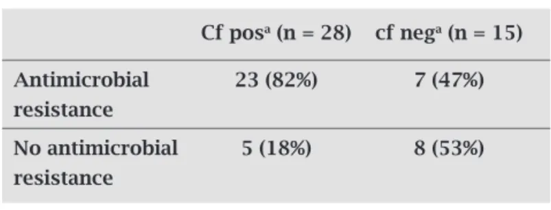
Finally, we found that there was a statistically signifi-cant correlation (Table 2) between expression of CFs and presence of antimicrobial resistance, since 82% (23/28) of the CF positive strains harbored antimicrobial resistance, in contrast to the CF negative strains which only were re-sistant in 47% (7/15) of the cases. This finding may suggest that resistance genes might be located on ETEC virulence plasmids. Presence of CFs has previously been found to correlate to disease37 and we found in this study that a high proportion of the ETEC strains that all were collected from hospitalized children expressed known CFs. Since these strains may also be more likely to also carry antibiotic re-sistance genes, our results further implicate that vaccina-tion is a better soluvaccina-tion than antimicrobial treatment.
ACKNOWLEDGMENTS
This work is part of the UMSA-IBMB “Diarrheal Disease Project” supported by the Swedish Agency for Research Eco-nomic Cooperation (SIDA). Support was also obtained from the Swedish Research Council, grant no. 6X-09084, Minis-terio de Ciencia e Innovación (Instituto de Salud Carlos III, Fondo de Investigación Sanitaria, REIPI- RD06/0008-1018 and PS09/01273) and Xunta de Galicia (07MRU036261PR, 09TAL007261PR, 2007/000044-0).
Claudia Rodas acknowledges the financial support from the Swedish Institute.
REFERENCES
1. Kosek M, Bern C, Guerrant RL. The magnitude of the global burden of diarrhoeal disease from studies published 1992-2000. Bull World Health Organ 2003; 81:197-204.
2. Parashar UD, Bresee JS, Glass RI. The global burden of diarrhoeal disease in children. Bull World Health Organ 2004; 81:236. 3. SNIS. Sistema Nacional de Información en Salud 2009. Bol
Minis Salud Pub 2009.
4. Qadri F, Svennerholm AM, Faruque AS, Sack RB. Enterotoxi-genic Escherichia coli in developing countries: epidemiology, microbiology, clinical features, treatment, and prevention. Clin Microbiol Rev 2005; 18:465-483.
5. Gaastra W, Svennerholm AM. Colonization factors of human enterotoxigenic Escherichia coli (ETEC). Trends Microbiol 1996; 4:444- 452.
6. Grewal H, Valvatine H, Bhan MK, van Dijk L, Gaastra W, Som-merfelt H. A new putative fimbrial colonization factor, CS19, of human enterotoxigenic Escherichia coli. Infect Immun 1997; 65:507-513.
7. Pichel MG, Binsztein N, Qadri F, Girón JA. Type IV longus pi-lus of enterotoxigenic Escherichia coli: occurrence and associa-tion with toxin types and colonizaassocia-tion factors among strains isolated in Argentina. J Clin Microbiol 2002; 40:694-697. 8. Gomez-Duarte OG, Chattopadhyay S, Weissman SJ, Giron JA,
Kaper JB, Sokurenko EV. Genetic diversity of the gene cluster encoding longus, a type IV pilus of enterotoxigenic Escherichia coli. J Bacteriol 2007; 189:9145-9149.
9. López-Vidal Y, Calva JJ, Trujillo A et al. Enterotoxins and ad-hesins of enterotoxigenic Escherichia coli: are they risk factors for acute diarrhea in the community? J Infect Dis 1990; 162:442-447. 10. Walker RI, Steele D, Aguado T. Analysis of strategies to suc-cessfully vaccinate infants in developing countries against en-terotoxigenic E. coli (ETEC) disease. Vac 2007; 25:2545-2566. 11. Svennerholm AM, Tobias J. Vaccines against enterotoxigenic
Escherichia coli. Expert Rev Vaccines 2008; 7:795-804. 12. Pratts G, Mirelis B, Llovet T. Antibiotic resistance trends in
en-teropathogenic bacteria isolated in 1985-1987 and 1995-1998 in Barcelona. Antimicrob Agents Chemother 2000; 44:1140-5. 13. Sjöling Å, Wiklund G, Savarino SJ, Cohen DI, Svennerholm
AM. Comparative analyzes of phenotypic and genotypic meth-ods for detection of enterotoxigenic Escherichia coli toxins and colonization factors. J Clin Microbiol 2007; 45:3295-3301. 14. Rodas C, Iniguez V, Qadri F, Wiklund G, Svennerholm AM,
Sjöling Å. Development of multiplex PCR assays for detection of enterotoxigenic Escherichia coli colonization factors and toxins. J Clinl Microbiol 2009; 47:1218-1220.
Table 2. Analysis of the relation of expression of colonization factors and antimicrobial resistance in the selected etec strains. The association between an-tibiotic resistance and presence of cf was considered to be statistically significant by the fishers exact test. (P < 0.0339)
Cf posa (n = 28) cf nega (n = 15)
Antimicrobial 23 (82%) 7 (47%) resistance
No antimicrobial 5 (18%) 8 (53%) resistance
Acf pos, colonization factor positive; cf neg, colonization
15. Guineé PAM, Jansen H, Wadstrom T, Sellwood R. Escheri-chia coli associated with neonatal diarrhoea in piglets and calves. In: de Leeuw PW, Guinee PAM (eds) Laboratory di-agnosis in neonatal calf and animal science, 13. The Neth-erlands, Martinus Nijhoff. 1981; 126-162.
16. Blanco J, González EA, Blanco M et al. Enterotoxigenic Es-cherichia coli associated with infant diarrhoea in Galicia, north-western Spain. J Med Microbiol 1991; 35:162-167. 17. National Committee for Clinical Laboratory Standards.
Performance Standards for Antimicrobial Disk Susceptibil-ity Tests—Eighth Edition: Approved Standard M2-A8. NC-CLS, Villanova, PA, USA 2002.
18. Blanco J, Blanco M, González EA et al. Serotypes and colo-nization factors of enterotoxigenic Escherichia coli isolated in various countries. Eur J Epidemiol 1993; 9:489-496. 19. Wolf MK. Occurrence, distribution, and associations of O
and H serogroups, colonization factor antigens, and tox-ins of enterotoxigenic Escherichia coli. Clin Microbiol Rev. 1997; 10:569-584.
20. Ansaruzzaman M, Bhuiyan NA, Begum YA et al.
Character-ization of enterotoxigenic Escherichia coli from diarrhoeal patients in Bangladesh using phenotyping and genetic pro-filing. J Med Microbiol 2007; 56:217-222.
21. Vicente AC, Teixeira LF, Iniguez-Rojas L et al. Outbreaks of cholera-like diarrhoea caused by enterotoxigenic Escheri-chia coli in the Brazilian Amazon Rainforest Trans R Soc Trop Med Hyg 2005; 99: 669-674.
22. Rivas M, Binsztein N, Basanta G et al. Antibody responses against Escherichia coli heat-labile toxin and colonization factor antigens I and II in Argentinean children. J Inf Dis 1995; 171: 1045-1049.
23. Harris AM, Chowdhury F, Begum YA et al. Shifting preva-lence of major diarrheal pathogens in patients seeking hos-pital care during floods in 1998, 2004, and 2007 in Dhaka, Bangladesh. Am J Trop Med Hyg 2008; 79: 708-714. 24. Steinsland H, Valentiner-Branth P, Grewal HM, Gaastra W,
Mølbak KK, Sommerfelt H. Development and evaluation of genotypic assays for the detection and characterization of enterotoxigenic Escherichia coli. Diag Microbiol Infec Dis 2003; 45:97-105.
25. Girón JA, Viboud GI, Sperandio V et al. Prevalence and associa-tion of the longus pilus structural gene (lngA) with colonizaassocia-tion factor antigens, enterotoxin types, and serotypes of enterotoxi-genic Escherichia coli. Infect Immun 1995; 63:4195-4198.
26. Gutiérrez-Cázarez Z, Qadri F, Albert MJ, Girón JA. Identifica-tion of enterotoxigenic Escherichia coli harboring longus type IV pilus gene by DNA amplification. J Clin Microbiol 2000; 38:1767-1771.
27. Nishimura LS, Girón JA, Nunes SL, Guth BE. Prevalence of enterotoxigenic Escherichia coli strains harboring the longus pilus gene in Brazil. J Clin Microbiol. 2002; 40:2606-2608. 28. Orskov F, Orskov I. Escherichia coli serotyping and disease in
man and animals. Can J Microbiol 1992; 38:699-704.
29. Åhrén CM, Svennerholm AM. Synergistic protective effect of antibodies against Escherichia coli enterotoxin and coloniza-tion factor antigens. Infect Immun 1982; 38:74-79.
30. Reis MH, Matos DP, de Castro AF, Toledo MR, Trabulsi LR. Relationship among enterotoxigenic phenotypes, serotypes, and sources of strains in enterotoxigenic Escherichia coli. In-fect Immun 1980; 28:24-27.
31. Sommerfelt H, Steinsland H, Grewal H et al. Colonization fac-tors of enterotoxigenic Escherichia coli isolated from children in North India. J Infec Dis 1996; 174:768-776.
32. Pacheco A, Guth BE, Soares KC, Nishimura L, de Almeida DF, Ferreira LC. Random amplification of polymorphic DNA re-veals serotype-specific clonal clusters among enterotoxigenic Escherichia coli strains isolated from humans. J Clin Microbiol 1997; 35:1521-1525.
33. Black RE, Levine MM, Clements ML, Cisneros L, Daya V.
Treatment of experimentally induced enterotoxigenic
Es-cherichia coli diarrhea with trimethoprim, trimethoprim-sul-famethoxazole, or placebo. Rev Infect Dis 1982; 4:540-545. 34. Benguigui Y. Atención Integrada a las Enfermedades
Preva-lentes de la Infancia. Bol Noticias IRA 2007; 33:2-5.
35. Vila J, Vargas M, Ruiz J. Quinolone resistance in Enterotoxi-genic Escherichia coli causing diarrhea in travelers to India in comparison with other geographical areas. Antimicrob Agents Chemother 2000; 44:1731-33.
36. Frisari L, Viggiano V, Pelagalli M. An open, controlled study of two non-absorbable antibiotics for the oral treatment of paedi-atric infectious diarrhoea. Curr Med Res1997; 14:39-45. 37. Qadri F, Das SK, Faruque AS et al. Prevalence of toxin types