Escherichia coli
and Molecular Change under Resistance
Selection
Wentao Guo., Haihong Hao., Menghong Dai, Yulian Wang, Lingli Huang, Dapeng Peng, Xu Wang,
Hailan Wang, Min Yao, Yawei Sun, Zhenli Liu, Zonghui Yuan*
National Reference Laboratory of Veterinary Drug Residues/MOA Key Laboratory of the Detection of Veterinary Drug Residues, Huazhong Agricultural University, Wuhan, Hubei, People’s Republic of China
Abstract
Quinoxaline 1, 4-dioxides (QdNOs) has been used in animals as antimicrobial agents and growth promoters for decades. However, the resistance to QdNOs in pathogenic bacteria raises worldwide concern but it is barely known. To explore the molecular mechanism involved in development of QdNOs resistance inEscherichia coli,6 strains selected by QdNOsin vitro
and 21 strains isolated from QdNOs-used swine farm were subjected to MIC determination and PCR amplification ofoqxA
gene. A conjugative transfer was carried out to evaluate the transfer risk of QdNOs resistant determinant. Furthermore, the transcriptional profile of a QdNOs-resistantE. coli(79O4-2) selectedin vitrowith its parent strain 79–161 was assayed with a prokaryotic suppression subtractive hybridization (SSH) PCR cDNA subtraction. The result showed that more than 95% (20/ 21) clinical isolates wereoqxApositive, while all the 6 induced QdNOs-resistant strains carried nooqxAgene and exhibited low frequency of conjugation. 44 fragments were identified by SSH PCR subtraction in the QdNOs-resistant strain 79O4-2. 18 cDNAs were involved in biosynthesis of Fe-S cluster (narH), protein (rpoA, trmD, truA, glyS, ileS, rplFCX, rpsH, fusA), lipoate (lipA), lipid A (lpxC), trehalose (otsA), CTP(pyrG) and others molecular. The 11 cDNAs were related to metabolism or degradation of glycolysis (gpmAand pgi) and proteins (clpX, clpA, pepN and fkpB). The atpADG and ubiB genes were associated with ATP biosynthesis and electron transport chain. The pathway of the functional genes revealed thatE. coli
may adapt the stress generated by QdNOs or develop specific QdNOs-resistance by activation of antioxidative agents biosynthesis (lipoate and trehalose), protein biosynthesis, glycolysis and oxidative phosphorylation. This study initially reveals the possible molecular mechanism involved in the development of QdNOs-resistance inE. coli, providing with novel insights in prediction and assessment of the emergency and horizontal transfer of QdNOs-resistance inE. coli.
Citation:Guo W, Hao H, Dai M, Wang Y, Huang L, et al. (2012) Development of Quinoxaline 1, 4-Dioxides Resistance inEscherichia coliand Molecular Change under Resistance Selection. PLoS ONE 7(8): e43322. doi:10.1371/journal.pone.0043322
Editor:Asad U. Khan, Aligarh Muslim University, India
ReceivedMay 31, 2012;AcceptedJuly 19, 2012;PublishedAugust 28, 2012
Copyright:ß2012 Guo et al. This is an open-access article distributed under the terms of the Creative Commons Attribution License, which permits unrestricted use, distribution, and reproduction in any medium, provided the original author and source are credited.
Funding:This article is supported by National Natural Science Foundation of China (31101856) and the Fundamental Research Funds for the Central Universities (2011QC005). The funders had no role in study design, data collection and analysis, decision to publish, or preparation of the manuscript.
Competing Interests:The authors have declared that no competing interests exist. * E-mail: [email protected]
.These authors contributed equally to this work.
Introduction
Quinoxaline-1,4-dioxides (QdNOs) have been introduced into animal production as potent antibacterial agents against most gram-positive and gram–negative pathogens since 1970s [1,2,3]. These drugs have been also widely used as growth promoters for decades [4,5]. Carbadox, maquindox and olaquindox are the well known members of QdNOs [6]. Cyadox is a new member of QdNOs which exhibits better antibacterial activity, lower toxicity and higher efficiency on animal growth promotion [7,8,9,10].
It is demonstrated that usage of olaquindox on pig farms may increase the emergence of olaquindox resistance in some pathogens [11,12]. The bacteria with resistance to olaquindox are generally co-resistant to ampicillin, tetracycline or chloram-phenicol [1,13]. For the mechanism of QdNOs resistance, it is only known that plasmid-encoded multidrug efflux pump OqxAB confers resistance to ampicillin, chloramphenicol, ciprofloxacin and olaquindox inE. coli[14,15]. This OqxAB located plasmid has
a high frequency of transfer among enterobacterial pathogens (Salmonella Typhimurium, Klebsiella pneumoniae, Kluyvera sp. and Enterobacter aerogenes, etc) [14,16]. It is rarely known how E. coli develop or acquire the QdNO’s resistance. For the concern of animal and human safety, it is required to be elucidated whether the selection pressure of QdNOs contributes the emergence of OqxAB and whether the resistant determinants would spread to other enterobacterial pathogens. During the development of QdNOs resistance, it is an urgent need to explore the novel molecular mechanisms involved in the emergency and transfer of QdNO’s resistance in pathogenic bacteria especiallyE. coli.
important way of horizontal transfer of some antibiotic resistance determinants in E. coli [18], the horizontal transfer of QdNOs-resistant determinant was then assessed by conjugative experi-ment. Since a prokaryotic SSH PCR cDNA subtraction approach developed by De Long is a technique that can be used to identify differentially expressed genes under given conditions [19], this approach was consequently carried out to investigate the novel mechanism beyond the OqxAB-mediated QdNOs resistance.
Materials and Methods
Drugs and Stock Solutions
Olaquindox (OLA), mequidox (MEQ), ampicillin (AMP), ceftiofur (CTF), tetracycline (TET), chlorotetracycline(CTE), chloramphenicol (CHL), florfenicol (FFC), trimethoprim (TMP), sulfamethoxazole (SMZ), ciprofloxacin hydrochloride (CIP) and colistin sulfate (CS) were supplied by China Institute of Veterinary Drug Control. Gentamicin (GEN), oxytetracycline (OTC), fura-zolidone (FZD) and rifampicin (RIF) were provided by National Institute for the Control of Pharmaceutical and Biological Products (Beijing, China). Carbadox (CAR) was purchased from Sigma Chemical Co. (St. Louis, MO) and cyadox (CYA) was kept in our lab. Stock solutions of the antimicrobials were prepared following the guidelines of the Clinical and Laboratory Standards Institute (CLSI, 2009) or the suppliers’ recommendations. Besides, stock solutions of CYA, CAR and FZD were prepared in DMSO; Stock solutions of OLA, MEQ and FFC were prepared in hot water; Stock solutions of CTF were prepared in phosphate buffer (pH 6.0, 0.1 mol/L). After vortexing, the solutions were sterilized by filtration through 0.22mm pore size filters (Millipore) before storage in aliquots at220uC. Solutions of ampicillin, tetracycline and oxytetracycline were prepared fresh on each occasion.
Strains
The porcineE. coli79–161 strain was purchased from China Institute of Veterinary Drug Control (Beijing, China). E. coli NK5449 (CGMCC NO. 1.1437) being resistant to rifampicin at 100mg/ml and as conjugation recipient was purchased from China General Microbiological Culture Collection Center (CGMCC, Beijing).E. coli ATCC25922 used as quality control in MIC test was kindly donated by South China Agricultural University (Guangzhou, China). All the strains were cultured at 37uC in Luria-Bertani (LB) broth medium in aerobic incubator.
In vitro Selection of High-level QdNOs Resistant Strains The selection of high-level QdNOs-resistant strains was performed with ancestor strainE. coli79–161 by serial transferring in LB broth containing two-fold increased concentrations of CYA or OLA. The selection concentrations of CYA and OLA ranged from 1/2MIC to 4MIC. Each step of transferring contained 5 passages of parent strain in broth with certain concentration of drug. Four increased concentrations of drug (1/2MIC, MIC, 2MIC and 4MIC) led to a total of 20 passages of parent strain. During each passage, an initial colony concentration of 104,105cfu/ml of parent strain was grown at 37uC for 24 h in
aerobic incubator. Cultures obtained at 5th, 10th, 15th and 20th passages were stored at 220uC. To estimate the stability of resistance, the 20th subcultures were serially transferred in drug-free LB broth for 10 times. For each passage, a drug-drug-free culture (growth control) and an uninoculated medium sterility control were included.
In vivo Isolation of ResistantE.colifrom Swine
On swine farm in Wuhan Huaguoshan Livestock Company, six groups of swine were breeding with drugs (Group 1 with no drug, group 2 with 50mg/ml olaquindox, group 3 with 50mg/ml chlorotetracycline, group 4 with 50mg/ml cyadox, group 5 with 150mg/ml cyadox and group 6 with 250mg/ml cyadox). The samples of anal swab were collected from each group of swine after 0, 45thand 100thday adding drugs to their basal diet. TheE. coli strains were isolated and identified by the selective agar (MacConkey Agar) and classic biochemical test following the microbiology laboratory guidebooks published by ministry of health in China (2003). Species identification was confirmed using an ABI 3130 system (Applied Biosystem, USA). 1,2 respectiveE.
colistrains selected from each of the six groups were subjected to MIC determination as below. The experimental procedures involving animals in the study were approved by the Animal Care Center, Hubei Academy of Medical Sciences. All efforts were made to minimize suffering of animals.
MIC Determination
MIC was determined by agar dilution antimicrobial suscepti-bility test according to the M7-A7 guideline of the Clinical and Laboratory Standards Institute (CLSI, 2006).Briefly, the density of E.coli suspensions was adjusted to equal the density of a 0.5 McFarland standard (16108CFU/ml). After 10 fold dilution by MH broth, 1,2mL of the diluted suspension was inoculated on prepared MH agar plates containing two-fold diluted concentra-tion of each drug. The final concentraconcentra-tion of strains was 104CFU/ point on drug-containing plates. The standard strain of E. coli ATCC25922 was used as quality control (QC) strain. Most of the QC range and susceptible breakpoint were cited from CLSI document M100-S19. The QC range of CIF was quoted from performance standard of cefotaxime and ceftriaxone. The QC range of FFC was quoted from performance standard of chloramphenicol. The resistance breakpoint of FZD (128mg/ml) was quoted from performance standard of furantoin. The resistance breakpoints of CTF, FFC and CAR were cited from published paper [12]. Olaquindox resistance has been defined with a breakpoint of 64mg/ml [1]. Acquisition of cyadox resistance was defined as an$4-fold increase of the MIC value comparing with its control parent strain [20].
PCR Amplification ofoqxAGenes
Colony PCR was proceeded to screen for oqxA genes in the selected E.coli strains. Based on the sequences of oqxA (GeneID 5962055) in E. coli, a pair of primers (OqxA-F 59 -GGGA-TAGTTTTAACGGTCGCATTG-39 and OqxA-R 59-TT CACGGGAGACGAGGTTGGT-39) was designed to amplify 266-bp oqxA gene fragment. The amplification conditions com-prised a denaturation step of 95uC for 5 min, followed by 32 cycles of 94uC for 30 s, 64uC for 30 s, 72uC for 1 min and a final extension for 10 min at 72uC. Amplicons were sequenced by Nanjing Genscript Biotechnology Limited Company (Nanjing, China).
following formula: CFU of transconjugant/CFU of donor. The donor, recipient and transconjugant were identified by comparison of their EcoRI RiboPrintH pattern with that in identification database [22]. Comparing toE. coliNK5449, the similarity of the RiboPrintHpattern was shown in percent.
Preparation of mRNA
Before isolation of total RNA, driver and tester stains were grown in 50-ml drug-free Luria-Bertani (LB) broth till log phase. Briefly, overnight cultures were 100 fold diluted in 50-ml of LB broth, the culture were grown at 37uC with 250 rpm shacking. When the density of culture reached to 0.6,0.8 OD600, the cells
were harvested by centrifugation of 1-ml aliquots in a micro-centriguge for 2 min. An RNeasy Protect Bacteria Mini Kit (Qiagen, Valencia, CA, USA) was used to lyse the cells and extract total RNA. The RNA was quantified by a light absorption at 260/ 280 nm. The purity and integrity of the RNA samples were verified by an electrophoresis in agarose gels using the 16S and 23S ribosomal bands as indicators. Samples of valid RNA were DNase treated with RQ1 RNase-free DNase (Promega, Madison, WI, USA). The DNA contamination was verified by PCR amplification ofrpoEgene using the primers in Table 1. Reactions containing pure RNA preparations would fail to yield a product. The mRNA was isolated using a MICROBExpress bacterial mRNA enrichment kit (Ambion, Austin, TX, USA). Every 5 preparations for each substrate were pooled before ethanol precipitation to obtain at least 2mg mRNA per preparation.
The mRNA quantity was measured by absorbance at 260 nm, and quality was assessed on agarose gel.
Prokaryotic SSH PCR cDNA Subtraction
cDNA synthesis, cDNA digestion, adaptor ligation, first and second hybridization, suppression PCR and clone screening were carried out following the protocols of De Long with slight modification on the PCR-select cDNA subtraction kit(Clontech) [19].
A RNA polymerase sigma factor rpoE(the primer in Table 1) was used to detect the quality of double-strand cDNA. The amplification conditions comprised a denatured step of 95uC for 5 min, followed by 32 cycles of 94uC for 30 s, 51uC for 30 s, and 72uC for 1 min and a final extension for 10 min at 72uC. Products were resolved by gel electrophoresis on a 2% (wt/vol) agarose gel. After second-strand cDNA synthesis, two cDNA synthesis reaction mixtures were pooled and purified with DNA fragment purifica-tion kit (Takara, Kyoto, Japan). Tester and driver cDNA were digested with Rsa I and purified also with DNA fragment purification kit (Takara).
To determine the efficiency of adapter ligation and subtraction, PCR amplification were performed according to the protocol in the PCR-Select cDNA subtraction kit by use of a housekeeping gene ribosomal protein S1 (rpsA) (the primer in Table 1). The amplification conditions comprised a denatured step of 95uC for 5 min, followed by cycles of 94uC for 30 s, 60uC for 30 s, and 72uC for 1 min and a final extension for 10 min at 72uC. For test
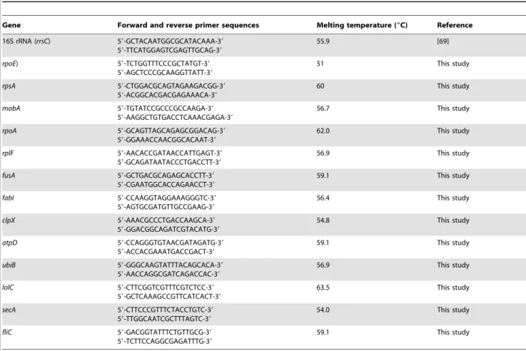
Table 1.Primers used in SSH PCR cDNA subtraction and RT-qPCR.
Gene Forward and reverse primer sequences Melting temperature (6C) Reference
16S rRNA (rrsC) 59-GCTACAATGGCGCATACAAA-39
59-TTCATGGAGTCGAGTTGCAG-39
55.9 [69]
rpoE) 59-TCTGGTTTCCCGCTATGT-39
59-AGCTCCCGCAAGGTTATT-39
51 This study
rpsA 59-CTGGACGCAGTAGAAGACGG-39
59-ACGGCACGACGAGAAACA-39
60 This study
mobA 59-TGTATCCGCCCGCCAAGA-39
59-AAGGCTGTGACCTCAAACGAGA-39
56.7 This study
rpoA 59-GCAGTTAGCAGAGCGGACAG-39
59-GGAAACCAACGGCACAAT-39
62.0 This study
rplF 59-AACACCGATAACCATTGAGT-39
59-GCAGATAATACCCTGACCTT-39
56.9 This study
fusA 59-GCTGACGCAGAGCACCTT-39
59-CGAATGGCACCAGAACCT-39
59.1 This study
fabI 59-CCAAGGTAGGAAAGGGTC-39
59-AGTGCGATGTTGCCGAAG-39
56.4 This study
clpX 59-AAACGCCCTGACCAAGCA-39
59-GGACGGCAGATCGTACATG-39
54.8 This study
atpD 59-CCAGGGTGTAACGATAGATG-39
59-ACCACGAAATGACCGACT-39
59.1 This study
ubiB 59-GGGCAAGTATTTACAGCACA-39
59-AACCAGGCGATCAGACCAC-39
56.9 This study
lolC 59-CTTCGGTCGTTTCGTCTCC-39
59-GCTCAAAGCCGTTCATCACT-39
63.5 This study
secA 59-CTTCCCGTTTCTACCTGTC-39
59-TTGGCAATCGCTTTAGTC-39
54.0 This study
fliC 59-GACGGTATTTCTGTTGCG-39
59-TCTTCCAGGCGAGATTTG-39
59.1 This study
of ligation efficiency, the cycles were 32. And for test of subtraction efficiency, the cycles were 18, 23, 28, 33 and 38.
After the PCR adaptor screen, 1, 2-clones were selected and submitted to Nanjing Genscript Biotechnology Limited Company (China) for sequencing directly. The sequences were aligned to available sequences in NCBI library by blastn algorithm (www. ncbi.nlm.nih.gov. BLAST). The homological genes were subjected into molecular annotation system (http://www.capitalbio.com/ support/mas) or KEGG library for gene function annotation and pathway analysis.
Reverse Transcription, Quantitative Real-time PCR (RT-qPCR)
4,5mg of DNase-treated total RNA (isolated as described above) was used to synthesize cDNA according the method of De Long [19]. Real-time PCR was carried out on Bio-Rad iQTM5 Multicolor Real-Time PCR Detection System, using SYBRH
Premix Ex TaqTM (Takara) in a total volume of 25ml: 12.5ml SYBRHPremix Ex TaqTM, 1.0ml template cDNA, 0.5ml (final concentration 0.2mM) or 0.25ml (final concentration 0.1mM) of each primer, and PCR-grade water to a final volume of 25ml. Samples without template were set as negative control. At least 3 replicates were analyzed for each gene. Primer sequences were presented in Table 1.The fold change of selected genes was normalized to fold change of reference gene (16S rRNA gene). The relative expression was calculated according to the formula 22ggCt[23], The data were considered for significant differences by one-way ANOVA using SPSS programme (P#0.05).
Results
Resistance Phenotype of Selected Strains
Six representative strains (79O4-1, 79O4-2, 79O4-3, 79C4-1, 79C4-2 and 79C4-3) were obtained after stepwise selection by two-fold ascended concentration of olaquindox and cyadox. The variations of MICs during the selection procedure were presented in Table 2. Before selection, strains were susceptible to all the antibiotics tested. After 20 passages, the MICs of growth-control strains (79,20) have no significant change. High-level olaquindox
resistant E. colistrains (79O4-1, 79O4-2 and 79O4-3) with MIC
$256mg/ml were selected under the pressure of olaquindox. Cyadox resistantE. colistrains (79C4-1, 79C4-2 and 79C4-3) with MIC$128mg/ml were obtained by stepwise selection of cyadox. Notably,E. colistrains selected by olaquindox exhibited reduced susceptibility to ampicillin, tetracycline, oxytetracycline, chloram-phenicol, florfenicol, trimethoprim and ciprofloxacin (see Table 2). All of the three strains (79O4-1, 79O4-2 and 79O4-3) selected by olaquindox were cross-resistant to the four QdNOs tested, while the three strains (79C4-1, 79C4-2 and 79C4-3) selected by cyadox were only resistant to carbadox and cyadox. Compared to cyadox, olaquindox might provide more potential in selection of multidrug resistance and cross-resistance inE.coli.
Before and after adding olaquindox, chlorotetracycline and cyadox into swine diet, 21 representative E.coli strains were isolated from 6 groups of swine by anal swab. From the data shown in Table 3, there is no significant change of the MIC before and after usage of QdNOs. Before usage of drug, the 7 E.coli strains selected at 0 day exhibited resistance to olaquindox with MIC $64mg/ml. Notably, the 3 E.coli strains from the control group is naturally resistant to olaquindox with MIC = 64mg/ml or 256mg/ml.
oqxAGene Mediated Resistance
All of the in vitro selected stains presented in Table 2 were negative foroqxAgene products, indicating that other mechanisms except for OqxAB mediated the resistance in the selected strains. However, 95% (20/21) E.coli strains isolated from swine farms were positive foroqxAgene (see Table 3).TheoqxApositive strains are isolated before and after the usage of QdNOs drugs on pig farm. No relationship can be observed between the occurrence of oqxAgene and usage of QdNOs drugs. The oqxAgene positive strains always exhibited resistance to olaquindox except for E100-4 strain which carriedoqxA gene but is susceptible to olaquindox (MIC = 32mg/ml).
Frequency of Conjugal Transfer
Frequencies of conjugal transfer for each strain were exempli-fied in Table 4. The conjugation frequencies of the threeE. coli donors were at 10210 CFU/donor. Before conjugation, the recipient is susceptible to the four QdNOs but resistant to rifampicin, the donors are resistant to the four QdNOs but susceptible to rifampicin. After conjugation, the transconjugants were resistant to both four QdNOs and rifampicin (see Table 4). The recipient and transconjugants share similar RiboPrintH
pattern with the similarity.90% (see Table 4), confirming that QdNOs-resistant determinant had been horizontally transferred by conjugation inE. colistrains.
Differentially Expressed Genes Determined by Prokaryotic SSH PCR cDNA Subtraction
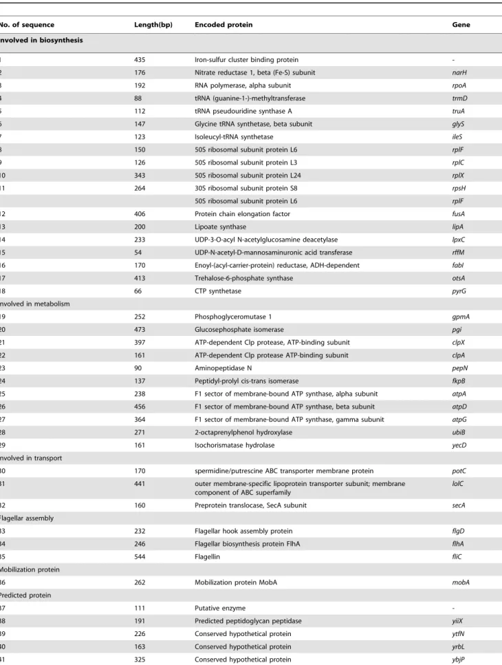
By prokaryotic SSH PCR cDNA subtraction, 44 unique fragments were found in the QdNOs resistant strain E. coli 79O4-2 comparing to its control strain E. coli 79,20. Among
these 44 sequences, 3 sequences were with no significant similarity found in the Genbank; 40 sequences were found in genome ofE. coli; 1 sequence was found in plasmid ofE. coli(mobA) (see Table 5). The homology search results showed that QdNOs-resistantE. coli alters the expression of 18 genes involved in biosynthesis, 11 genes associated with metabolism, 3 genes encoding transporters, 3 genes involved in flagellar assembly, 1 gene encoding mobilization protein and 5 genes associated with hypothetical proteins (see Table 5).
In the 18 genes involved in biosynthesis, the number 1 gene encodes a iron-sulfur cluster binding protein, the number 2 gene (narH) encodes a nitrate reductase I. The number 3 to 12 genes (rpoA, trmD, truA, glyS, ileS, rplFCX, rpsH and fusA) encode RNA polymerase, tRNA-methyltransferase, tRNA synthase, tRNA synthase and protein chain elongation factor respectively. The number 13 to18 genes (lipA, lpxC, rffM, fabI, otsA and pyrG) are related to biosynthesis of lipoate, lipid A, enterobacterial common antigen, fatty acid, trehalose and cytidine triphosphate respectively (see Table 5 and Figure 1).
In the 11 genes involved in metabolism, the number 19 and 20 genes (gpmA and pgi) participate in glycogen degradation, the number 21 to 24 genes (clpX, clpA, pepN and fkpB) take part in degradation of damaged protein or protein modulation, the number 25 to 29 genes (atpA, atpD, atpG, ubiB and yecD) contribute to electron transport and ATP biosynthesis (see Table 5 and Figure 1).
Table 2.The MICs to 16 antibiotics of the strains selected by two-fold ascended concentrations of olaquinodox or cyadoxin vitro.
strains MIC to different antibiotics (mg/ml)
AMP CTF GEN TET OT CHL FFC TMP SMZ FZD CIP CS OLA MEQ CAR CYA
selected by olaquinodox
79O4-1 16* 1 0.25 64#
32#
8 8 8* 8 8 0.031* 1 256#
128# 64#
.128#
79O4-2 16* 2* 0.25 64# 32# 32# 32# 8* 8 8 0.031* 1
.256#
.128# 128#
.128#
79O4-3 8 #0.5 0.5* 64#
32#
8 8 4* 8 16 0.031* 1 256#
128# 32#
.128# selected by
cyadox
79C4-1 2 #0.5 0.25 32# 32# 2 2 1 8 8 0.015 1 32 32 64#
.128#
79C4-2 2 #0.5 0.25 32#
32#
4 4 1 8 8 0.015 1 16 16 8 .128#
79C4-3 4 #0.5 0.5* 32#
32#
8 8 1 8 4 0.015 1 32 32 64#
.128# Growth control
strain
79,20 4 #0.5 0.125 2 2 4 4 1 8 8 0.008 1 16 16 16 32
QC standards
ATCC25922 2 0.125 0.5 2 2 2 4 2 8 8 0.008 1 8 4 4 16
QC range 2–8 0.03–0.12f 0.25–1 0.5–2 0.5–2 2–8 2–8 0.5–2 8–32 4–16h 0.004– 0.015
0.25–1 NA NA NA NA
Breakpoint 32 8 16 16 16 32 16 16 512 128 4 NA 64 4MIC 32 4MICk
Note:. * MIC ascends 4-fold or more, but not reaching breakpoint; #
MIC reach breakpoint. NA Not available. doi:10.1371/journal.pone.0043322.t002
Table 3.Olaquindox MICs andoqxAcontaining in the clinical strains isolated from pigs before and after usage of growth promoters.
Time for drug
addition (days) group
Usage of Drugs and concentration
in swine diet strains
Olaquindox MIC (mg/ml)
OqxA (+positive, -negative)
0 day 1 control E0-1 64 +
2 50mg/ml olaquindox E0-2 64 +
3 50mg/ml chlorotetracycline E0-3 128 +
4 50mg/ml cyadox E0-4 64 +
5 150mg/ml cyadox E0-5 128 +
6 250mg/ml cyadox E0-6-1 128 +
6 250mg/ml cyadox E0-6-2 128 +
After 45 days 1 control E45-1 256 +
2 50mg/ml olaquindox E45-2 256 +
3 50mg/ml chlorotetracycline E45-3 128 +
4 50mg/ml cyadox E45-4 256 +
5 150mg/ml cyadox E45-5 128 +
6 250mg/ml cyadox E45-6-1 64 +
6 250mg/ml cyadox E45-6-2 64 +
After 100 days 1 control E100-1 256 +
2 50mg/ml olaquindox E100-2 256 +
3 50mg/ml chlorotetracycline E100-3 128 +
4 50mg/ml cyadox E100-4 32 +
5 150mg/ml cyadox E100-5 64
-6 250mg/ml cyadox E100-6-1 128 +
6 250mg/ml cyadox E100-6-2 256 +
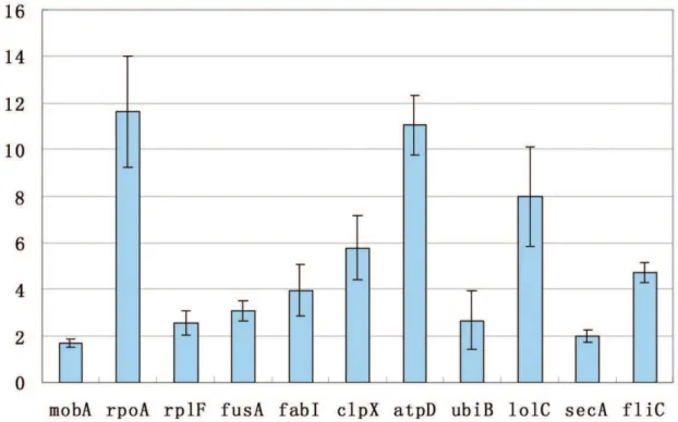
Up-regulation of 11 respresentative genes (mobA,rpoA,rplF,fusA, fabI, clpX, atpD, ubiB, lolC, secA and fliC) was confirmed by RT-qPCR. Fold change of the 11 genes is in the range of 1.7 to 11 fold of over-expression with the P value much less than 0.05, indicating that the methodology of prokaryotic SSH PCR cDNA subtraction is reliable to identify differentially expressed genes (see Figure 2).
Discussion
Resistance Development Under QdNOs
Although the resistant E. colistrains were obtained from step-wise selection by QdNOs drugs in vitro, there is no significant change of susceptibility of theE.colistrains isolated from pigs farm before and after the usage of QdNOs drugs. It was revealed that the incidence and level of olaquindox resistance in the coliform floras isolated from pigs and pig farms was complex and largely fluctuated, the possible origin of olaquindox-resistant strains may be related to the biotypes of the drug-resistant sub-populations [24]. During the usage of olaquindox as feed additive on commercial pig farms from 1982 to 1984, the monitoring result about the development of olaquindox resistance showed that there was a low but not significant increasing incidence and level of resistance to olaquindox on drug-using farms [25]. Conclusively, it is unreliable to connect the emergency of QdNOs resistance to the usage of QdNOs drugs only by series passage in vitro. The resistance ofE.colistrains isolated from the control group of pigs may due to the complexity of clinical environment.
Compared the MICs of QdNOs in the three strains (79O4-1, 79O4-2 and 79O4-3) selected by olaquindox to the three strains (79C4-1, 79C4-2 and 79C4-3) selected by cyadox, olaquindox appears to possess more potency in selection of cross-resistance to QdNOs, suggesting that long-term use of cyadox could be safer than olaquindox. The difference in the development of cross-resistance induced by olaquindox and cyadox may attribute to the discrepancy in the chemical characteristics or structure of these two drugs [2].
Similar to previous studies of resistant phenotype inE.colistrains isolated from pigs and pig farms, the QdNOs resistant strains selected in our study also exhibited multi-drug resistance against tetracycline and chloramphenicol [24,25,26]. Both the result of previous studies and our study revealed that multidrug resistance in most of clinical isolates may due to the multidrug efflux pump OqxAB [14,15]. The OqxAB located plasmid was reported to has a high frequency (1024 CFU/recipient) of transfer among enterobacterial pathogens including S.Typhimurium and K.pneumo-niae, etc[14,16]. The E100-4 strain (see table 3) carried the oqxA gene but is susceptible to olaquindox, indicating that some strains may acquire theoqxAgene from the horizontal transfer in clinical environment. However, different with previous studies, the six QdNOs resistant strains selected by in vitro passage ((79O4-1, 79O4-2, 79O4-3, 79C4-1, 79C4-2 and 79C4-3) were negative in OqxAB pump (data not shown ) and showed much lower frequency of horizontal transfer (see table 4) with the ratio of transconjugant to donor only in a range of 2.5,4.0610210CFU/
donor, suggesting that other mechanism beyond OqxAB might confer the multidrug resistance and lower transfer risk in the selected strains in our study. A prokaryotic SSH PCR cDNA subtraction was thus carried out to find the novel molecular mechanism involved in QdNOs resistance.
Differently Expressed Genes Involved in the QdNOs Resistance
Based on networks of the drug-target induced cell death pathway found recently [27], it is supposed that the QdNOs
Table 5.The differentially expressed fragments identified in the QdNOs resistant strainE. coli79O4-2.
No. of sequence Length(bp) Encoded protein Gene
Involved in biosynthesis
1 435 Iron-sulfur cluster binding protein
-2 176 Nitrate reductase 1, beta (Fe-S) subunit narH
3 192 RNA polymerase, alpha subunit rpoA
4 88 tRNA (guanine-1-)-methyltransferase trmD
5 112 tRNA pseudouridine synthase A truA
6 147 Glycine tRNA synthetase, beta subunit glyS
7 123 Isoleucyl-tRNA synthetase ileS
8 150 50S ribosomal subunit protein L6 rplF
9 126 50S ribosomal subunit protein L3 rplC
10 343 50S ribosomal subunit protein L24 rplX
11 264 30S ribosomal subunit protein S8 rpsH
50S ribosomal subunit protein L6 rplF
12 406 Protein chain elongation factor fusA
13 200 Lipoate synthase lipA
14 233 UDP-3-O-acyl N-acetylglucosamine deacetylase lpxC
15 54 UDP-N-acetyl-D-mannosaminuronic acid transferase rffM
16 170 Enoyl-(acyl-carrier-protein) reductase, ADH-dependent fabI
17 413 Trehalose-6-phosphate synthase otsA
18 66 CTP synthetase pyrG
Involved in metabolism
19 252 Phosphoglyceromutase 1 gpmA
20 473 Glucosephosphate isomerase pgi
21 397 ATP-dependent Clp protease, ATP-binding subunit clpX
22 161 ATP-dependent Clp protease ATP-binding subunit clpA
23 90 Aminopeptidase N pepN
24 137 Peptidyl-prolyl cis-trans isomerase fkpB
25 238 F1 sector of membrane-bound ATP synthase, alpha subunit atpA
26 456 F1 sector of membrane-bound ATP synthase, beta subunit atpD
27 364 F1 sector of membrane-bound ATP synthase, gamma subunit atpG
28 271 2-octaprenylphenol hydroxylase ubiB
29 161 Isochorismatase hydrolase yecD
Involved in transport
30 170 spermidine/putrescine ABC transporter membrane protein potC
31 441 outer membrane-specific lipoprotein transporter subunit; membrane
component of ABC superfamily
lolC
32 160 Preprotein translocase, SecA subunit secA
Flagellar assembly
33 232 Flagellar hook assembly protein flgD
34 246 Flagellar biosynthesis protein FlhA flhA
35 544 Flagellin fliC
Mobilization protein
36 262 Mobilization protein MobA mobA
Predicted protein
37 111 Putative enzyme
-38 191 Predicted peptidoglycan peptidase yiiX
39 226 Conserved hypothetical protein ytfN
40 163 Conserved hypothetical protein yrbL
41 325 Conserved hypothetical protein ybjP
Figure 1. The hyperthetical mechanism of cell death induced by QdNOs and genes involved in the development of Quinoxaline resistance.The primary drug (red triangle)-target (unknown target) interaction initiate a metabolic feedback dependent on tricarboxylic acid (TCA)
cycle, stimulate the oxidation of NADH through the electron transport chain, promote superoxide (O22) formation, damage the structure of Fe-S
clusters, lead to the formation of hydroxyl radicals (OH2); and finally damage DNA, lipids or proteins and contributes to the cell death. The
overexpressed genes found in our study (in the box and cycle) were involved into this process. The overexpression of some genes were induced in this drug-target cell death pathway (The dotted arrow), the others may inhibit the formation free radicals or prevent the damage by free radical (dotted line with a bar).
doi:10.1371/journal.pone.0043322.g001
Figure 2. Fold change of 11 up-regulated genes determined by RT-qPCR.The X-axis was the 11 representative genes (mobA,rpoA,rplF,fusA,
fabI,clpX,atpD,ubiB,lolC,secAandfliC) which were selected from all the 44 differentially expressed genes. The Y-axis was the relative fold change of
binding to unknown target may initiate a metabolic feedback dependent on the tricarboxylic acid (TCA) cycle, stimulate the oxidation of NADH through the electron transport chain; promote superoxide (O22) formation; damage the structure of Fe-S clusters, lead to the formation of hydroxyl radicals (OH2); and finally damage DNA, lipids or protein and contributes to the death of E.coli(see Figure 1).
In the differently expressed genes, the iron-sulfur cluster binding protein encoding gene (gene number 1) participates in Fe-S cluster assembly. The nitrate reductase 1 encoding gene (narH) contribute to the electron transfer for Fe-S cluster [28,29]. Thus, overex-pression of number 1 and 2 genes (narH) may compensate Fe-S cluster damage by assembly of Fe-S cluster (see figure 1).
TherpoAis a DNA-direct RNA polymerase which can interact with other proteins (rplF, rplC, rplX, rpsH and others) and plays importance role in transcriptional regulation [30,31]. The trmD, truA,glyS and ileSare essential enzymes for tRNA modification and aminoacyl-tRNA synthesis which also play essential role on the protein synthesis [32]. ThefusAgene encoding for a translational regulator which is essential for translational elongation [33,34]. The overexpression of the translational regulator fusAand some ribosomal proteins involved in translational process may compen-sate protein synthesis for oxidative damage by QdNOs and contribute to the multidrug resistance against some protein target drug of tetracycline and chloramphenicol.
The number 13 gene (lipA) encodes lipoate synthase (LS) which is involved in the biosynthesis of Lipoic acid [35,36,37]. Lipoic acid is a protein-bound dithiol- containing cofactor which is commonly required for the energy metabolism in E. coli [37]. Recent studies also show that lipoic acid is a free radical scavenger and a potent antioxidant [38,39,40]. The number 17 gene (otsAB) encodes trehalose-6-phosphate synthase which is responsible for trehalose biosynthesis [41,42,43]. It has been shown that trehalose can protect proteins and cellular membranes from inactivation or denaturation caused by a variety of stress conditions, including desiccation, dehydration, heat, cold or oxidation [42,43,44]. The accumulation of trehalose can protect Saccharomyces cerevisiaefrom oxidative damage and enhance resistance of bacterial to H2O2, indicating that trehalose may be a good free radical scavenger [45]. Therefore, the overexpression of lipA and otsA gene may promote production of two free radical scavenger (lipoic acid and trehalose) which may protect QdNOs-resistant E. coli from oxidative damage and result in QdNOs-resistance. There is no significant change of the expression ofoxyRorsoxRSregulon, so this mechanism of antioxidative protection mediated by lipoic acid and trehalose may be independent on the regulation of oxyRor soxRS[46].
The number 19 gene (phosphoglyceromutase 1, gpmA) and number 20 gene (glucosephosphate isomerase,pgi) are involved in glycolysis which generates a small quantity of ATP and degrades glucose into pyroracemic acid and acetyl-CoA for tricarboxylic acid cycle (TCA) under aerobic growth condition. These two enzymes encoded by gpmA and pgi gene are also important for central carbon metabolism (CCM) pathway which may influence the fidelity of DNA replication in E.coli [47,48]. The 2-octaprenylphenol hydroxylase (ubiB), F1 sector of membrane-bound ATP synthasea (atpA), b (atpD) and c(atpG) subunit are involved in ATP biosynthesis, electron transport chain and oxidative phosphorylation [49].
Based on the function annotation and pathway analysis, the Clp, one of the key ATP-dependent proteases inE. coli, is required during some stress conditions [50,51,52,53]. The PepN, a major aminopeptidase in E. coli, can degrade amino acids into small molecular for TCA cycle [54]. The Clp protease cleave damaged
proteins into large peptides and amino acids which are further degraded by PepN [55]. The fkpB, peptidyl-prolyl cis-trans isomerase, participates in several biochemical processes including protein folding, receptors signaling, protein trafficking and transcription [56]. Therefore, up-regulation of Clp proteases (21-ClpX and 22-ClpA), aminopeptidase pepN (number 23 gene)and protein folding chaperones fkpB (number 24 gene)may play important roles in regulating levels of specific proteins and in eliminating or modifying damaged or abnormal proteins [57].
Considering the drug-target induced cell death pathway and function analysis of the differentially expressed genes, the upregulation of some genes may be response for some process of cell death pathway, but others may compensate or inhibit that pathway. See figure 1, the two genes involved in glycolysis for TCA cycle (19-gpmA and 20-pgi), four genes related to ATP biosynthesis and electron transport chain (25-atpA, 26-atpD, 27-atpG and 28-ubiB) and four genes associated with protein degradation and modulation (21-clpX, 22-clpA, 23-pepN and 24-fkpB) might be response for the metabolic feedback, hyperactiva-tion of electron transport chain and protein damage, respectively. However, the two genes involved in Fe-S cluster assembly (1-gene and 2-narH) and several genes associated with protein biosynthesis may compensate the Fe-S cluster damage and protein damage respectively (see Figure 1). The overexpression of genes involved in biosynthesis of lipoic acid and trehalose (13-lipA and ostA) may eliminate superoxide formation and hydroxyl radical production (see Figure 1). The role oflipAandostAon clearage of free radical and development of QdNOs resistance would be investigated in our next plan, because unpublished data obtained in our lab recently revealed that QdNOs are endowed with producing free radical inE.colicell.
Conclusions
Results of the present study suggest that olaquindox possess more potency in selection of multi-drug resistant and cross-resistantE. colistrains. The induced QdNOs-resistant determinant shows lower frequencies of conjugation than previous reports in clinical isolates. More than 90% clinical isolates carryoqxAgene, but it is negative in ourin vitroselected QdNOs-resistant strains, indicating that there might be other mechanism conferring resistance. By SSH PCR subtraction, 44 genes were differentially expressed in the QdNOs-resistant strain 79O4-2 selected by olaquindox in vitro. The overexpression of genes involved in glycolysis for TCA cycle (gpmA and pgi) and genes involved in ATP biosynthesis and electron transport chain (atpADG and ubiB) may due to the metabolic feedback and hyperactivation of electron transport chain, respectively. The genes involved in Fe-S cluster assembly (1-gene andnarH) and genes involved in biosynthesis of lipoic and trehalose (lipAandostA) may play an important role in clearage of free radical and prevention of Fe-S damage. The genes involved in protein biosynthesis (rpoA, trmD, truA, glyS, ileS, rplF, rplC, rplX, rpsH, rplF and fusA) and genes involved in protein degration and modulation may participate in compensation of the protein damage (see Figure 1). The other genes including genes encoding transporter, flagellar, lipid A and mobilization protein may be essential for the growth and physiological functions of
QdNOs-resistant E. coli.Conclusively, most of these genes have previously been implicated in resistance or tolerance to superoxide stress, suggesting that their altered expression may be a part of a protective response in QdNOs-resistant E. coli [68]. Likewise, other identified changes in gene expression may affect the growth and transfer capacity of QdNOs resistance. Further studies are required to investigate the roles of these genes in development of QdNOs resistance by technique of gene knockout and transcrip-tomics or proteomics. In addition, the molecular mechanism involved in the discrepancy of resistance development selected by olaqindox and cyadox is necessary to be investigated in future, because these information will be useful for the designation of new member of versatile QdNOs.
Acknowledgments
We thank S. K. De Long for providing valuable suggestions while performing the prokaryotic SSH PCR cDNA subtraction approach.
Author Contributions
Conceived and designed the experiments: WG MD HH XW ZY. Performed the experiments: WG HW MY YS. Analyzed the data: WG HH MD DP ZY. Contributed reagents/materials/analysis tools: ZY YW LH ZL. Wrote the paper: WG HH MD ZY.
References
1. Sørensen AH, Hansen LH, Johannesen E, Sørensen SJ (2003) Conjugative plasmid conferring resistance to olaquindox. Antimicrobial Agents and Chemotherapy 47: 798–799.
2. Carta A, Corona P, Loriga M (2005) Quinoxaline 1,4-dioxide: a versatile scaffold endowed with manifold activities. Curr Med Chem 12: 2259–2272. 3. Ganley B, Chowdhury G, Bhansali J, Daniels JS, Gates KS (2001)
Redox-activated, hypoxia-selective DNA cleavage by quinoxaline 1,4-di-N-oxide. Bioorg Med Chem 9: 2395–2401.
4. Beutin L, Preller E, Kowalski B (1981) Mutagenicity of quindoxin, its metabolites, and two substituted quinoxaline-di-N-oxides. Antimicrob Agents Chemother 20: 336–343.
5. Zou J, Chen Q, Tang S, Jin X, Chen K, et al. (2009) Olaquindox-induced genotoxicity and oxidative DNA damage in human hepatoma G2 (HepG2) cells. Mutat Res 676: 27–33.
6. Ding MX, Yuan ZH, Wang YL, Zhu HL, Fan SX (2006) Olaquindox and cyadox stimulate growth and decrease intestinal mucosal immunity of piglets orally inoculated with Escherichia coli. J Anim Physiol Anim Nutr (Berl) 90: 238–243.
7. Ding MX, Wang YL, Zhu HL, Yuan ZH (2006) Effects of cyadox and olaquindox on intestinal mucosal immunity and on fecal shedding of Escherichia coli in piglets. J Anim Sci 84: 2367–2373.
8. Huang L, Wang Y, Tao Y, Chen D, Yuan Z (2008) Development of high performance liquid chromatographic methods for the determination of cyadox and its metabolites in plasma and tissues of chicken. J Chromatogr B Analyt Technol Biomed Life Sci 874: 7–14.
9. He Q, Fang G, Wang Y, Wei Z, Wang D, et al. (2006) Experimental evaluation of cyadox phototoxicity to Balb/c mouse skin. Photodermatol Photoimmunol Photomed 22: 100–104.
10. Fang G, He Q, Zhou S, Wang D, Zhang Y, et al. (2006) Subchronic oral toxicity study with cyadox in Wistar rats. Food Chem Toxicol 44: 36–41.
11. Akwar HT, Poppe C, Wilson J, Reid-Smith RJ, Dyck M, et al. (2008b) Associations of antimicrobial uses with antimicrobial resistance of fecal Escherichia coli from pigs on 47 farrow-to-finish farms in Ontario and British Columbia. Can J Vet Res 72: 202–210.
12. Akwar HT, Poppe C, Wilson J, Reid-Smith RJ, Dyck M, et al. (2008) Prevalence and patterns of antimicrobial resistance of fecal Escherichia coli among pigs on 47 farrow-to-finish farms with different in-feed medication policies in Ontario and British Columbia. Can J Vet Res 72: 195–201.
13. Dunlop RH, McEwen SA, Meek AH, Clarke RC, Black WD, et al. (1998) Associations among antimicrobial drug treatments and antimicrobial resistance of fecal Escherichia coli of swine on 34 farrow-to-finish farms in Ontario, Canada. Prev Vet Med 34: 283–305.
14. Hansen LH, Johannesen E, Burmolle M, Sorensen AH, Sorensen SJ (2004) Plasmid-encoded multidrug efflux pump conferring resistance to olaquindox in Escherichia coli. Antimicrob Agents Chemother 48: 3332–3337.
15. Sorensen AH, Hansen LH, Johannesen E, Sorensen SJ (2003) Conjugative plasmid conferring resistance to olaquindox. Antimicrob Agents Chemother 47: 798–799.
16. Hansen LH, Jensen LB, Sorensen HI, Sorensen SJ (2007) Substrate specificity of the OqxAB multidrug resistance pump in Escherichia coli and selected enteric bacteria. J Antimicrob Chemother 60: 145–147.
17. Martinez JL, Baquero F, Andersson DI (2011) Beyond serial passages: new methods for predicting the emergence of resistance to novel antibiotics. Curr Opin Pharmacol 11: 439–445.
18. Gomis-Ruth FX, Coll M (2006) Cut and move: protein machinery for DNA processing in bacterial conjugation. Curr Opin Struct Biol 16: 744–752. 19. De Long SK, Kinney KA, Kirisits MJ (2008) Prokaryotic suppression subtractive
hybridization PCR cDNA subtraction, a targeted method to identify differentially expressed genes. Appl Environ Microbiol 74: 225–232. 20. Drago L, De Vecchi E, Nicola L, Legnani D, Prenna M, et al. (2005) In vitro
selection of resistance to clarithromycin in Streptococcus pneumoniae clinical isolates. J Chemother 17: 161–168.
21. Yuan YM, Hu XM, Liu HZ, Hansen BM, Yan JP, et al. (2007) Kinetics of plasmid transfer among Bacillus cereus group strains within lepidopteran larvae. Arch Microbiol 187: 425–431.
22. Kim YR, Batt CA (2008) Riboprint and virulence gene patterns for Bacillus cereus and related species. J Microbiol Biotechnol 18: 1146–1155.
23. Bustin SA (2000) Absolute quantification of mRNA using real-time reverse transcription polymerase chain reaction assays. J Mol Endocrinol 25: 169–193. 24. Hedges AJ, Linton AH (1988) Olaquindox resistance in the coliform flora of pigs
and their environment: an ecological study. J Appl Bacteriol 64: 429–443. 25. Linton AH, Hedges AJ, Bennett PM (1988) Monitoring for the development of
antimicrobial resistance during the use of olaquindox as a feed additive on commercial pig farms. J Appl Bacteriol 64: 311–327.
26. Ohmae K, Yonezawa S, Terakado N (1981) R plasmid with carbadox resistance from Escherichia coli of porcine origin. Antimicrob Agents Chemother 19: 86– 90.
27. Kohanski MA, Dwyer DJ, Collins JJ (2010) How antibiotics kill bacteria: from targets to networks. Nat Rev Microbiol 8: 423–435.
28. Blasco F, Guigliarelli B, Magalon A, Asso M, Giordano G, et al. (2001) The coordination and function of the redox centres of the membrane-bound nitrate reductases. Cell Mol Life Sci 58: 179–193.
29. Rothery RA, Blasco F, Weiner JH (2001) Electron transfer from heme bL to the [3Fe-4S] cluster of Escherichia coli nitrate reductase A (NarGHI). Biochemistry 40: 5260–5268.
30. Butland G, Peregrin-Alvarez JM, Li J, Yang W, Yang X, et al. (2005) Interaction network containing conserved and essential protein complexes in Escherichia coli. Nature 433: 531–537.
31. Rudd KE (2000) EcoGene: a genome sequence database for Escherichia coli K-12. Nucleic Acids Res 28: 60–64.
32. Zengel JM, Lindahl L (1994) Diverse mechanisms for regulating ribosomal protein synthesis in Escherichia coli. Prog Nucleic Acid Res Mol Biol 47: 331– 370.
34. Katunin VI, Savelsbergh A, Rodnina MV, Wintermeyer W (2002) Coupling of GTP hydrolysis by elongation factor G to translocation and factor recycling on the ribosome. Biochemistry 41: 12806–12812.
35. Ollagnier-de Choudens S, Fontecave M (1999) The lipoate synthase from Escherichia coli is an iron-sulfur protein. FEBS Lett 453: 25–28.
36. Cicchillo RM, Booker SJ (2005) Mechanistic investigations of lipoic acid biosynthesis in Escherichia coli: both sulfur atoms in lipoic acid are contributed by the same lipoyl synthase polypeptide. J Am Chem Soc 127: 2860–2861. 37. Cronan JE, Zhao X, Jiang Y (2005) Function, attachment and synthesis of lipoic
acid in Escherichia coli. Adv Microb Physiol 50: 103–146.
38. Moreira PI, Harris PL, Zhu X, Santos MS, Oliveira CR, et al. (2007) Lipoic acid and N-acetyl cysteine decrease mitochondrial-related oxidative stress in Alzheimer disease patient fibroblasts. J Alzheimers Dis 12: 195–206. 39. Goraca A, Skibska B (2008) Beneficial effect of alpha-lipoic acid on
lipopolysaccharide-induced oxidative stress in bronchoalveolar lavage fluid. J Physiol Pharmacol 59: 379–386.
40. Goraca A, Piechota A, Huk-Kolega H (2009) Effect of alpha-lipoic acid on LPS-induced oxidative stress in the heart. J Physiol Pharmacol 60: 61–68. 41. Kaasen I, McDougall J, Strom AR (1994) Analysis of the otsBA operon for
osmoregulatory trehalose synthesis in Escherichia coli and homology of the OtsA and OtsB proteins to the yeast trehalose-6-phosphate synthase/phosphatase complex. Gene 145: 9–15.
42. Styrvold OB, Strom AR (1991) Synthesis, accumulation, and excretion of trehalose in osmotically stressed Escherichia coli K-12 strains: influence of amber suppressors and function of the periplasmic trehalase. J Bacteriol 173: 1187– 1192.
43. Strom AR, Kaasen I (1993) Trehalose metabolism in Escherichia coli: stress protection and stress regulation of gene expression. Mol Microbiol 8: 205–210. 44. Elbein AD, Pan YT, Pastuszak I, Carroll D (2003) New insights on trehalose: a
multifunctional molecule. Glycobiology 13: 17R-27R.
45. Benaroudj N, Lee DH, Goldberg AL (2001) Trehalose accumulation during cellular stress protects cells and cellular proteins from damage by oxygen radicals. J Biol Chem 276: 24261–24267.
46. Lushchak VI (2001) Oxidative stress and mechanisms of protection against it in bacteria. Biochemistry (Mosc) 66: 476–489.
47. Maciag M, Nowicki D, Szalewska-Palasz A, Wegrzyn G (2012) Central carbon metabolism influences fidelity of DNA replication in Escherichia coli. Mutat Res 731: 99–106.
48. Maciag M, Nowicki D, Janniere L, Szalewska-Palasz A, Wegrzyn G (2011) Genetic response to metabolic fluctuations: correlation between central carbon metabolism and DNA replication in Escherichia coli. Microb Cell Fact 10: 19. 49. Senior AE, Nadanaciva S, Weber J (2002) The molecular mechanism of ATP
synthesis by F1F0-ATP synthase. Biochim Biophys Acta 1553: 188–211. 50. Damerau K, St John AC (1993) Role of Clp protease subunits in degradation of
carbon starvation proteins in Escherichia coli. J Bacteriol 175: 53–63. 51. Laskowska E, Kuczynska-Wisnik D, Skorko-Glonek J, Taylor A (1996)
Degradation by proteases Lon, Clp and HtrA, of Escherichia coli proteins aggregated in vivo by heat shock; HtrA protease action in vivo and in vitro. Mol Microbiol 22: 555–571.
52. Schweder T, Lee KH, Lomovskaya O, Matin A (1996) Regulation of Escherichia coli starvation sigma factor (sigma s) by ClpXP protease. J Bacteriol 178: 470–476.
53. Zhou Y, Gottesman S (1998) Regulation of proteolysis of the stationary-phase sigma factor RpoS. J Bacteriol 180: 1154–1158.
54. Chandu D, Nandi D (2003) PepN is the major aminopeptidase in Escherichia coli: insights on substrate specificity and role during sodium-salicylate-induced stress. Microbiology 149: 3437–3447.
55. Kumar A, Nandi D (2007) Characterization and role of Peptidase N from Salmonella enterica serovar Typhimurium. Biochem Biophys Res Commun 353: 706–712.
56. Kang CB, Hong Y, Dhe-Paganon S, Yoon HS (2008) FKBP family proteins: immunophilins with versatile biological functions. Neurosignals 16: 318–325. 57. Maurizi MR (1992) Proteases and protein degradation in Escherichia coli.
Experientia 48: 178–201.
58. Anderson JK, Smith TG, Hoover TR (2010) Sense and sensibility: flagellum-mediated gene regulation. Trends Microbiol 18: 30–37.
59. Yakushi T, Masuda K, Narita S, Matsuyama S, Tokuda H (2000) A new ABC transporter mediating the detachment of lipid-modified proteins from mem-branes. Nat Cell Biol 2: 212–218.
60. Narita S, Tanaka K, Matsuyama S, Tokuda H (2002) Disruption of lolCDE, encoding an ATP-binding cassette transporter, is lethal for Escherichia coli and prevents release of lipoproteins from the inner membrane. J Bacteriol 184: 1417–1422.
61. Narita S, Kanamaru K, Matsuyama S, Tokuda H (2003) A mutation in the membrane subunit of an ABC transporter LolCDE complex causing outer membrane localization of lipoproteins against their inner membrane-specific signals. Mol Microbiol 49: 167–177.
62. Segers K, Anne J (2011) Traffic jam at the bacterial sec translocase: targeting the SecA nanomotor by small-molecule inhibitors. Chem Biol 18: 685–698. 63. Krehenbrink M, Edwards A, Downie JA (2011) The superoxide dismutase SodA
is targeted to the periplasm in a SecA-dependent manner by a novel mechanism. Mol Microbiol 82: 164–179.
64. Liang X, Lee CJ, Chen X, Chung HS, Zeng D, et al. (2011) Syntheses, structures and antibiotic activities of LpxC inhibitors based on the diacetylene scaffold. Bioorg Med Chem 19: 852–860.
65. Vaughan S (2010) Assembly of the flagellum and its role in cell morphogenesis in Trypanosoma brucei. Curr Opin Microbiol 13: 453–458.
66. Peed L, Parker AC, Smith CJ (2010) Genetic and functional analyses of the mob operon on conjugative transposon CTn341 from Bacteroides spp. J Bacteriol 192: 4643–4650.
67. Varella Coelho ML, Ceotto H, Madureira DJ, Nes IF, Bastos Mdo C (2009) Mobilization functions of the bacteriocinogenic plasmid pRJ6 of Staphylococcus aureus. J Microbiol 47: 327–336.
68. Pomposiello PJ, Bennik MH, Demple B (2001) Genome-wide transcriptional profiling of the Escherichia coli responses to superoxide stress and sodium salicylate. J Bacteriol 183: 3890–3902.