FONSECA, C. T.1 and ALVES, J. B.1,2
1Department of Morphology, Institute of Biological Sciences, Federal University of Minas Gerais, Belo Horizonte, MG, Brazil
2Pontifícia Universidade Católica de Minas Gerais, Av. Dom José Gaspar, 500, Prédio 46, Bairro Coração Eucarístico, CEP 30535-901, Belo Horizonte, MG, Brazil
Correpondence to: José Bento Alves, Department of Morphology, Institute of Biological Sciences, Federal University of Minas Gerais, C. P. 486, CEP 31270-901, Belo Horizonte, MG, Brazil,
e-mail: alvesjb@mono.icb.ufmg.br
Received July 3, 2003 – Accepted February 6, 2004 – Distributed February 28, 2006 (With 5 figures)
ABSTRACT
The formation of incisors and canines in marsupials of D. albiventris was studied at various stages of development. Seventy-six specimens, with ages varying from 0 to 100 days, were used in this investigation. Serial sections of the maxilla were obtained in the transverse plane and stained with hematoxylin and eosin. Histological analyses were made to verify the pattern of teeth development, as well as their chronology of eruption. The period of time from birth to 100 days comprised the entire process of teeth development, from epithelial bud formation to early eruption of the teeth. Oral epithelium thickening gave rise to the functional incisors and canines. In addition, a secondary dental lamina emerged in different phases of development in the outer epithelium of incisors and canines, which degenerated when it reached the bud stage. No evidence of deciduous dentition was observed. The results of this investigation suggest that secondary dental lamina represents remnants of a primitive condition in which secondary dentition used to be present.
Keywords: Didelphis albiventris, dental development, incisors, canines.
RESUMO
Desenvolvimento dental de Didelphis albiventris (Marsupialia). I. Incisivos e caninos
Estudou-se o desenvolvimento dos dentes incisivos e caninos em 76 amostras de Didelphis albiventris
com idade entre 0 e 100 dias. Cortes transversais, seriados de 6 µm de espessura foram obtidos da região da maxila, corados com Hematoxilina e Eosina e analisados ao microscópio de luz. Verificou-se que o período estudado abrange todo o desenvolvimento dental, desde a fase de iniciação da interação epitélio/ mesenquima até a completa formação e erupção dos incisivos e caninos. O espessamento do epitélio oral dá origem aos incisivos e caninos funcionais, enquanto o epitélio dental externo do órgão dental origina uma lâmina dental secundária, a qual sofre degeneração, quando o dente alcança o estágio de botão. Não há vestígios de dentição decídua. Sugere-se que a lâmina dental secundária é remanescente de uma condição primitiva na qual ocorria dentição secundária.
INTRODUCTION
Teeth, like other organs, arise from a reciprocal interaction between the epithelium and the mesenchyme, which promotes cellular organization and differentiation, resulting in histogenesis and organogenesis. A continuous epithelium/mesenchyme interaction occurs during tooth development, making this organ one of the best models for differentiation studies. However, the morphogenesis of organs requires precise control of cell proliferation and differentiation in time and space. This intricate control of patterns and shapes during organogenesis is clearly evidenced in the evolutionary diversity of shapes found in mammalian molars (Jernvall, 1995; Jernvall et al., 1996).
Multiple signaling molecules are expressed in the developing tooth germ and the interaction among these molecules mediates and stimulates tissue interactions. Signaling molecules belonging to the TGFß super family (BMPs, FGF, WNT and HH families) are evidenced in tooth germs at different stages and act as morphogenetic signals mediating the different steps of inductive interactions (Thesleff & Sahlberg, 1996).
A problem frequently encountered while attempting to link differences in gene expression developmental patterns bearing different morpho-logies is the complexity of the morphological patterns. This complexity can be a challenge when analyzing gene expression data, as well as inferring patterning mechanisms involved in morphological evolution (Gilbert & Sarkar, 2000; Weiss & Fullerton, 2000).
The teeth of modern mammals have evolved from the presumed ancestral dentition comprising three incisors, one canine, four premolars, and three molars in each dental quadrant, which appeared in two successive generations (Stock et al., 1997).
Mice have only one generation of functional teeth, which are reduced in number, morphologically specific and highly specialized. This remarkable specialization is an advantage of the mouse odontogenesis model.
The Didelphidae family is a highly interesting group because of its biological characteristics. The semi-embryonic state of the marsupial at birth provides the greatest research potential for studying tooth formation, offering one of the few
opportunities of this process for direct observation among mammals without a placental barrier and with minimal maternal metabolic influence (Jurjeski, 1974). In Didelphis, true organogenesis takes place during the last 3.5 days of pregnancy, corresponding to the phase in which vitelline placenta associates with uterine epithelium (Krause & Cutts, 1986).
Marsupials have been considered excellent models for ontogenetic studies owing to their short intra-uterine development. The major part of their embryogenesis occurs during the lactating period, which, in D. albiventris, lasts about 3 months. During the first 90 days of life, the young animal is attached to the mother’s nipple in the pouch, enabling its free manipulation without surgical intervention.
However, interpretation of marsupial dentition is complicated by the presence of small, orally positioned, and non-functional teeth in relation to the functional incisors and canine during the early stages of tooth development. The homology of the so-called “prelacteal teeth” is a source of controversy in the literature (Leche, 1895). Understanding of Marsupial dentition is further complicated by the presence of lingual downgrowths of epithelium located next to the developing teeth. The meaning of these lingual downgrowths is also a subject of controversy. Some authors, such as Kukental (1891), regarded them as representatives replacing series of teeth, while others (Wilson & Hill, 1897) consider the downgrowths a residual dental lamina.
Although several biological aspects of
Didelphis have been studied, reports on tooth
development and chronology of eruption in
Didelphis albiventris are scanty. Therefore, the purpose of this study is to establish the chronology of development for incisors and canines in this species and to check for the presence of a successive dentition as the first step in using Didelphis
albiventris as a model in signaling pathways during
epithelium/mesenchyme interactions.
MATERIAL AND METHODS
habitat and maintained in captivity until the animals were born. The specimens were then collected according to Table 1, removed from their mothers’ nipples in the pouch, weighed and measured, anesthetized with ether, and decapitated. The heads of the specimens were fixed in Bouins fluid for 24 h at room temperature. After fixation, the heads of the animals between 1 and 15 days of age were prepared for histological evaluation without decalcification. However, the heads of the marsupials over fifteen days of age were decalcified using Planck Rychols solution for one week (Maeda et al., 1986).
All the specimens were dehydrated using graded ethanol solutions, embedded in paraffin, and serially sectioned at 6 µm in the transverse plane. Lastly, the slides were stained with H.E. or Gomori Trichromic.
All the procedures performed in this experiment were previously approved by the Brazilian Institute of Natural Environment and Renewable Resources (“Instituto Brasileiro de Meio Ambiente e de Recursos Naturais Renováveis” – IBAMA, permit # 076/94 and 076/95).
RESULTS
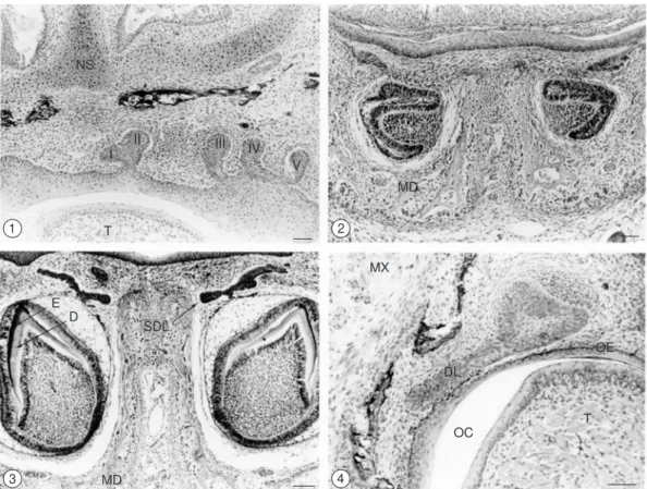
Dental lamina formation is one of the first events to take place during tooth development. This process begins with a thickening of the oral epithelium invaginating in the underlying mesenchyme. The five upper incisors of
D. albiventris were found to originate from two tooth-forming areas of these invaginations. Both first and second upper incisors derived from an anterior area, which was small and narrow. In contrast, all other incisors originated from a posterior area, which was larger and expanded. Between the 6th
and 7th day subsequent to birth, the third, fourth and
fifth upper incisors were bud-shaped and showed individual dental laminas connecting them to the oral epithelium (Fig. 1). Inversely, each of the lower incisors presented not only its own forming area but also its own dental lamina. The lower first incisor
showed its concavity turned towards the midline until it reached the bell stage, and the secondary dental lamina was visible as a thickening of the outer epithelium (Fig. 2). A thin to moderately thick layer of dentin and enamel, located between the epithelial cap and the underlying stroma cell, was visible by day 40, when the incisors presented their definitive orientation (Fig. 3).
The dental lamina of the upper incisors underwent a degenerative process on the 14th day,
whereas that of the lower incisors initiated this process on the 13th day. Finally, the developing teeth
became fully disconnected from the oral epithelium on the 19th day. However, all the incisors showed
a secondary dental lamina that arose at different stages and began to degenerate after reaching the bud stage (Fig. 3).
Canine teeth development occurred faster than other teeth, except for the lower first molar and upper second molar, which unfolded simultaneously. The upper canine dental lamina was thicker than that observed during the development of other teeth, and was positioned obliquely in relation to the oral epithelium (Fig. 4). Both dental lamina and secondary dental lamina degenerated on the 30th day. The canines were also the first teeth
to reach the oral epithelium (Fig. 5), which took place around day 70.
Dentin and amelogenesis were observed in the young animals inside the pouch at about 20 days of age for canines and at 30 days for third incisors. However, first, second, fourth and fifth incisors were visible 30 to 40 days after birth (Tables 2 and 3).
Tables 2 and 3 show the histological stages of developing incisors and canines in
Didelphis albiventris according to the following developmental stages:
I) Dental lamina; II) Bud shaped;
III) Intermediate phase between bud and cup stages;
IV) Cup shaped;
TABLE 1
Specimens collected at each age (in days).
AGE 0 1 2 3 4 5 6 7 8 9 10 14 20 30 40 50 60 70 80 90 100
NS
I
T
D
SDL
MD E
II III
IV V
MD
MX
DL
OC
OE
T 1
3
2
4
Figs. 1-4 — 1) Frontal section through the maxillary process of 6-day-old animals. The first (I) and second (II) incisor germs were visible in the same tooth-forming area. The third (III), fourth (IV) and fifth (V) incisors showed individual dental lamina connecting them to the oral epithelium. Nasal septum (NS); Tongue (T). Bar = 100 µm. H.E; 2) Frontal section through the mandibular process of 20-day-old animals. Note the first (I) incisor germ in the bell stage, showing its concavity turned towards the midline. Mandibule (MD). Bar = 100 µm. H.E; 3) Frontal section through the mandibular process at 40 days of age. The first (I) incisor in the crown formation stage showed a thin to moderately thick layer of dentin (D) and enamel (E). The secondary dental lamina (SDL) arose in the outer epithelium (OE), Mandibule (MD). Bar = 100 µm. H.E; and 4) Frontal section through the maxillary process of 7-day-old animals. Note the upper canine germ in the cap stage, showing a dental lamina (DL) obliquely positioned in relation to the oral epithelium (OE). Oral cavity, Tongue (T), MX – Maxillar. One-day-old animals. Bar = 100 µm. H.E.
Fig. 5 — Frontal longitudinal section through the completely formed upper canine, showing its proximity to the oral epithelium (OE). Forty-day-old animals. Bar = 100 µm. H.E.
OE
TABLE 2
Dental development stages in upper incisors and canines from D. albiventris.
First incisor Second incisor Third incisor Fourth incisor Fifth incisor Canine
Day 0 I I I I I I
Day 1 to 5 I I II II II III
Day 6 to 7 II II II II II IV B
Day 8 II II III III III IV C
Day 10 II III IVB IVB IVB VI A
Day 14 II IV A IV C IV B IV C VI A
Day 20 IV A IV A IV C IV C IV C VII B*
Day 30 IV A IV C VII A* VI B* IV C VIII A
Day 40 VII C* VII C* VII C VII C VII C* VIII B Day 50 VIII A VIIIA VIII A VIII A VIII A VIII B Day 60 VIII A VIII C VIII A VIII A VIII A VIII C Day 70 VIII B VIII C VIII D VIII D VIII D X
Day 80 VIII E X X X X X
Day 90 X X X X X X
Day 100 X X X X X X
*Animal’s age, tooth, and developmental stage in specimens where the secondary dental lamina was observed.
IVa) Nondifferentiated dental organ; IVb) Dental organ in differentiation ; and IVc) Differentiated dental organ.
V) Intermediate stage between cap and bell stages;
VI) Bell shaped;
VIa) Dental lamina connected to the oral epithelium;
VIb) Dental lamina in degenerative process;
TABLE 3
Dental development stages in lower incisors and canine from D. albiventris.
First incisor Second incisor Third Incisor Fourth incisor Canine
Day 0 I I I I I
Day 1 to 5 II II II II III
Day 6 to 7 II III III III IV B
Day 8 III III III III IV C
Day 10 IV B IV A IV B IV B V
Day 14 V IV C V IV C VI A
Day 20 VI A IV C VI A IV C VII B*
Day 30 VII C* VII B VII C* VII B* VII B**
Day 40 VII C VII C* VII C VII C VII C Day 50 VIII A VIII A VIII A VIII B VIII A Day 60 VIII C VIII C VIII D VIII D VIII C Day 70 VIII E VIII E VIII E VIII E X
Day 80 X X X VIII E X
Day 90 X X X X X
Day 100 X X X X X
* Animal’s age, tooth, and developmental stage in specimens where the secondary dental lamina was observed
VII) Crown formation; VIIa) Dentinogenesis ;
VIIb) Dentinogenesis and initial signs of amelogenesis at the tip of the tooth;
VIIc) Dentin formation completed and enamel present on at least 1/3 of the tooth crown;
VIII) Rizogenesis stage;
VIIIa) Epithelial root sheath without odontoblast differentiation; VIIIb) Epithelial root sheath with
odontoblast differentiation; VIIIc) Pre-dentin formation; VIIId) Dentin formation;
VIIIe) Cement and periodontal fibers; IX) Tip of the cuspid reaches oral epithelium; X) Eruptive stage: tooth already seen in the oral
cavity.
DISCUSSION
Most dental development studies reported in the literature estimated the age of animals based on growth lines, according to the specimen’s body weight and length. Weight is the parameter which presents the largest standard deviation, regarding its inverse relation to the number of animals by brood (Dezone et al., 1984). The ages of all specimens of Didelphis albiventris used in this study were
recorded. Histological evaluations were made of serial sections of the dental arches from each age group studied.
The dental formula of adult Didelphis albiventris is:
I C PM M 4
5
1 1
3 3
4 4
Before tooth development in D. albiventris, two oral epithelium thickenings invaginated into the underlying ectomesenchyme of the maxillary region, and all the functional upper incisors developed from these invaginations. Both first and second maxillary incisors (I1 and I2) originated
from the anterior thickened area. However, the posterior region of the invaginations gave rise to third, fourth and fifth maxillary incisors (I3, I4 and
I5). Conversely, each of the four incisors in the
lower jaw had its own forming area and its own dental lamina. These findings are similar to those observed in other marsupials.
Setonix brachyurus also showed also two
tooth-forming areas. The anterior tooth-forming area was subdivided into two regions: the first located anteriorly, and the second posteriorly. The first area was smaller and narrower, giving rise to the first non-functional maxillary incisor (i1). Inversely, the second region was larger and
lingually expanded, thus originating the maxillary first functional incisor (I1). The posterior
tooth-forming area consisted of three thickenings: an anterior thickening, which gave rise to the second functional incisor (I2); a mid-buccal one, from
which the second non-functional incisor developed (i2); and a posterior one, which originated the third
functional incisor (I3). According to their buccal
or lingual positions, and their early development and calcification, non-functional teeth formed a primary series of teeth, while the functional dental elements comprised a secondary or permanent series (Berkovitz, 1968). In P. eugenii, two non-functional incisors (i) were buccally positioned in relation to the functional incisors (I) in both jaws. They also developed and calcified before the functional dental elements (Berkovitz, 1972).
In S. virginae, the formation of small
epithelial cap-nodules at the end of the short primary dental lamina stalks has been reported in 5 to 10-day-old animals in the pouch. These structures gave rise to the mandibular first and third deciduous incisors (dI1-3), as well as to the deciduous canine (dC). A notable improvement in differentiation and formation of a thin to moderately thick dentinal arc was observed at around the 10th day. This structure was located between the
epithelial cap and the underlying mesenchyme cells of the lower second deciduous incisor (dI2). Between the 25th and 30th day, this dentinal arc was
still associated with the small dI2 cap, whereas the deciduous rudiments were less distinct for most of the anterior teeth in both jaws (Luckett & Woolley, 1996).
Dentin formation initiated on day 20 for canines, on day 30 for third incisors and between the day 30 and 40 for the other incisors. In contrast, while studying incisor development in P. eugenii,
Berkovitz (1972) noted that dentinogenesis occurred on day 52.
According to the findings of this investigation, between days 0 and 100, all five incisors and canines were completely developed and were visible in the oral cavity of both jaws. The earliest penetration of the oral epithelium for the canine was on around day 70, followed by second, third, fourth and fifth incisors on day 80. The first and the fourth incisors were the last teeth to erupt in the maxilla and in the mandible, respectively (Table 3). These observations are in agreement with the results reported by Luckett & Wolley (1996), who described incomplete first incisor eruption in
Sminthopswis virginae between days 79-81.
In Perameles gunii, it has been noted that two incisor germs did not develop in the lower jaw. Instead, they degenerated and were considered the so-called “absorptive teeth”. Inversely, all five incisors developed in the maxilla and were considered part of the deciduous dentition (Wilson and Hill, 1897). Dependorf (1898) considered the “absorptive teeth” part of a pre-lacteal dentition, whereas Müller (1969) suggested they were part of the same dentition as the other teeth. While studying
Didelphis albiventris and S. brachiurus, Kühental
(1891) and Berkovitz (1966), respectively, found no absorptive teeth. In fact, they found the dental laminas were positioned lingually to the developing tooth germs. The former author believed these teeth were remnants of the secondary dentition, while the latter interpreted them as remnants of the dental lamina free edge rather than a substitutive set of teeth.
The genus Didelphis has conserved the dental formula of its ancestral marsupials, while other species of these animals have undergone different reductions in their dental number. The authors of the present study support the theory that the non-functional absorptive teeth and the prelacteal dental elements are phylogenetic degeneration processes of the original teeth, as reported by Leche (1895), Wilson & Hill (1897), Dependorf (1898), and Muller (1969).
Gray short-tailed opossums (Monodelphis
domestica) reportedly exhibited monophyodonty
and have about 50 teeth (dental formula: 5134/4134). In these animals, the tooth germ of the maxillary and mandibular deciduous canines, as well as the deciduous maxillary first incisor, had a successor dental lamina and a replacement tooth germ. Tooth germs of both deciduous mandibular first incisor and deciduous maxillary fourth incisor had developed up to the bud stage by day 12, but were reduced by day 18 (Kozawa et al., 1998).
The results of this study showed that thickening of the oral epithelium (primary dental lamina) gave rise to the functional incisors and canines in D.albiventris. In addition, a secondary
dental lamina emerged in different developmental stages in the outer epithelium of the incisors and canines, undergoing degeneration when it reached the bud stage. These observations suggest that the first dentition prevailed as the permanent dentition. The findings of the present investigation are congruent with those reported by Kukental (1891) and Fosse (1994), who described these dental laminae as remnants of a primitive condition when secondary dentition used to be present.
Acknowledgments — This research was supported by FAPEMIG, CAPES and CNPq (Brazil).
REFERENCES
BERKOVITZ, B. K. B., 1966, The Homology of the Premolar Teeth in Setonix brachiururs (Macropodidae: Marsupialia).
Arch. of Oral Biol., 11: 1371-84.
BERKOVITZ, B. K. B., 1968, The Early Development of the Incisor Teeth of Setinix brachiurus. (Macropodidae: marsupialia) With Special Reference to the Prelacteal Teeth. Arch. of Oral Biol., 13: 171-190.
BERKOVITZ, B. K. B., 1972, Tooth Development in Protem-nodon eugenii. J. Dent. Res., 52: 1467-73.
DEPENDORF, T., 1898, Entwicklungsgeschichte des Zahnsystems der Marsupialer. Denkschr. Med. Natturw. Ges 6: 243-402. (Retracted by G. Fosse, S. Risnes. In:
Archives of Oral Biology, 17: 829-38, 1972)
DEZONE, M. F. M., CARREIRA, J. C. A. & FRANCO, A. M. R., 1984, Estudo do desenvolvimento extra-uterino de Didelphis marsupialis e estabelecimento de uma tabela de classes etárias (Marsupialia, Didelphidae). Congresso Brasileiro de Zoologia, 11: 363-64.
FOSSE, G., 1994, Development of the Teeth in a Pouch-Young Speci1men of Antechinus stuartii and a Pouch-Young Specimen of Sinanthopsis crassicaudata. Dasyuridae: Marsupialia. Arch. of Oral Biol., 14: 207-18.
JERNVALL, J., 1995, Mammalian molar cusp patterns: Development mechanisms of diversity. Acta Zool. Fennica, 198: 1-61.
JERNVALL, J., HUNTER, J. P. & FORTELIUS, M., 1996, Molar tooth diversity, disparity, and ecology in Cenozoic ungulate radiations. Science, 274: 1489-1492.
JURJESKI-JUNIOR, W., 1974, The Opossum (D. virginiana
Kerr) as a Biomedical Model. Lab. Animal Sci., 24: 376-425.
KOZAWA, Y., IWASA, Y. & MISHIMA, H., 1998, Degeneration of tooth germ in the developing dentition of the gray short-tailed opossum (Monodelphis domestica). Eur. J. Oral Sci., 106: 509-12.
KRAUSE, W. J. & CUTTS, J. H., 1986, Scanning Electron Microscopic Observations on Developing Opossum Embryos: days 9 through 12. Anat. Anz., 161: 11-21. KÜKENTAL, W., 1891, Das Gebiss von Didelphis.
Anatomischer Anzeiger, 6: 658-66. (Retracted by
G. Fosse, S. Risnes. In: Archives of Oral Biology,
17: 829-838, 1972).
LECHE, W., 1895, Zur Entwichlungsgeschichte des Zahnsys-tems der Saugethiere. Bibl. Zool., 7: 1-160.
LUCKETT, P. W. & WOOLLEY, P. A., 1996, Ontogeny and homology of the dentition in Dasyurid Marsupials:
Development in Sminthopsis virginiae. J. Mammal. Evol., 3: 327-364.
MAEDA, T., IWANAGA, T., FUJITA, T. & KOBAYASHI, S., 1986, Immunohistochemical. Demonstration of Nerves in the Predentin and Dentin of Human Third Molars With the Use of an Antiserum Against Neurofilament Protein (NFP).
Cell and Tiss. Res., 243: 469-75.
MÜLLER, K., 1969, Über die Zahnentwicklung bei Perameles.
Geg. Morphol. Jahr., 61: 457-88. (Retracted by G. Fosse, S. Risnes. In: Archives of Oral Biology, 17: 829-38, 1972).
STOCK, D. W., WEISS, K. M. & ZHAO, Z., 1997, Patterning of the mammalian dentition in development and evolution.
BioEssays, 19: 481-490.
THESLEFF, I. & SAHLBERG, C., 1996, Growth factors as inductive signals regulating tooth morphogenesis. Sem. Cell Dev. Biol., 7: 185-193.
WEISS, K. M. & FULLERTON, S. M., 2000, Phenogenetic drift and the evolution of genotype-phenotype relationships
Theor. Popul. Biol., 57: 187-195.
WILSON, J. T. & HILL, J. P., 1897, Observations Upon the Development and Succession of the Teeth in Perameles.
Quart. J. of Microsc. Sci., 39: 427-88. (Retracted by G.
Fosse, S. Risnes. In: Archives of Oral Biology,7: 829-38,