Association between the increase in brain
temperature and physical performance at
different exercise intensities and protocols
in a temperate environment
A.C. Kunstetter, S.P. Wanner, L.G. Madeira, C.F. Wilke, L.O.C. Rodrigues and N.R.V. Lima
Laborato´rio de Fisiologia do Exercı´cio, Departamento de Educac¸a˜o Fı´sica, Escola de Educac¸a˜o Fı´sica, Fisioterapia e Terapia Ocupacional, Universidade Federal de Minas Gerais, Belo Horizonte, MG, Brasil
Abstract
There is evidence that brain temperature (Tbrain) provides a more sensitive index than other core body temperatures in
determining physical performance. However, no study has addressed whether the association between performance and increases in Tbrainin a temperate environment is dependent upon exercise intensity, and this was the primary aim of the present
study. Adult male Wistar rats were subjected to constant exercise at three different speeds (18, 21, and 24 m/min) until the onset of volitional fatigue. Tbrainwas continuously measured by a thermistor inserted through a brain guide cannula. Exercise induced a
speed-dependent increase in Tbrain, with the fastest speed associated with a higher rate of Tbrainincrease. Rats subjected to
constant exercise had similar Tbrainvalues at the time of fatigue, although a pronounced individual variability was observed
(38.7-41.76C). There were negative correlations between the rate of Tbrainincrease and performance for all speeds that were studied.
These results indicate that performance during constant exercise is negatively associated with the increase in Tbrain, particularly
with its rate of increase. We then investigated how an incremental-speed protocol affected the association between the increase in Tbrainand performance. At volitional fatigue, Tbrainwas lower during incremental exercise compared with the Tbrainresulting
from constant exercise (39.3±0.3 vs40.3±0.16C; P,0.05), and no association between the rate of Tbrain increase and
performance was observed. These findings suggest that the influence of Tbrainon performance under temperate conditions is
dependent on exercise protocol.
Key words: Brain cortex; Fatigue; Hyperthermia; Speed; Thermoregulation; Treadmill running.
Introduction
Homeothermic animals maintain their core body tem-perature (Tcore) within narrow limits, even when exposed to
a wide range of environmental temperatures. This ability to tightly control Tcoreis important for the maintenance of body
homeostasis, and large deviations in Tcorefrom the narrow
limits suggest the existence of a pathological condition (1). However, marked changes in Tcoredo not occur exclusively
in sick animals. For instance, hyperthermic states are also observed in healthy, homeothermic animals while exercising.
Physical exercise accelerates the rate of heat produc-tion and leads to a rapid increase in Tcore, which may reach
values above 406C, depending on exercise intensity/ duration and ambient temperature (2,3). It is well estab-lished that exercise-induced hyperthermia negatively
affects the ability to sustain prolonged physical efforts through mechanisms that have not yet been elucidated. In particular, marked increases in the temperature of the brain, one of the sites where Tcoreis measured, have been
associated with high rates of perceived exertion (4) and alterations in cerebral function (5).
The temperatures measured in specific body compart-ments are not homogeneous and do not respond in a similar way (particularly regarding their time course) to several arousing stimuli (6). Indeed, there is evidence that brain temperature (Tbrain) is a more sensitive index than
other temperatures of the body core in determining physical performance (7) and thermoeffector activity (8). Unfortunately, the temperature of brain structures cannot be directly measured in humans during exercise. In this
Correspondence: S.P. Wanner, Laborato´rio de Fisiologia do Exercı´cio, Departamento de Educac¸a˜o Fı´sica, Escola de Educac¸a˜o Fı´sica, Fisioterapia e Terapia Ocupacional, UFMG, Av. Antoˆnio Carlos, 6627, 31270-901 Belo Horizonte, MG, Brasil. E-mail: samuelwanner@eeffto.ufmg.br
context, rodent experiments are essential to increase our understanding of the alterations in the central nervous system that may regulate performance or explain the central impairments caused by hyperthermia. So far, most of the experiments that measured Tbrainin exercising rats
were performed in hot environments at a constant treadmill speed (9-12) or compared the magnitude of the hyperther-mia induced by similar exercise intensity at two different ambient temperatures (13,14). As expected, high values of Tbrain were associated with reduced performance in the
heat (12), and the Tbrainincrease was exaggerated during
treadmill running in a warm compared with a cool environ-ment (14) or a temperate environenviron-ment (13). To our surprise, no study has evaluated whether Tbrain is associated with
physical performance at different exercise intensities and protocols in a temperate environment, and this was the primary aim of the present study. Our hypothesis was that the influence of Tbrainon performance would be less evident
during incremental-speed exercise and during high-inten-sity, constant exercise, since rats exercise at intensities close to their maximum aerobic capacity in both conditions and, therefore, fatigue would be more influenced by metabolic and cardiovascular adjustments.
Furthermore, we investigated whether performance in a temperate environment was associated more with an absolute, critical Tbrain or with dynamic changes in
tem-perature (i.e., the rate of Tbrainincrease). Finally, we also
investigated whether the brain lesions caused by the chronic implantation of a guide cannula or acute thermistor insertion for measuring Tbrainaffected performance.
Material and Methods
Animals
Twenty-two adult male Wistar rats weighing 250-350 g were used in all experiments. The rats were housed in collective cages and, after the surgical procedure, they were housed individually. The animals were maintained in a room with controlled light (5:00 am to 7:00 pm) and temperature (24±16C) conditions, and water and rat chow were providedad libitum. All experimental proce-dures were approved by the Ethics Committee of the Universidade Federal de Minas Gerais for the Care and Use of Laboratory Animals (protocol #076/2011) and conducted in accordance with the policies described in the Committee’s Guiding Principles Manual.
Experimental protocols
Two sets of experiments were conducted to achieve the goals of this study. The first set was performed to investigate the effects of exercise intensity (evaluated by changing the treadmill speed) on the association between the exercise-induced increase in Tbrain and physical
performance. Initially, a brain guide cannula was surgically implanted in the rats (n=9). After recovering from this surgery, the animals were familiarized with running on a
treadmill (5-day protocol) and then subjected to three constant-speed exercises. The order of the exercise trials followed a Latin square design to prevent the repetition of trials from interfering with the results. An interval of at least 2 days was given between trials. On a different day following the three constant exercises, the rats were subjected to a control experiment that consisted of measuring Tbrainunder
resting conditions.
The second set of experiments was conducted to investigate whether the exercise protocol interfered with the association between Tbrain increase and physical
performance, and whether the procedures that we used to measure Tbrainaffected performance. Another group of
rats (n=13) was used in this set of experiments. After being familiarized with running on a treadmill (5-day protocol), all animals were subjected to an incremental-speed exercise protocol, and 1 day after this exercise trial, they were divided into two groups: a group that had a guide cannula implanted into the brain (operated group) and another group that did not undergo surgery (control group; the animals were only transferred from a collective to an individual cage). Five days after the surgical procedure (or the transfer to an individual cage), animals from both groups were familiarized with running on a treadmill for two additional days. The rats were then subjected to two incremental exercise sessions, and operated animals were evaluated with or without the insertion of a thermistor through the guide cannula. Tbrainwas measured during the
trial in which the thermistor was inserted. The order of the incremental exercises (i.e., with and without the insertion of the thermistor) was randomized, and an interval of at least 2 days was given between these experimental trials. The reason for conducting incremental exercises for the second set of experiments was because performance has a lower variation coefficient in this protocol compared with constant exercise (15).
Brain guide cannula implantation
A guide cannula was implanted into the brain to allow for the insertion of a thermistor that enabled the measurement of the right brain cortex temperature.
Familiarization with exercise on a treadmill
The rats were gradually introduced to exercise on a treadmill for small animals (Gaustec Magnetismo, Brazil) over 5 consecutive days before initiation of the experiments. Each daily session consisted of running at a constant speed of 18 m/min and a 5% inclination for 5 min (17). During the familiarization sessions, the animals were encouraged to run by light electrical stimulation (0.5 mA). The purpose of this familiarization procedure was to show the animals which direction to run, thereby minimizing their exposure to the electrical stimulation during the experimental trials (17). Prior to each familiarization session, the rats were extensively handled by the experimenter, who also briefly restrained the animals and then simulated the insertion of a thermistor.
Experimental trials
The experiments were performed between 10:00 am and 4:00 pm, and care was taken to subject the same animal to the assigned experimental trials at the same time of day.
Constant-speed exercise. On the day of the experi-ments, the rats were taken to the experimental room, and a thermistor (Beta Therm Corp., USA) for measuring Tbrain
was inserted into the brain through the guide cannula. The animals were not anesthetized or sedated for this procedure and did not exhibit any signs of pain or discomfort. The rats were then placed on a treadmill with the inclination always set to 5% and subjected to a running exercise session until volitional fatigue at one of the following treadmill speeds: 18 m/min (S18), 21 m/min (S21), or 24 m/min (S24). Volitional fatigue was defined as the point at which the animals were no longer able to maintain pace with the treadmill, even when being stimulated by light electrical shocks (0.5 mA) for 10 s (17).
Incremental-speed exercise. The incremental exercise was performed at a constant inclination (5%) with a starting speed of 10 m/min. The treadmill speed was increased by 1 m/min every 3 min until volitional fatigue (18). The procedures used to measure Tbrain and to determine
volitional fatigue during the incremental exercise sessions were similar to those used during the constant exercise sessions.
Control experiments. Control experiments were performed with resting rats. The animals were allowed to move freely in their home cages, and their Tbrain was
measured for 60 min after a stabilization period.
Measurements
Total exercise time (TET) was measured from the beginning of exercise until volitional fatigue. The Tbrainwas
measured using a thermistor that was inserted into the brain and connected to a multimeter (Fluke, 289 FVF, Brazil). The multimeter recorded resistance values that were converted into temperature values using the Steinhart-Hart equation. In the exercise trials, Tbrain was
recorded every minute throughout the running period,
whereas in the control trial, Tbrain was measured each
minute during 60 min of resting. The ambient temperature was recorded every 4 min using a thermocouple (Yellow Spring Instruments, YSI 409B, USA) fixed to the ceiling of the acrylic chamber that contained the treadmill (during the exercise trials). In the resting experiments, the same thermocouple was placed next to the animal’s home cage. The ambient temperature was maintained at 25.2±0.26C during all experimental trials by the use of air conditioning. According to our previous experiments, rats exhibit a mean heat loss index of 0.23 when resting in the same experimental setup for 60 min and with the ambient temperature set at 25-266C (19). The heat loss index data suggest that the environmental conditions used in the present study were within the thermoneutral zone (20).
Calculations
The rate of Tbrainincrease was calculated by dividing
the change in Tbrain by the TET. The maximum speed
(Smax) attained during the incremental-speed exercise
sessions was calculated by modifying the equation proposed for the calculation of the maximal power output (21): Smax=S1++(S26t/180), where S1 is the speed
reached in the last completed stage, S2 is the increment in the treadmill speed at each stage, andtis the time spent (in seconds) in the uncompleted stage.
Verification of the position of the guide cannula At the end of the experiments, the rats were deeply anesthetized intraperitoneally with 120 mg/kg ketamine and 15 mg/kg xylazine and perfused with 150 ml 0.9% NaCl, followed by 300 ml 10% formalin solution. After the perfusion, the brain was removed, post-fixed by immer-sion in formalin for 24 h at 46C, and ultimately transferred to a 30% sucrose solution maintained at 46C for at least 2 days. The brain tissues were then frozen at ––186C and cut into 50-mm slices using a cryostat microtome (Leica Microsystems, Germany). Brain slices were stained with cresyl violet acetate (0.5 g/100 mL) and examined under a light microscope (Studar, Poland). The position of the thermistors was determined by comparing the lesioned areas present on the slides with coronal drawings in the Paxinos and Watson atlas.
Statistical analysis
Data are reported as means±SE. In the first set of experiments, the changes in Tbrainvalues were compared
between exercise intensities and time points using two-way analysis of variance (ANOVA) with repeated mea-sures, followed by the post hoc Tukey test. One-way ANOVA with repeated measures followed by thepost hoc
Duncan test was used to compare the TET and rate of Tbrain increase between the three exercise trials. The
association between the TET and rate of Tbrainincrease
and thermoregulatory parameters (initial Tbrain,
exercise-induced changes in Tbrain, and Tbrainat the end of effort)
was assessed using Spearman correlation coefficient. The curves describing the percentage of rats that were still running at a given exercise time were compared between the experimental trials using the log-rank test (22).
In the second set of experiments, the maximal treadmill speeds were compared between groups (operated and control) and between the three incremental tests using two-way ANOVA followed by thepost hoc Duncan test. The changes in Tbrain were compared between exercise
protocols (incremental or constant speed) and time points using two-way ANOVA followed by thepost hocTukey test. In all analyses, the significance level was set at P,0.05.
Results
First set of experiments
Histological analysis. Figure 1A shows schematic drawings indicating the location of the thermistor tips (defined by the deepest lesion observed in the brain tissue). In three animals, the thermistor tips were observed in ventral regions of the right frontal cortex (coordinates ranging from 5 to 7 mm under the skull), including the lateral orbital cortex (LO) and the agranular insular cortex (AIV). However, in four other rats, the thermistor was
inserted into dorsal regions of the right frontal cortex (i.e., from 2 to 3 mm under the skull), including the primary motor cortex (M1), the frontal area 3 (Fr3), and the primary somatosensory cortex (SIJ). In two animals, the thermistor was inserted into the caudate putamen nucleus (CPu). With regard to the anterior-posterior (AP) coordinates, the thermistor tips were observed at more rostral levels in five animals (3-4.2 mm from the bregma), while the tips were located more caudally in the other four animals (2.16-2.76 mm from the bregma). With respect to the medial-lateral (ML) coordinates, the thermistor tips were observed at similar coordinates (2.5-3.3 mm from the bregma).
Figure 1B shows representative photomicrographic images of brain coronal slices from two rats, including one rat in which the thermistor was inserted into the frontal cortex and another in which the thermistor was inserted into the CPu. Despite the large differences observed in the location of the thermistor tips in the 15 animals, we did not observe any correlation between the AP coordinates and the thermoregulatory parameters evaluated (initial Tbrain, exercise-induced change in Tbrain, and Tbrainat the
end of effort). With regard to the depth of the thermistor tips, we observed a correlation between the dorso-ventral (DV) coordinates and initial Tbrain (r=0.650, P=0.050;
Figure 1C), as previously observed by other investigators (11,23). However, the DV location of the thermistor was not associated with Tbrain at the end of effort (r=0.550,
P=0.111) or the exercise-induced change in Tbrain
(r=0.183, P=0.612). Considering that the animals had similar changes in Tbrainduring exercise irrespective of the
DV location of the thermistor, we analyzed all of the animals as a single group.
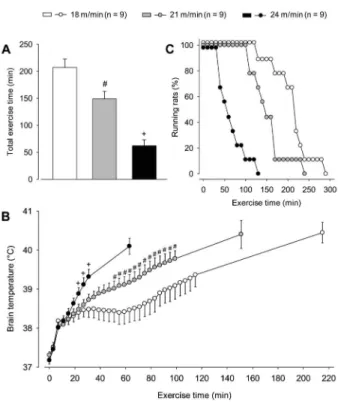
Physical performance and brain temperature. As expected, the treadmill speed greatly influenced perfor-mance; specifically, TET was shorter during exercise at
24 m/min compared with the two other running speeds, and it was shorter at 21 m/min compared with 18 m/min (S24: 62±11 min vs S21: 149±14 min vs S18: 207± 15 min, P,0.001, Figure 2A). As illustrated in Figure 2C, the curve that represents the percentage of rats that were still running at given exercise time points was shifted to the left during the S24 trial compared with the S21 (P,0.01, log-rank test) and S18 (P,0.01, log-rank test) trials. For example, when all animals stopped running at 24 m/min, 89% and 67% of them were still running at 18 and 21 m/ min, respectively.
The rats that were allowed to move freely in their home cages at,256C had an average Tbrainof 37.18±0.096C.
In contrast, treadmill running caused marked increases in Tbrainat all exercise intensities studied (Figure 2B). Tbrain
was already increased at the fifth minute of exercise compared with the preexercise values (minute 5: 37.80±0.106C vs minute 0: 37.22±0.086C, P,0.05; pooled data for the three exercise speeds). During this initial phase of running, rats exhibited the highest rates of Tbrain increase (0.116C/min until the 8th minute), which
were independent of exercise intensity. Afterward, Tbrain
continued to increase at slower rates, and exercise-induced hyperthermia became dependent on running speed. Higher values of Tbrain were observed for the
highest exercise intensity compared with the other two intensities beginning at 23 min after the exercise had begun (S24: 38.88±0.166C vs S18: 38.40±0.166C vs
S21: 38.48±0.146C; P,0.05), and this enhanced running hyperthermia persisted until the 31st minute of exercise (S24: 39.32±0.196C vs S18: 38.48±0.206C vs S21: 38.73±0.146C, P,0.05). Moreover, Tbrainwas higher from
the 50th until the 100th minute of exercise at 21 m/min compared with these time points at a speed of 18 m/min. Interestingly, a plateau in Tbrainwas observed only for the
slowest speed from the 60th until the 100th minute and then, after 40 min of stable recordings, Tbrain markedly
increased for a second time. Despite the differences mentioned above, there were no differences in Tbrain
between the three exercise intensities when the animals voluntarily terminated their effort (S18: 40.45±0.266Cvs
S21: 40.31±0.286CvsS24: 40.10±0.206C). However, it is important to note that pronounced individual variability in Tbrainwas observed at this time (38.7-41.76C).
Figure 2. Total exercise time (panel A) and cortical brain temperature (panel B) of rats subjected to constant-speed exercise sessions on a treadmill at three different speeds (18, 21, and 24 m/min). Data are reported as means±SE and were analyzed using one-way (panel A) or two-way (panel B) ANOVA. #P
,0.05 compared with 18 m/min;+P,0.05 compared with 18
and 21 m/min. Panel C, Curves representing the maximum exercise duration tolerated by rats subjected to the constant exercise sessions. Data are reported as the number of rats that continued to run at specific time points at each treadmill speed. These data were analyzed using the log rank test.
Exercise intensity had a significant effect on the rate of Tbrainincrease. When exercise was performed at 24 m/min,
the rate of increase was greater compared with that resulting from the other two exercise speeds (S24: 0.058±0.0096C/ minvsS18: 0.017±0.0026C/minvsS21: 0.021±0.0026C/ min; P,0.001). In fact, a negative correlation between rate of increase in Tbrainand TET was observed for each speed
that was studied (Figure 3) and when all speeds were analyzed together (r=––0.80, P,0.001).
Second set of experiments
Histological analysis. In this set of experiments, histological analysis was important for determining the location of the thermistor tips and verifying the extension of brain lesions caused by chronic implantation of the guide cannula and acute insertion of the thermistor.
The schematic drawings presented in Figure 4 show the brain areas lesioned by implantation of the guide cannula and insertion of the thermistor. In four animals, the lesions were observed at DV coordinates ranging from 5 to 7 mm under the skull, reaching ventral regions that included the LO, AIV, the piriform cortex, the endopiriform nuclei, and Layer 3 of the cortex. However, in the other three rats, the lesions were restricted to dorsal regions (2.5-4 mm under the skull). With respect to the AP coordinates, the lesions were present at more rostral levels in four animals (3-4.2 mm from the bregma), while the lesions were concentrated more caudally in the other three animals (2.16-2.76 mm from the bregma). Although there were differences observed between the DV and AP coordinates, the lesions induced by the guide cannula and thermistor were observed at similar ML coordinates
(2.2-3.5 mm from the bregma).
Physical performance and brain temperature. All animals were initially subjected to incremental exercise to determine their maximal treadmill speed before being allocated to groups (i.e., control and operated). The groups were matched for performance measured during incremental exercise; therefore, the maximal treadmill speed attained was not different between the two groups Figure 4.Lines showing the contours of brain lesions caused by
the implantation of a guide cannula and insertion of a thermistor are drawn in coronal cortical sections taken from the Paxinos and Watson atlas (16). The numbers above the schematic of each cortical section indicate the distance (in mm) between the section plane and bregma.
Figure 5. Maximum treadmill speed attained during the three incremental-speed exercise sessions. Initial exercise was con-ducted prior to any surgical procedure in both groups. Animals with a brain guide cannula (operated group) were tested with or without thermistor insertion in a counterbalanced order. The control animals (not operated) were subjected to second and third incremental exercise sessions. Data are reported as means±SE and were analyzed using two-way ANOVA.
(control: 27.9±1.3 m/minvs operated: 28.6±0.9 m/min, P=0.917).
The control group allowed verification of whether learning or training-associated effects could increase running performance during the second or third incremen-tal exercise sessions. No differences were observed in the maximal treadmill speeds attained by animals in the control group between the three incremental exercise sessions (first: 27.9±1.3 m/min vs second: 29.8±2.4 m/min vs
third: 27.8±2.0 m/min; P=0.917). However, the main objective of this second set of experiments was to evaluate whether the procedures required to measure Tbrainwould
affect running performance. It was observed that the maximal speed attained during incremental exercise was not affected by chronic brain lesions resulting from implantation of a guide cannula or acute lesions resulting from thermistor insertion (first exercise: 28.6±0.9 m/min
vscannula: 28.6±1.4 m/minvsthermistor: 29.1±1.4 m/ min; Figure 5, P=0.977).
Tbrain was measured during incremental exercise
performed after the thermistor had been inserted into the brain. Incremental exercise also produced an increase in Tbrainthat was lower than that observed during constant
exercise at 24 m/min from the 19th min (S24: 38.63± 0.146C vs incremental exercise: 38.08±0.196C) until volitional fatigue (Figure 6). Moreover, differences in Tbrain between incremental exercise and exercise at
21 m/min were observed from the 30th minute (S21: 38.74±0.156C vs incremental exercise: 38.18±0.246C) until the end of the running period. At volitional fatigue, Tbrain was lower during incremental exercise compared
with the three constant exercise sessions (incremental exercise: 39.30±0.256C,vsS18: 40.45±0.266C,vsS21: 40.31±0.286C, vs S24: 40.10±0.206C; P,0.05). There was no association between the rate of Tbrainincrease and
physical performance during incremental exercise (r=0.30, P=0.501), and pronounced individual variability in Tbrainat the time of fatigue was again observed
(38.6-39.86C).
Discussion
The main finding of this study was that the exercise-induced increase in Tbrain of rats subjected to
constant-speed exercise was associated with their performance at all three intensities that were studied. In contrast, such association was not observed during incremental exer-cise, indicating that the influence of Tbrain increase on
performance is dependent on the running protocol. Moreover, Tbrainrecorded at the time of fatigue was lower
during incremental exercise compared with the three constant exercise sessions, with no significant differences observed among the latter. Last, the experimental procedures that we used to measure Tbrain of running
rats did not affect their performance.
During the initial phase of exercise up to the eighth
minute, rats exhibited the highest rates of Tbrainincrease,
which were independent of the treadmill speed (Figure 2B). This marked Tbrain increase was likely caused by animal
handling (a stressful procedure required to insert the thermistor) and/or by the lower rate of heat loss compared with the rate of heat production generally observed at the beginning of exercise [cutaneous heat dissipation usually increases within 8 to 10 min of exercise under temperate conditions (15)]. Indeed, the former hypothesis helps to understand why the initial Tbrainincrease was unspecific to
the treadmill speeds studied, whereas the second hypoth-esis is supported by observations that a steady-state Tcore
(rectal or abdominal) was only attained following the increase in tail skin temperature during low-intensity exercise (24,25).
After the eighth minute of exercise, the increase in Tbrain
was clearly different among the three running speeds (Figure 2B). At 18 m/min, equilibrium between the rates of heat production and heat loss was attained and, there-fore, a plateau in Tbrainwas observed. This steady-state
condition lasted approximately 40 min and was followed by a second, clear increase in Tbrain, a response that was
possibly a consequence of gradual reduction in running economy (which may have enhanced heat production). On the other hand, a temperature plateau was never observed at 21 and 24 m/min. We suggest that these intensities provoked elevated rates of heat production that had overcome the rat’s ability to dissipate heat, and this made Tbrain increase constantly during exercise. Interestingly,
this temperature behavior was also reported in studies that measured the colonic temperature of rats subjected to high-intensity exercise (24,26).
Physical exercise intensity was directly associated with the rate of Tbrain increase in rats subjected to treadmill
running at the same temperate ambient temperature; specifically, the faster treadmill speed (24 m/min) induced greater rates of increase compared with the slower speeds (18 and 21 m/min). The dependence of hyperthermia level on exercise intensity was not observed for running mice (27), but it agrees with observations for exercising humans (28,29) and exercising rodents that had their abdominal or rectal temperature measured (25,30). That exercising mice present different physiological responses from other species was also reported in a recent investigation focusing on their running-induced ventilatory responses (31), which may lead ultimately to a distinctive pattern of evaporative heat loss from the respiratory tract. These findings indicate the existence of interspecies differences in mechanisms underlying exercise hyperthermia and that rat experiments may be a more interesting tool than mouse experiments for studying some aspects of human thermo-regulation during exercise.
On average, rats subjected to constant exercise fatigued with Tbrainabove 406C, irrespective of the treadmill
speed (Figure 2B). Similar Tbrain values were previously
environments (10,12,14). In this sense, the existence of a critical Tcoreof approximately 40-416C that limits physical
performance of exercising humans and rodents has already been suggested (10,12,32). Marked increases in Tbrainalter cerebral function, as evidenced by decreases in
electroencephalogram activity of the frontal cortical area (4,5) and inhibition of the cortical areas responsible for motor activation (3). These hyperthermia-mediated changes in brain function may have also played an important role in determining volitional fatigue in the rats in our study that were subjected to constant exercise in a temperate environment. Indeed, evidence exists in the literature consistent with the hypothesis that central alterations may be involved in fatigue, even when exercise is performed in the absence of severe environmental thermal stress (33).
Although mean Tbrainwas approximately 40.1-40.56C
when the animals terminated their effort at the three speeds studied, the individual values ranged from 38.7 to 41.76C, which corresponds to a nearly 36C variation among the animals. This observation is a strong argument against the existence of a critical, absolute Tcorethat limits
prolonged performance. The current observations support those who argue that dynamic changes in Tcore, instead of
an absolute value, will regulate physical performance (30,34), because we observed negative correlations between TET and the rate of Tbrainincrease for each of
the three exercise intensities that were studied.
The running protocol also affected exercise-induced hyperthermia. Rats presented lower Tbrain values during
the incremental protocol compared with the constant protocols in which they ran at 21 and 24 m/min (Figure 6). This influence of the running protocol on the increase in Tbrain may be a consequence of differences in the
evolution of exercise intensity, which is an inherent characteristic of each running protocol. During the initial stages of the incremental exercise, the workload per-formed by the animals and, consequently, the rate of heat production was low (e.g., it takes 33 and 42 min for rats to begin running at 21 and 24 m/min, respectively). How-ever, even when animals achieved high speeds during the final stages of incremental exercise, their Tbrainremained
lower in this running protocol.
The animals terminated the incremental exercise with lower Tbrainvalues compared with the values derived from
the three constant exercise sessions (Figure 6), and no association between performance and the rate of Tbrain
increase was observed. These findings suggest that non-thermal factors, most likely metabolic factors, are more important for regulating fatigue than thermoregulation during an incremental exercise. It is important to note that exercise fatigue is a complex brain-regulated phenom-enon that involves the integration of afferent information arising from several physiological systems (35). Depending on the characteristics of the exercise, some physiological responses are more important than others in
determining its interruption. For example, during the final stages of incremental exercise, animals are usually exercising at intensities close to their maximum aerobic capacity. In this case, it is likely that the ability of the cardiovascular system to specifically increase coronary and skeletal muscle blood flow is more of a determinant for physical performance than thermoregulatory responses (36). Therefore, our results suggest that incremental exercise in a temperate environment is not an adequate protocol for investigating the mechanisms by which high Tbrainvalues limit prolonged performance.
Another aim of the present study was to investigate whether the brain lesions caused by the chronic guide cannula implantation or acute thermistor insertion would affect the Smaxattained during incremental exercise. These
experiments were particularly important because the rats in the first set of experiments traveled a greater distance before being fatigued than animals tested under the same conditions in our previous studies (15), suggesting that the brain lesions somehow inhibited the feeling of fatigue. The lesions were concentrated in the frontal cortical area, including the primary motor cortex, the lateral orbital cortex, frontal area 3, and the caudate/putamen nucleus, which are involved in the planning, organization, and initiation of movement. There is evidence that lesioning the frontal cortical area produces deficits in motor behavior control (37) and that activity in frontal areas is associated with the motivation for voluntary wheel running (38). Furthermore, Nonneman and Corwin (39) observed increased voluntary wheel-running activity in adult, lesioned rats. In contrast with these early data, our results indicate that lesions promoted by the guide cannula or thermistor do not change motor behavior or the motivation to run and, consequently, the physical performance measured in rats subjected to incremental treadmill running (Figure 5). Our findings also indicate that the high performance of the rats used in the initial protocol was not a consequence of brain cortex lesions. The differences in performance between studies may be explained by intrinsic variations in the exercise capacity of rats, including those from the same strain (18), or differences in the criteria adopted by different experi-menters for determining fatigue.
In conclusion, the increase in Tbrainof rats subjected to
constant-speed exercise was dependent on treadmill speed. Although our experiments were conducted in a temperate environment, the Tbrain increase impacted
performance negatively, with the rate of increase being apparently more of a determinant than the absolute values of temperature. In contrast to our hypothesis, the influence of Tbrain on performance during constant exercise was
always observed, regardless of the exercise intensity. On the other hand, performance was not associated with the rate of increase in Tbrain during incremental exercise,
indicating that the role of Tbrain in determining
of exercising rats did not affect their physical performance.
Acknowledgments
We acknowledge Ubirajara Fumega for excellent technical assistance that helped us to measure brain temperature. We also would like to thank Dr. Christiano
Antoˆnio Machado-Moreira for very constructive comments regarding the revised manuscript. Research supported by FAPEMIG (#APQ-02032-11), CNPq (#APQ 483067/2011-3), and the Pro´-Reitoria de Pesquisa of Universidade Federal de Minas Gerais. A.C. Kunstetter, L.G. Madeira, and C.F. Wilke were recipients of fellowships from CAPES.
References
1. Romanovsky AA. Thermoregulation: some concepts have changed. Functional architecture of the thermoregulatory system.Am J Physiol Regul Integr Comp Physiol2007; 292: R37-R46, doi: 10.1152/ajpregu.00668.2006.
2. Galloway SD, Maughan RJ. Effects of ambient temperature on the capacity to perform prolonged cycle exercise in man. Med Sci Sports Exerc1997; 29: 1240-1249, doi: 10.1097/ 00005768-199709000-00018.
3. Nybo L, Nielsen B. Hyperthermia and central fatigue during prolonged exercise in humans. J Appl Physiol2001; 91: 1055-1060.
4. Nybo L, Nielsen B. Perceived exertion is associated with an altered brain activity during exercise with progressive hyperthermia.J Appl Physiol2001; 91: 2017-2023. 5. Ftaiti F, Kacem A, Jaidane N, Tabka Z, Dogui M. Changes in
EEG activity before and after exhaustive exercise in sedentary women in neutral and hot environments. Appl Ergon2010; 41: 806-811, doi: 10.1016/j.apergo.2010.01.008. 6. Kiyatkin EA. Brain temperature fluctuations during physio-logical and pathophysio-logical conditions.Eur J Appl Physiol2007; 101: 3-17, doi: 10.1007/s00421-007-0450-7.
7. Caputa M, Feistkorn G, Jessen C. Effects of brain and trunk temperatures on exercise performance in goats. Pflugers Arch1986; 406: 184-189, doi: 10.1007/BF00586681. 8. Gisolfi CV, Mora F.The hot brain: survival, temperature,
and the human body. Cambridge: The Massachusetts Institute of Technology; 2000.
9. Caputa M, Kamari A, Wachulec M. Selective brain cooling in rats resting in heat and during exercise.J Thermal Biol 1991; 16: 19-24, doi: 10.1016/0306-4565(91)90046-5. 10. Fuller A, Carter RN, Mitchell D. Brain and abdominal
temperatures at fatigue in rats exercising in the heat. J Appl Physiol1998; 84: 877-883, doi: 10.1063/1.368150. 11. Walters TJ, Ryan KL, Belcher JC, Doyle JM, Tehrany MR,
Mason PA. Regional brain heating during microwave exposure (2.06 GHz), warm-water immersion, environmental heating and exercise.Bioelectromagnetics1998; 19: 341-353, doi: 10.1002/(SICI)1521-186X(1998)19:6,341::AID-BEM2.
3.0.CO;2-1.
12. Walters TJ, Ryan KL, Tate LM, Mason PA. Exercise in the heat is limited by a critical internal temperature. J Appl Physiol2000; 89: 799-806.
13. Walters TJ, Ryan KL, Tehrany MR, Jones MB, Paulus LA, Mason PA. HSP70 expression in the CNS in response to exercise and heat stress in rats.J Appl Physiol1998; 84: 1269-1277.
14. Hasegawa H, Piacentini MF, Sarre S, Michotte Y, Ishiwata T, Meeusen R. Influence of brain catecholamines on the development of fatigue in exercising rats in the heat. J Physiol 2008; 586: 141-149, doi: 10.1113/jphysiol.2007.
142190.
15. Pires W, Wanner SP, Lima MR, Fonseca IA, Fumega U, Haibara AS, et al. Physical exercise performance in temperate and warm environments is decreased by an impaired arterial baroreflex.PLoS One 2013; 8: e72005, doi: 10.1371/journal.pone.0072005.
16. Paxinos G, Watson C. The rat brain in stereotaxic coordinates. 6th edn. Waltham: Academic Press; 2007. 17. Guimaraes JB, Wanner SP, Machado SC, Lima MR, Cordeiro
LM, Pires W, et al. Fatigue is mediated by cholinoceptors within the ventromedial hypothalamus independent of changes in core temperature. Scand J Med Sci Sports 2013; 23: 46-56, doi: 10.1111/j.1600-0838.2011.01350.x. 18. Primola-Gomes TN, Campos LA, Lauton-Santos S,
Balthazar CH, Guatimosim S, Capettini LS, et al. Exercise capacity is related to calcium transients in ventricular cardiomyocytes. J Appl Physiol2009; 107: 593-598, doi: 10.1152/japplphysiol.91218.2008.
19. Lima MR, Pires W, Fonseca IA, Fonseca CG, Martinelli PM, Wanner SP, et al. Chronic sympathectomy of the caudal artery delays cutaneous heat loss during passive heating. Neurosci Lett 2013; 537: 11-16, doi: 10.1016/j.neulet. 2013.01.013.
20. Romanovsky AA, Ivanov AI, Shimansky YP. Selected contribution: ambient temperature for experiments in rats: a new method for determining the zone of thermal neutrality. J Appl Physiol2002; 92: 2667-2679.
21. Kuipers H, Verstappen FT, Keizer HA, Geurten P, van Kranenburg G. Variability of aerobic performance in the laboratory and its physiologic correlates.Int J Sports Med 1985; 6: 197-201, doi: 10.1055/s-2008-1025839.
22. Wanner SP, Garami A, Pakai E, Oliveira DL, Gavva NR, Coimbra CC, et al. Aging reverses the role of the transient receptor potential vanilloid-1 channel in systemic inflamma-tion from anti-inflammatory to proinflammatory.Cell Cycle 2012; 11: 343-349, doi: 10.4161/cc.11.2.18772.
23. Kiyatkin EA, Brown PL, Wise RA. Brain temperature fluctuation: a reflection of functional neural activation.Eur J Neurosci2002; 16: 164-168, doi: 10.1046/j.1460-9568.2002. 02066.x.
24. Wilson NC, Gisolfi CV, Farber J, Hinrichs DK. Colonic and tail-skin temperature responses of the rat at selected running speeds. J Appl Physiol Respir Environ Exerc Physiol1978; 44: 571-575.
25. Tanaka H, Yanase M, Nakayama T. Body temperature regulation in rats during exercise of various intensities at different ambient temperatures. Jpn J Physiol 1988; 38: 167-177, doi: 10.2170/jjphysiol.38.167.
Exerc Physiol1984; 57: 1872-1877.
27. Wanner SP, Costa KA, Soares AD, Cardoso VN, Coimbra CC. Physical exercise-induced changes in the core body temperature of mice depend more on ambient temperature than on exercise protocol or intensity. Int J Biometeorol 2013 [ahead of print].
28. Saltin B, Hermansen L. Esophageal, rectal, and muscle temperature during exercise.J Appl Physiol1966; 21: 1757-1762.
29. Greenhaff PL. Cardiovascular fitness and thermoregulation during prolonged exercise in man.Br J Sports Med1989; 23: 109-114, doi: 10.1136/bjsm.23.2.109.
30. Rodrigues LO, Oliveira A, Lima NR, Machado-Moreira CA. Heat storage rate and acute fatigue in rats.Braz J Med Biol Res 2003; 36: 131-135, doi: 10.1590/S0100-879X2003000 100018.
31. Iwase M, Izumizaki M, Tsuchiya N, Homma I. Dopamine D1 receptors control exercise hyperpnoea in mice.Exp Physiol 2013; 98: 491-500, doi: 10.1113/expphysiol.2012.068312. 32. Gonzalez-Alonso J, Teller C, Andersen SL, Jensen FB,
Hyldig T, Nielsen B. Influence of body temperature on the development of fatigue during prolonged exercise in the heat.J Appl Physiol1999; 86: 1032-1039.
33. Racinais S, Girard O, Micallef JP, Perrey S. Failed excitability of spinal motoneurons induced by prolonged running exercise.J Neurophysiol 2007; 97: 596-603, doi:
10.1152/jn.00903.2006.
34. Tucker R, Marle T, Lambert EV, Noakes TD. The rate of heat storage mediates an anticipatory reduction in exercise intensity during cycling at a fixed rating of perceived exertion. J Physiol 2006; 574: 905-915, doi: 10.1113/jphysiol. 2005.101733.
35. Lambert EV, St Clair GA, Noakes TD. Complex systems model of fatigue: integrative homoeostatic control of periph-eral physiological systems during exercise in humans.Br J Sports Med 2005; 39: 52-62, doi: 10.1136/bjsm.2003. 011247.
36. Noakes TD. Physiological models to understand exercise fatigue and the adaptations that predict or enhance athletic performance.Scand J Med Sci Sports2000; 10: 123-145, doi: 10.1034/j.1600-0838.2000.010003123.x.
37. Uylings HB, Groenewegen HJ, Kolb B. Do rats have a prefrontal cortex?Behav Brain Res2003; 146: 3-17, doi: 10.1016/j.bbr.2003.09.028.
38. Rhodes JS, Garland T Jr, Gammie SC. Patterns of brain activity associated with variation in voluntary wheel-running behavior. Behav Neurosci 2003; 117: 1243-1256, doi: 10.1037/0735-7044.117.6.1243.