Ische m ic pre co nditio ning re duce s
pe riphe ral o xidative dam age asso ciate d
with brain ische m ia in rats
1Departamento de Bioquímica, Instituto de Ciências Básicas da Saúde,
Universidade Federal do Rio Grande do Sul, Porto Alegre, RS, Brasil
2Setor de Bioquímica, Departamento de Q uímica, Centro de Ciências
Naturais e Exatas, Universidade Federal de Santa Maria, Santa Maria, RS, Brasil S.S. Frassetto1,
M.R.C. Schetinger2,
A. Webber1, J.J.F. Sarkis1
and C.A. Netto1
Abstract
Brain ischemia followed by reperfusion causes neuronal death related to oxidative damage. Furthermore, it has been reported that subjects suffering from ischemic cerebrovascular disorders exhibit changes in circulating platelet aggregation, a characteristic that might be impor-tant for their clinical outcome. In the present investigation we studied tert-butyl hydroperoxide-initiated plasma chemiluminescence and thiol content as measures of peripheral oxidative damage in naive and preconditioned rats submitted to forebrain ischemia produced by the 4-vessel occlusion method. Rats were submitted to 2 or 10 min of global transient forebrain ischemia followed by 60 min or 1, 2, 5, 10 or 30 days of reperfusion. Preconditioned rats were submitted to a 10-min ischemic episode 1 day after a 2-10-min ischemic event (2 + 10 10-min), followed by 60 min or 1 or 2 days of reperfusion. It has been demonstrated that such preconditioning protects against neuronal death in rats and gerbils submitted to a lethal (10 min) ischemic episode. The results show that both 2 and 10 min of ischemia cause an increase of plasma chemiluminescence when compared to control and sham rats. In the 2-min ischemic group, the effect was not present after reperfusion. In the 10-min ischemic group, the increase was present up to 1 day after recirculation and values returned to control levels after 2 days. However, rats preconditioned to ischemia (2 + 10 min) and reperfusion showed no differences in plasma chemiluminescence when compared to controls. We also analyzed plasma thiol content since it has been described that sulfhydryl (SH) groups significantly contribute to the antioxidant capacity of plasma. There was a signifi-cant decrease of plasma thiol content after 2, 10 and 2 + 10 min of ischemia followed by reperfusion when compared to controls. We conclude that ischemia may cause, along with brain oxidative damage and cell death, a peripheral oxidative damage that is reduced by the preconditioning phenomenon.
Co rre spo nde nce
C.A. Netto
Departamento de Bioquímica Instituto de Ciências Básicas da Saúde, UFRGS
Rua Ramiro Barcelos, 2600 (anexo) 90035-003 Porto Alegre, RS
Brasil
Fax: + 55-51-316-5535 E-mail: alexneto@ vortex.ufrgs.br
Research supported in part by FINEP and CNPq. S.S. Frassetto was the
recipient of a CAPES fellowship.
Received February 2, 1999 Accepted July 26, 1999
Ke y wo rds
·Brain ischemia
·Ischemic preconditioning
·O xidative damage
·Hydroperoxide-initiated chemiluminescence
·Plasma thiols
Intro ductio n
Neuronal damage following transient brain ischemia is mediated by various mecha-nisms, among which reactive oxygen
trans-genic mice overexpressing superoxide dis-mutase exhibit less edema and smaller in-farcts when subjected to ischemia (2). Fur-thermore, damage to the brain is also ob-served during reperfusion after ischemia (when the oxygen supply is restored to the brain) and may be attributed primarily to the presence of reactive oxygen species that can induce oxidative stress (3). The sources of reactive oxygen species include: a) products from the arachidonic acid cascade, b) me-tabolism of xanthine by xanthine oxidase (4),and c) release of excitatory amino acids (5).
Besides the effects on the central nervous system, recent studies have reported that peripheral events also occur after ischemic cerebrovascular disease, such as changes of platelet surface glycoproteins (6), platelet activation and erythrocyte aggregation in the circulation (7,8), and diffusion of nitric ox-ide (NO) produced by neurons into the blood-stream (9). Although these investigators have shown that ischemic cerebrovascular dis-ease induces peripheral effects, it has not been shown if a probable oxidative damage is related to these effects. Salvemini and Botting (10) have suggested that in ischemic heart disease platelet activation is related to oxidative damage. Thus, superoxide anions (O2·-) enhance platelet activation and aggre-gation in the circulation, while NO is able to inhibit the response of platelets to superox-ide (11).
The results of the present study suggest that brain ischemia may cause, along with brain oxidative damage and cell death, a peripheral oxidative damage that is probably related to important alterations such as microthrombus formation during this dis-ease. However, it has been proposed that brain ischemic preconditioning (when brief periods of ischemia precede longer and inju-rious ischemic episodes) is responsible for attenuation of neural ischemic injury (12). Thus, our aim was also to establish a rela-tionship between ischemic preconditioning
and a possible protection against the state of peripheral oxidative damage. To approach this question, we first measured the tert -butyl hydroperoxide-initiated chemilumines-cence emission in plasma of naive and pre-conditioned rats submitted to forebrain is-chemia and reperfusion. This assay is based on the determination of stable products or by-products of the reaction chain of lipid peroxidation, a process that follows the pro-duction of oxygen free radicals in biological systems, and has been applied to detect the existence of oxidative damage associated with experimental pathological situations (13).We next examined the effect of brain ischemia and preconditioning on plasma thi-ols because extracellular sulfhydryl (SH) groups have been proposed to serve as scav-enging antioxidants (14) and are generally highly effective in reducing ischemia-reper-fusion injury (15). Our hypothesis was that brain ischemia leads to a peripheral oxida-tive damage and that ischemic precondition-ing protects against this effect.
Mate rial and Me tho ds
Tert-butyl hydroperoxide was obtained from Aldrich Chemicals Co. (Milwaukee, WI, USA) and 5,5-dithiobis-(2-nitrobenzoic acid) (DTNB) was obtained from Sigma Chemical Co. (St. Louis, MO, USA). All other reagents were of analytical grade.
Brain ische mia and pre co nditio ning
later the ties were tightly clamped for 2 or 10 min (single-ischemic rats). Variable periods of reperfusion were then allowed, i.e., 60 min or 1, 2, 5, 10 and 30 days. The precondi-tioned group (double-ischemic rats) received 10-min ischemia 1 day after a 2-min ische-mic episode (referred to as 2' + 10'), fol-lowed by 60 min or 1 and 2 days of reperfu-sion. Rats that did not lose the righting reflex or that convulsed during the ischemia or reperfusion period were discarded. Sham-operated and intact rats were used as con-trols. Animals were killed by decapitation and blood was collected into citrated tubes. All animal use procedures were approved by the local Animal Care Committee.
Plasm a iso latio n
Plasma was obtained from fresh citrated rat blood. Red blood cells and buffy coat were removed by centrifugation at 200 g for 5 min at room temperature and platelets were removed by plasma centrifugation at 200 g for 20 min. The supernatant was used for subsequent experiments.
D e te rm inatio n o f plasm a te rt-butyl
hydro pe ro xide -initiate d che m ilum ine sce nce
Chemiluminescence emission was meas-ured with an LKB Rack Beta liquid scintilla-tion spectrometer model 1215 (LKB-Produkter AB, Bromma, Sweden). Plasma samples in glass vials were kept in the dark up to the moment of assay and determina-tions were carried out in a dark room in order to avoid vial phosphorescence activated by fluorescent light. When tert-butyl hydroper-oxide was added to plasma the emission increased with time to reach a maximal level after 30 min. Thus, we analyzed chemilumi-nescence emissions 30 min after the addition of tert-butyl hydroperoxide. Assay condi-tions as described by Flecha and colleagues (18) were: 1 to 1.5 mg/ml of plasma protein in a reaction medium consisting of 120 mM
KCl,30 mM phosphate buffer, pH 7.4, plus 3 mM tert-butyl hydroperoxide, in a final volume of 3 ml. Results are reported as counts per second (cps)/mg protein.
D e te rminatio n o f plasma thio l gro ups
Plasma SH groups (which originate pre-dominantly from plasma proteins) were meas-ured spectrophotometrically after reaction with DTNB (19). Absorbance at 412 nm was measured after 20-min incubation at room temperature. Plasma SH groups were calcu-lated using an absorptivity of 13600 cm/M.
Pro te in de te rm inatio n
Protein was determined by the Coomassie blue method (20)with bovine serum albu-min used as standard.
Statistical analysis
Data were analyzed by one-way and two-way analysis of variance. Post hoc tests in-cluded the Duncan procedure for multiple intergroup comparisons. P values less than 0.05 were considered to be significant.
Re sults and D iscussio n
Effe cts o f single ische m ic e piso de s and
pre co nditio ning to brain ische mia o n
plasma te rt-butyl hydro pe ro xide -initiate d
che m ilum ine sce nce and plasm a thio ls
be-cause the brief episode of ischemia that be-causes no neuronal death induces tolerance to the longer ischemic episode (21). Plasma hydro-peroxide-initiated chemiluminescence of rats submitted to 2- and 10-min brain ischemia was significantly increased by about 40-50% when compared to the photoemission of plasma from control rats (Figures 1A and 2A). In contrast to 2- or 10-min ischemic episodes alone, plasma from preconditioned rats (submitted to the double-ischemic epi-sode) did not show an increase in emission when compared to control groups or to the
group that was submitted to only 2-min is-chemia followed by 1-day reperfusion (Fig-ure 3A). The fact that single ischemic epi-sodes (2 or 10 min) show an increase in plasma chemiluminescence emission implies that brain ischemia may induce peripheral oxidative damage. Since brain ischemia in-creased plasma hydroperoxide-initiated chemiluminescence, we next examined its effects on plasma SH concentration. The thiols in plasma make a significant contribu-tion to the peroxyl-radical scavenging ca-pacity of plasma (21). Brain ischemia (2 or 10 min) and the double-ischemic episode (2' + 10') did not cause alteration of plasma SH groups (Figures 1B, 2B and 3B).
Effe cts o f re pe rfusio n afte r single ische m ic
e piso de s and pre co nditio ning o n plasma
te rt-butyl hydro pe ro xide -initiate d
che milumine sce nce and plasma thio ls
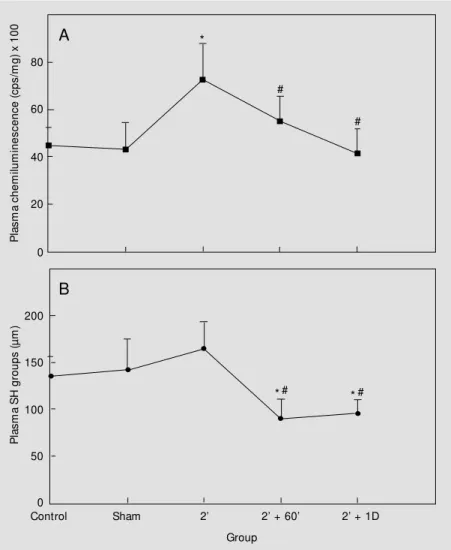
Figure 1A illustrates the effect of 60-min and 1-day reperfusion after 2-min brain is-chemia on plasma chemiluminescence. As can be seen, reperfusion significantly de-creased chemiluminescence emission to con-trol levels when these data were compared to 2-min ischemia. Compared to the effect of reperfusion after 2-min ischemia on plasma thiols (Figure 1B), 60-min and 1-day reper-fusion resulted in a significant decrease in SH groups to levels below the control and 2-min ischemia values. On the other hand, 60-min and 1-day reperfusion after 10-60-min brain ischemia still resulted in a 30-40% increase in plasma chemiluminescence emission (Fig-ure 2A). Values for plasma chemilumines-cence of rats submitted to 2-, 5-, 10- and 30-day reperfusion after 10-min ischemia were similar to controls (Figure 2A). Similar to the results of reperfusion after 2-min is-chemia, reperfusion (60 min, 1, 2, 5 and 10 days) after 10-min ischemia also reduced plasma SH when compared to data from control and 10-min ischemia without reper-fusion (Figure 2B). Although the effect of
P
la
s
m
a
c
h
e
m
ilu
m
in
e
s
c
e
n
c
e
(
c
p
s
/m
g
)
x
1
0
0
80
60
20
0 40
P
la
s
m
a
S
H
g
ro
u
p
s
(
µ
m
) 200
150
50
0 100
Control Sham 2’ 2’ + 60’ 2’ + 1D
Group
A
B
*
*# *#
#
#
Figure 1 - Effect of 2-min (2' ) transient forebrain ischemia and reperfusion in rats on tert -butyl hydroperoxide-initiated chemiluminescence of plasma (A) and plasma thiols (B). Rats w ere submitted to 2-min (2' ) ischemia follow ed by 60 min or 1 day (D) of reperfusion. Data represent the mean ± SD for six to nine different animals. * P<0.05 compared to control groups. #P<0.05 compared to the 2-min (2' ) ischemic group (Duncan multiple range test).
reperfusion after 10-min ischemia on the reduction of plasma thiols was observed up to 10 days, values for plasma SH groups after 30-day reperfusion were not signifi-cantly different from controls (Figure 2B), probably because protein thiols (albumin) have a half-life of 20 days (14).
A possible interpretation of these results is that reperfusion after brain ischemia may also induce peripheral alterations in plasma. The results from animals reperfused after 10-min ischemia indicate that there is a rela-tionship between plasma oxidative damage and brain reperfusion when this injurious ischemic episode occurs, since 60-min and 1-day reperfusion still maintain the increase in chemiluminescence emission caused by 10-min ischemia even when plasma thiols are decreased (Figure 2A). In contrast, re-perfusion after a brief ischemic episode (2 min) may lead to the utilization of plasma thiols as antioxidants and, consequently, to a decrease of chemiluminescence emission to control values (Figure 1A). Thus, it seems that the decrease of plasma chemilumines-cence emission to control levels during longer periods of reperfusion after 10-min ischemia (Figure 2A) may also be related to the utili-zation of plasma thiols (Figure 2B) as a long-term peripheral antioxidant control against possible oxidative damage. Thus, the inter-esting aspects of the results illustrated in Figure 2A and B are the ability of plasma to recover from oxidative damage and the re-generation of thiols after 30-day reperfu-sion, even after reduction of about 50% of thiol levels after 5-day reperfusion.
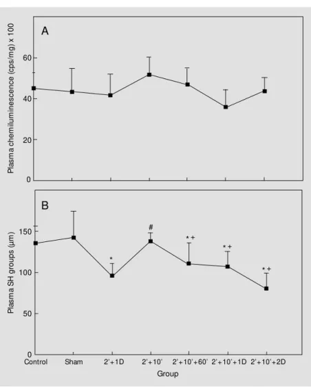
The effect of 60 min, 1 and 2 days of reperfusion on plasma of rats submitted to a 10-min brain ischemic episode after precon-ditioning (2' + 10')is demonstrated in Figure 3A and B. As shown in Figure 3A, when chemiluminescence emission was measured, plasma of rats submitted to 10-min injurious ischemia after a preconditioning time (2' + 1-day reperfusion)was not significantly dif-ferent from controls or from the
precondi-Figure 2 - Effect of 10-min (10' ) transient forebrain ischemia and reperfusion in rats on tert -butyl hydroperoxide-initiated chemiluminescence of plasma (A) and plasma thiols (B). Rats w ere submitted to 10-min (10' ) ischemia follow ed by 60 min or 1, 2, 5, 10 and 30 days (D) of reperfusion. Data represent the mean ± SD for six to eight different animals. * P<0.05 compared to control groups. #P<0.05 compared to the 10-min (10' ) ischemic group (Duncan
multiple range test). SH, Sulfhydryl.
tioned group. Reperfusion after precondi-tioning to brain ischemia did not change plasma chemiluminescence emission (Fig-ure 3A). Plasma thiol content was meas(Fig-ured under the same conditions of reperfusion after preconditioning to brain ischemia used to measure plasma chemiluminescence emis-sion. Figure 3B shows that preconditioning (2' + 1-day reperfusion) significantly de-creased plasma SH groups when compared to control. On the other hand, 10-min is-chemia after preconditioning (2' + 10') was able to maintain plasma SH groups at control
P
la
s
m
a
c
h
e
m
ilu
m
in
e
s
c
e
n
c
e
(
c
p
s
/m
g
)
x
1
0
0
80
60
20
0 40
P
la
s
m
a
S
H
g
ro
u
p
s
(
µ
m
) 200
150
50
0 100
Control Sham 10’ 10’+60’
Group
A
B
10’+1D 10’+2D 10’+5D 10’+10D 10’+30D
*
# *
#
#
*# *#
*# *#
levels. Since 2-min ischemia followed by 1-day reperfusion induces tolerance to a longer and injurious ischemic episode (10 min), we suggest that in this situation plasma SH groups were decreased as sacrificial antioxidants (14), probably to protect more important targets such as DNA and lipids against oxi-dative damage. Thus, these thiols are dam-aged but there are so many in plasma that the effect might be biologically nonsignificant (14). Probably, the plasma of rats submitted to 2 min of ischemia followed by 1 day of
reperfusion and to the double-ischemic epi-sode did not show an increase in chemilumi-nescence emission because plasma SH groups were consumed in the first situation. Never-theless, the results for plasma of rats submit-ted to 60 min or 1 and 2 days of reperfusion after preconditioning to brain ischemia (2' + 10') indicate that in this situation reperfusion causes a loss of SH groups (Figure 3B).
By comparing Figure 3A and B with others, we suggest that the permanence of plasma chemiluminescence emission at trol levels may be associated with the con-sumption of plasma thiols. Interestingly, it is clear that plasma was protected from oxida-tive damage by a longer ischemic episode (10 min) and reperfusion when brain ische-mic preconditioning occurred and, probably, when thiols were decreased (Figure 3A and B). The results illustrated in Figure 3A can be compared to the results in Figure 2A that shows a significant increase of plasma chemi-luminescence emission when rats were sub-mitted to 10-min ischemia and reperfusion without the protection of the precondition-ing time.
The findings of our study are that brain ischemia and reperfusion produce peripher-al peripher-alterations demonstrated in plasma that are probably related to an increased forma-tion of reactive oxygen species. Furthermore, it seems that brain ischemic precondition-ing, as a phenomenon responsible for the protection of the central nervous system against injurious ischemic episodes and re-perfusion (21), also reduced plasma oxida-tive damage as assessed by chemilumines-cence emission. In any case, the determina-tion of plasma thiols may indicate a reladetermina-tion- relation-ship between the consumption of SH groups and the permanence of plasma chemilumi-nescence emission at control levels. This relationship could be seen when rats were submitted to reperfusion after 2-min (Figure 1A and B) or 10-min ischemia (Figure 2A and B) and mainly after the preconditioning time followed by 10-min ischemia and
re-P
la
s
m
a
c
h
e
m
ilu
m
in
e
s
c
e
n
c
e
(
c
p
s
/m
g
)
x
1
0
0
60
20
0 40
P
la
s
m
a
S
H
g
ro
u
p
s
(
µ
m
) 150
50
0 100
Control Sham 2’+1D 2’+10’+60’
Group
A
B
2’+10’+1D 2’+10’+2D 2’+10’
*
#
*+
*+
*+
Figure 3 - Effect of brain ischemic preconditioning and reperfusion in rats on tert-butyl hydroperoxide-initiated chemiluminescence of plasma (A) and plasma thiols (B). Rats w ere submitted to 2-min (2' ) ischemia follow ed by 1-day (D) reperfusion (2' + 1D) and 10-min (10' ) ischemia 1 day after a 2-min ischemic episode (2' + 10' ). Rats w ere also submitted to 60 min or 1 and 2 days of reperfusion after the double-ischemic episode (2' + 10' ). Data represent the mean ± SD for six to nine different animals. * P<0.05 compared to the control group. #P< 0.05 compared to the (2' + 1D) group. +P< 0.05 compared to the (2' + 10' ) group
perfusion (Figure 3A and B), which suggests that reperfusion is related to the reduction of thiols and, probably, to normalization of plasma chemiluminescence emission as much after single ischemic episodes as after double-ischemic episodes (preconditioning). Thus, the results may indicate that peripheral oxi-dative damage is associated with brain is-chemia and reperfusion and that ischemic preconditioning induces protective effects. Considering the literature and our results, it seems that brain ischemia and precondition-ing cause injurious and protective effects, respectively, both in the central nervous sys-tem and in the bloodstream, that could be a possible peripheral indicator of central events.
It has been suggested that neuronal dam-age in brain ischemia and reperfusion is partly due to oxidative damage caused by free radical formation (22). Free radicals are extremely reactive compounds that can react with lipids, enzymes (23,24) or DNA to produce various harmful effects (25). Re-cent studies have shown the simultaneous production of NO and superoxide anion (O2·-) within reperfused ischemic cerebral tissue that might lead to the formation of cytotoxic peroxynitrite or an oxidant derived from it (26). Neuronal produced peroxyni-trite may diffuse to the bloodstream (26), where it induces oxidative damage by its effects in decreasing the levels of
antioxi-dants such as thiol groups (27,28). For this reason, thiols have been regarded as sacrifi-cial antioxidants in plasma (14), playing a role in the decreased mortality from coro-nary heart disease (29). We suggest that after brain ischemia, ischemic preconditioning and reperfusion, the reduction of thiols is prob-ably related to a protection against plasma oxidative damage. Thus, the reduction of thiols in plasma is related to their antioxida-tive capacity, since oxidaantioxida-tive damage re-vealed by chemiluminescence emission has been considered to be a consequence of reactive oxygen species generation (18). Strubelt and colleagues (30) demonstrated that thiols are related to protection against liver ischemia and hypoxia-reoxygenation-induced oxidative damage.
Since neuronal death is greatly reduced when ischemic preconditioning precedes an injurious ischemic episode and reperfusion (21), we suggest that sacrifice reactions of plasma SH groups with reactive oxygen spe-cies lead to oxidative reduction of thiols, an event implying the reduction of plasma oxi-dative damage by preconditioning. In con-clusion, although much work is needed to determine which oxidants participate in the development of plasma injury during brain ischemia, the present results confirm the hypothesis that preconditioning reduces pe-ripheral oxidative damage associated with brain ischemia and reperfusion.
Re fe re nce s
1. Watanabe T, Yuki S, Egaw a M & Nishi H (1994). Protective effects of M CI-186 on cerebral ischemia: possible involvement of free radical scavenging and antioxidant actions. Journal of Pharmacology and Ex-perimental Therapeutics,268: 1597-1604. 2. Kinouchi H, Epstein CJ, M izui T, Carlson E, Chen SF & Chan PH (1991). Attenua-tion of focal cerebral ischemic injury in transgenic mice overexpressing CuZn su-peroxide dismutase. Proceedings of the National Academy of Sciences, USA, 88:
11158-11162.
3. Globus M YT, Busto R, Lin B, Schnippering H & Ginsberg M D (1995). Detection of free radical activity during transient global ischemia and recirculation: effects of intraischemic brain temperature modula-tion. Journal of Neurochemistry, 65: 1250-1256.
4. Traystman RJ, Kirsch JR & Koehler RC (1991). Oxygen radical mechanism of brain injury follow ing ischemia and reper-fusion. Journal of Applied Physiology, 71
(Suppl): 1185-1195.
5. Yang C, Lin N, Tsai P, Liu L & Kuo J (1996). In vivo evidence of hydroxyl radi-cal formation induced by elevation of ex-tracellular glutamate after cerebral is-chemia in the cortex of anesthetized rats.
Free Radical Biology and M edicine, 20: 245-250.
33: 497-500.
7. Isaka Y, Kimura K, Uehara A, Hashikaw a K, M ieno M , M atsumoto M , Handa N, Nakabayashi S, Imaizumi M & Kamada T (1989). Platelet aggregability and in vivo
platelet deposition in patients w ith ische-mic cerebrovascular disease. Evaluation by indium-111-platelet scintigraphy. Throm-bosis Research,56: 739-749.
8. Tanahashi N, Tomita M , Kobari M , Takeda H, Yokoyama M , Takao M & Fukuuchi Y (1996). Platelet activation and erythrocyte aggregation rate in patients w ith cerebral infarction. Clinical Hem orheology, 16: 497-505.
9. Kumura E, Kosaka H, Shiga T, Yoshimine T & Hayakaw a T (1994). Elevation of plasma nitric oxide end products during focal cerebral ischemia and reperfusion in the rat. Journal of Cerebral Blood Flow and M etabolism, 14: 487-491.
10. Salvemini D & Botting R (1993). M odula-tion of platelet funcodula-tion by free radicals and free-radical scavengers. Trends in Pharmacological Sciences, 14: 36-42. 11. Sneddon JM & Vane JR (1988).
Endotheli-um-derived relaxing factor reduces plate-let adhesion to bovine endothelial cells.
Proceedings of the National Academy of Sciences, USA, 85: 2800-2804.
12. Losano G, Gattullo D & Pagliaro P (1996). M yocardial, neural and vascular aspects of ischemic preconditioning. Life Sci-ences,59: 1185-1192.
13. Llesuy S, M ilei J, Gonzalez Flecha B & Boveris A (1990). M yocardial damage in-duced by doxorubicins: hydroperoxide-ini-tiated chemiluminescence and morpholo-gy. Journal of Free Radicals in Biology and M edicine,8: 259-264.
14. Halliw ell B (1988). Albumin - an important extracellular antioxidant? Biochemical
Phar-macology, 37: 569-571.
15. Das DK & M aulik N (1994). Antioxidant effectiveness in ischemia-reperfusion tis-sue injury. M ethods in Enzymology, 233: 601-610.
16. Pulsinelli WA, Brierley M D & Plum F (1982). Temporal profile of neuronal dam-age in a model of transient forebrain is-chemia. Annals of Neurology, 1: 491-498. 17. Netto CA, Hodges H, Sinden JD, Le Peillet E, Kershaw T, Sow inski P, M eldrum BS & Gray JA (1993). Effects of fetal hippocam-pal field grafts on ischaemic-induced defi-cits in spatial navigation in the w ater maze. Neuroscience,54: 69-92. 18. Flecha BG, Llesuy S & Boveris A (1991).
Hydroperoxide-initiated chem ilum ines-cence: an assay for oxidative stress in biopsies of heart, liver, and muscle. Free Radical Biology and M edicine, 10: 93-100. 19. Hu M (1994). M easurement of protein thiol groups and glutathione in plasma. M ethods in Enzymology,233: 380-385. 20. Bradford M M (1976). A rapid and
sensi-tive method for the quantification of mi-crogram quantities of protein utilizing the principle of protein dye binding. Analytical Biochemistry, 72: 248-254.
21. Kitigaw a K, M atsumoto M , Tagaya M , Hata R, Ueda H, Niinobe M , Handa N, Fukunaga R, Kimura K, M ikoshiba K & Kamada T (1990). Ischemic tolerance phe-nomenon found in the brain. Brain Re-search, 528: 21-24.
22. Halliw ell B (1992). Reactive oxygen spe-cies and the central nervous system. Jour-nal of Neurochemistry, 59: 1609-1623. 23. Vietta M , Frassetto SS, Battastini AM O,
Klein AB, M oreira C, Dias RD & Sarkis JJF (1996). Sensitivity of ATPase-ADPase ac-tivities from synaptic plasma membranes of rat forebrain to lipid peroxidation in
vi-tro and the protective effect of vitamin E.
Neurochemical Research, 21: 299-304. 24. Frassetto SS, Dias RD & Sarkis JJF (1997).
Free radical-induced inhibition of ATP di-phosphohydrolase activity (EC 3.6.1.5) from rat blood platelets. Biochemistry and M olecular Biology International, 41: 161-168.
25. Siesjo BK, Agardh C-D & Bengtsson F (1989). Free radicals and brain damage.
Cerebrovascular and Brain M etabolism Review s, 1: 165-211.
26. Kum ura E, Yoshim ine T, Iw at suki K, Yamanaka K, Tanaka S, Hayakaw a T, Shiga T & Kosaka H (1996). Generation of nitric oxide and superoxide during reperfusion after focal cerebral ischemia in rats. Amer-ican Journal of Physiology, 270 (Cell Phys-iology, 39): C748-C752.
27. Van der Vliet A, Smith D, O’Neill CA, Kaur H, Darley-Usmar V, Cross CE & Halliw ell B (1994). Interactions of peroxynitrite w ith human plasma and its constituents: oxi-dative damage and antioxidant depletion.
Biochemical Journal, 303: 295-301. 28. Vásquez-Vivar J, Santos AM , Junqueira
VBC & Augusto O (1996). Peroxynitrite-mediated formation of free radicals in hu-man plasma: EPR detection of ascorbyl, albumin-thiol and uric acid-derived free radicals. Biochemical Journal, 314: 869-876.
29. Luoma PV, Nayha S, Sikkila K & Hassi J (1995). High serum alpha-tocopherol, al-bumin, selenium and cholesterol, and low mortality from coronary heart disease in northern Finland. Journal of Internal M edi-cine, 237: 49-54.