Magne sium chlo ride alo ne o r in
co m binatio n with diaze pam fails to
pre ve nt hippo cam pal dam age fo llo wing
transie nt fo re brain ische m ia
1Departamento de Farmácia e Farmacologia, Centro de Ciências da Saúde, and 2Departamento de Ciências Morfofisiológicas, Centro de Ciências Biológicas,
Universidade Estadual de Maringá, Maringá, PR, Brasil H. Milani1,
E.R. Lepri2,
F. Giordani1 and
L.A. Favero-Filho1
Abstract
In the central nervous system, magnesium ion (Mg2+) acts as an
endogenous modulator of N-methyl-D-aspartate (NMDA)-coupled calcium channels, and may play a major role in the pathomechanisms of ischemic brain damage. In the present study, we investigated the effects of magnesium chloride (MgCl2, 2.5, 5.0 or 7.5 mmol/kg),
either alone or in combination with diazepam (DZ), on ischemia-induced hippocampal cell death. Male Wistar rats (250-300 g) were subjected to transient forebrain ischemia for 15 min using the 4-vessel occlusion model. MgCl2 was applied systemically (sc) in single (1x, 2
h ischemia) or multiple doses (4x, 1, 2, 24 and 48 h post-ischemia). DZ was always given twice, at 1 and 2 h post-ischemia. Thus, ischemia-subjected rats were assigned to one of the following treatments: vehicle (0.1 ml/kg, N = 34), DZ (10 mg/kg, N = 24), MgCl2
(2.5 mmol/kg, N = 10), MgCl2 (5.0 mmol/kg, N = 17), MgCl2 (7.5
mmol/kg, N = 9) or MgCl2 (5 mmol/kg) + DZ (10 mg/kg, N = 14). Seven days after ischemia the brains were analyzed histologically. Fifteen minutes of ischemia caused massive pyramidal cell loss in the subiculum (90.3%) and CA1 (88.4%) sectors of the hippocampus (P<0.0001, vehicle vs sham). Compared to the vehicle-treated group,
all pharmacological treatments failed to attenuate the ischemia-in-duced death of both subiculum (lesion: 86.7-93.4%) and CA1 (lesion: 85.5-91.2%) pyramidal cells (P>0.05). Both DZ alone and DZ + MgCl2 reduced rectal temperature significantly (P<0.05). No animal death was observed after drug treatment. These data indicate that exogenous magnesium, when administered systemically post-ischemia even in different multiple dose schedules, alone or with diazepam, is not useful against the histopathological effects of transient global cerebral ischemia in rats.
Co rre spo nde nce
H. Milani
Departamento de Farmácia e Farmacologia, CCS
Universidade Estadual de Maringá Av. Colombo, 5790
87020-900 Maringá, PR Brasil
Fax: + 55-44-263-5116 E-mail: milani@ dff.uem.br
Research supported by CNPq and UEM.
Received February 25, 1998 Accepted July 15, 1999
Ke y wo rds
·Cerebral ischemia ·Hippocampal lesion ·CA1 cell loss ·Magnesium chloride ·Diazepam
Intro ductio n
Transient global cerebral ischemia re-sults in hippocampal cell death both in ani-mal models (1,2) and in humans, for ex-ample, victims of reversible cardiac arrest (3). Increased intracellular calcium seems to play an important role in the pathophysiol-ogy of delayed neuronal death caused by cerebral ischemia. Ischemia-induced calcium influx is coupled to the activation of voltage-sensitive and agonist receptor-gated calcium channels, mainly the N-methyl-D-aspartate (NMDA) type of glutamate receptor (4,5). Under resting conditions, the NMDA-coupled channel is normally blocked by Mg2+
(6,7). However, during excessive depolar-ization, such as occurs during ischemia, this Mg2+ blockade is released (5). There is
evi-dence of a relationship between the reduc-tion or loss of the Mg2+ blockade of the
NMDA response and persistent, neuronal hyperexcitability, which evolves to struc-tural changes in the dendrites and finally to hippocampal cell death (8). Such neuronal overactivation may be related to the cause rather than the effect of ischemia-induced neurodegeneration since it can be detected long before cell loss (9). The role of endog-enous Mg2+ in modulating calcium influx
through the NMDA glutamate receptor makes it a candidate for neuroprotective therapy.
The neuroprotective potential of Mg2+
has been studied in different animal models of brain damage. Positive findings have been obtained in adult rodent models of in vitro
anoxic challenge (10), in vivo glutamate neu-rotoxicity (11), spinal cord ischemia (12), focal ischemia following occlusion of the middle cerebral artery (13,14) and, more extensively, in models of traumatic, mechan-ical brain injury (for a review, see Ref. 15). The effects of Mg2+ in attenuating delayed
hippocampal cell death have been rarely in-vestigated in in vivo animal models of tran-sient global cerebral ischemia. Only a single study has attempted to evaluate the
neuro-protective potential of Mg2+ after systemic
delivery in adult rats subjected to transient forebrain ischemia. The intravenous injec-tion of MgCl2 before ischemia not only failed
to prevent CA1 hippocampal cell loss but aggravated the histological outcome (16). Clearly, further studies are necessary to pro-vide more conclusive data on the effects of Mg2+ in animal models of transient global
cerebral ischemia, including different treat-ment protocols.
The aim of the present study was to ex-amine the effects of MgCl2 administered
systemically using different schedules, ei-ther alone or in combination with diazepam (DZ), after ischemia. The 4-vessel occlusion (4-VO) model was used, thus extending find-ings to a more widely studied animal model of transient forebrain ischemia. The testing of the MgCl2 + DZ combination is due to the
fact that benzodiazepines potentiate the cell membrane hyperpolarization induced by ac-tivation of the GABA receptor/ion channel, thus inhibiting action potentials elicited by depolarization. Since ischemia-induced neu-ronal depolarization reduces the normal Mg2+
blockade of the NMDA-coupled calcium channel observed under resting conditions, we hypothesized that this effect of ischemia might be attenuated by the neuronal depres-sant action of DZ, thus facilitating the cal-cium blocking action of magnesium, inde-pendently of whether DZ alone may provide neuroprotection (17).
Mate rial and Me tho ds
Subje cts
Male Wistar rats weighing 250-300 g at the beginning of the surgical procedures were used. The animals were maintained in groups of 4-5 in plastic cages (39 x 33 x 16 cm) at a controlled temperature (22 ± 1oC), on a 12-h
Prin-ciples for Research Animal Utilization pub-lished by the Brazilian Society for Neurosci-ence and Behavior.
Ische m ia
Transient forebrain ischemia was induced using the 4-VO method (18) with the minor modifications described in a previous study (19). Under ether anesthesia plus the local application of 2% xylocaine, the vertebral arteries were electrocoagulated bilaterally and the carotid arteries were loosely snared with a silk thread. Five to six hours later, the thread was tightened for a period of 15 min. Loss of the righting reflex within 2 min of carotid occlusion, unresponsiveness to a gentle touch, mydriasis and tonic extension of the paws were considered to indicate ef-fective ischemia. Animals which recovered the righting reflex during the occlusion pe-riod or presented convulsions were excluded. After releasing the carotids, those animals which did not recover the red eye color within 2 min were also excluded. Core tem-perature, monitored by a rectal thermistor inserted to a depth of 6 cm, was maintained between 37o and 38oC throughout occlusion
and during the first hours of reperfusion by placing the rats in a warming box at 30oC
when necessary. Control rats were submit-ted to the same manipulations without occlu-sion of the vertebral and carotid arteries.
D rug tre atm e nt
MgCl2 (2.5, 5.0 or 7.5 mmol/kg, sc) was
given either as a single dose (1x) or in mul-tiple doses (4x). Diazepam (10 mg/kg, ip) was always given twice, 1 and 2 h after initiating reperfusion (17). According to the treatment schedule the animals were assigned to one of the following groups: G1: sham-operated; G2: ischemia + vehicle (saline 0.9%, 0.2 ml/100 g body weight, sc, 4x); G3: ischemia + DZ (2x, 1 and 2 h after reperfu-sion); G4: ischemia + MgCl2 (2.5 mmol/kg;
4x; 1, 2, 24 and 48 h after reperfusion); G5: ischemia + MgCl2 (5.0 mmol/kg; 1x, 2 h
after reperfusion); G6: ischemia + MgCl2
(5.0 mmol/kg, 4x, 1, 2, 24 and 48 h after reperfusion); G7: ischemia + MgCl2 (7.5
mmol/kg; 4x; 1, 2, 24 and 48 h after reperfu-sion); G8: ischemia + DZ (2x; 1 and 2 h after reperfusion) + MgCl2 (5.0 mmol/kg; 1x; 2 h
after reperfusion), and G9: ischemia + DZ (2x; 1 and 2 h after reperfusion) + MgCl2
(5.0 mmol/kg, 4x, 1, 2, 24, and 48 h after reperfusion).
MgCl2.7 H2O was purchased from Sigma
Chemical Co., St. Louis, MO, USA; Diaz-epam® was a commercial preparation from
Roche Pharmaceutic Inc., São Paulo, Brazil.
Histo lo gical analysis
On the 7th day after ischemia, the ani-mals were anesthetized deeply with ether and perfused transcardiacally with 0.9% sa-line followed by Bouins fixative solution at a rate of 20 ml/min for 7-10 min. The brain was kept in situ for 1 h at 1-2oC and then
removed and kept in the same fixative for 3 days. Eight to twelve coronal sections were taken from each brain at levels correspond-ing to 3.0-4.0 mm posterior to bregma, and stained with hematoxylin-eosin. Three sec-tions were chosen for each bilateral count. Thus, 6 fields were counted per rat and the number of apparently intact cells per animal was reported as the mean of the 6 fields. Fields were chosen by centering the 400X microscopic field on the medial portion of the subiculum and CA1 sectors. The identity of treatment groups was unknown to the examiner during the histological analysis.
D ata analysis
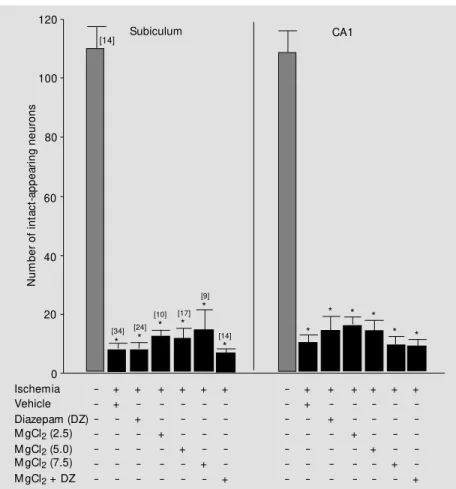
ef-15 min of ischemia caused a profound loss of pyramidal neurons in the subiculum (F6,113 =
100.7; P<0.0001) and CA1 (F6,113 = 65.9;
P<0.0001) sectors of the hippocampus in all groups (Figure 1). The degree of cell loss ranged from 86.7 to 94.0% in the subiculum and from 85.3 to 91.6% in the CA1 sectors. When given in different amounts and in single or multiple doses MgCl2 did not
pro-vide a neuroprotective effect against is-chemia-induced pyramidal cell loss in the subiculum and CA1 hippocampal sectors compared to the vehicle-treated group (Fig-ure 1) (P>0.05, Duncans multiple range test). The treatment with diazepam alone, given 1 and 2 h after reperfusion, also did not exert a neuroprotective effect. These results for the single drug protocol did not change with the MgCl2 plus diazepam combination.
No difference was observed among the vari-ous pharmacological treatments.
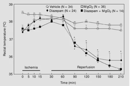
MgCl2 caused a consistent and
appar-ently dose-dependent loss in muscle tonus, indicating that the substance was well ab-sorbed. However, no rat died or required artificial respiration. Profound sedation was observed in rats given DZ alone or MgCl2 +
DZ. Compared to the vehicle-treated group, there was a slight but significant reduction in core temperature after treatment with ei-ther DZ alone or MgCl2 + DZ (P<0.05)
(Figure 2). This effect may have been due to the action of DZ since there was no difference between the vehicle- and MgCl2
-treated groups or the DZ and MgCl2 + DZ
groups.
D iscussio n
The present study demonstrates that the systemic application of increasing doses of MgCl2 employing either a single or multiple
administration schedule does not provide neuroprotection against delayed hippocam-pal cell death induced by transient forebrain ischemia in the adult rat. This situation is unaltered when DZ is added to MgCl2, even
Figure 1 - Effect of M gCl2 (2.5, 5.0 or 7.5 mmol/kg, sc) either alone or in combination w ith
diazepam (DZ) (M gCl2 + DZ, 5 mmol/kg and 10 mg/kg, ip, respectively) on hippocampal
pyramidal cell loss after 15-min transient forebrain ischemia. The effect of 5.0 mmol/kg M gCl2 w as tested after a single dose (1x, 2 h post-ischemia) or multiple (4x, 1, 2, 24 and 48
h post-ischemia) doses. The 2.5 and 7.5 mmol/kg M gCl2 doses w ere tested after multiple
administration (4x) only. The animals treated w ith a single dose (1x) or multiple (4x) doses of
M gCl2 (5.0 mmol/kg) w ere pooled, as w ere the groups treated w ith M gCl2 (1x) + DZ or
M gCl2 (4x) + DZ. Diazepam (10 mg/kg), alone or in combination w ith M gCl2, w as alw ays
given tw ice (1 and 2 h after reperfusion). Bars and vertical lines represent the mean and
SEM . The numbers w ithin brackets indicate the sample size. * P<0.0001 vs the
sham-operated group.
fect appeared, Duncans multiple range test was performed to determine differences be-tween groups. The effect of the pharmacolo-gical treatments on rectal temperature was analyzed by MANOVA with repeated meas-ures followed by the Newman-Keuls test. In the case of a significant group effect, the Student t-test was used to compare the re-spective groups for a given interval.
Re sults
Compared to the sham-operated group,
N
u
m
b
e
r
o
f
in
ta
c
t-a
p
p
e
a
ri
n
g
n
e
u
ro
n
s
120
100
80
60
40
20
0
[14] Subiculum CA1
- + + + + + +
- + - - - -
-- - + - - -
-- - - + - -
-- - - - + -
-- - - +
-- - - +
Diazepam (DZ) M gCl2 (2.5) M gCl2 (5.0) M gCl2 (7.5) M gCl2 + DZ
[34] [24] [10] [17]
[9]
[14]
* * * * *
*
*
* * *
* *
- + + + + + +
- + - - - -
-- - + - - -
-- - - + - -
-- - - - + -
-- - - +
-- - - +
though a modest fall in rectal temperature was recorded.
The lack of a neuroprotective effect of hypothermia is consistent with findings show-ing that hypothermia may prevent cell death when occurring during the intra-ischemic or immediate (5 min) post-ischemic periods, but not after longer (30 min) post-ischemic periods (for a review, see Refs. 17,20). In the present study, hypothermia appeared only in the groups treated with DZ or DZ + MgCl2
60 min following reperfusion (Figure 2). Our data for magnesium are consistent with a previous study which not only failed to demonstrate a neuroprotective effect of MgCl2, but detected worsening of
ischemia-induced hippocampal cell loss (16). This effect of MgCl2 was not observed in the
present experiments. A possible explanation may be the difference in the methodology used to produce transient cerebral ischemia in the two studies. In Blairs experiments, animals were subjected to 10 min ischemia by a combination of intravenous trimetha-phan, bilateral carotid artery occlusion and simultaneous central venous exsanguination, which required that the animal be main-tained under anesthesia. Thus, the onset and maintenance of ischemia was judged on the basis of an isoelectric electroencephalogram (EEG). As noted by others, this parameter is not an adequate index of the severity of ischemia since isoelectricity of the EEG may occur in the presence of electrophysiologi-cal activity, and therefore considerable blood flow, in the hippocampus (21). In the present study, we used the 4-VO model in which animals are not anesthetized during or after ischemia. Thus, clinical criteria such as loss of the righting reflex, mydriasis, lack of re-sponsiveness to touch, and tonic extension of the paws were used; these acute symp-toms are indicative of severe forebrain is-chemia (21). Unlike the present model, in that used by Blair et al. (16) mild ischemia may have been produced leading to sub-maximal hippocampal cell loss; this may
account for the aggravating effect of MgCl2.
In the present study, however, the almost complete loss of pyramidal cells may have masked the aggravating effect of MgCl2, had
it occurred.
Hyperglycemia induced by MgCl2 may
be the cause of the aggravating effect of this compound on the histological outcome of ischemia (16). It is well established that pre-ischemic hyperglycemia worsens the out-come of transient ischemia. Although plasma glucose was not measured after the adminis-tration of MgCl2 in the present experiment, it
is unlikely that hyperglycemia may have masked the neuroprotective properties of magnesium. In fact, Blair et al. (16) have found that while insulin prevents worsening of the histological changes caused by MgCl2,
it does not change the extent of hippocampal cell loss caused by ischemia in comparison to the control group. This suggests that the failure of MgCl2 to counteract the
histologi-cal changes resulting from ischemia was not due to the masking effect of hyperglycemia. In a model of focal ischemia, however,
insu-R
e
c
ta
l
te
m
p
e
ra
tu
re
(
oC
)
39
38
37
36
35
Vehicle (N = 34) Diazepam (N = 24)
M gCl2 (N = 36)
Diazepam + M gCl2 (N = 14)
Ischemia Reperfusion
* *
* * *
* * * * *
0 5 10 15 30 60 90 120 150 180 210
Time (min)
Figure 2 - Effect of M gCl2 and diazepam alone or in combination on rectal temperature. The groups assigned to the M gCl2 (2.5, 5.0 or 7.5 mmol/kg) treatments, either as a single dose or as multiple doses, w ere pooled since core temperatures did not differ among the
individual groups. The groups treated w ith both substances w ere also pooled, i.e., M gCl2
lin treatment potentiated the effect of MgCl2
in reducing infarct size (14). Additionally, in the present study, MgCl2 was administered 1
h post-ischemia, an interval during which adequate circulation would be restored avoid-ing exacerbated anaerobic metabolism of glucose with elevated lactate formation. Thus, in the present study, the lack of a neuropro-tective effect after the systemic administra-tion of MgCl2 cannot be explained by the
detrimental influence of possible MgCl2
-in-duced hyperglycemia.
In the study of Blair et al. (16), MgCl2
was given in a single dose prior to ischemia. These authors suggested that the failure of such treatment to protect against ischemia might have resulted from the lack of a maintained elevation of Mg2+ concentration
in the extracellular fluid beyond the acute insult, and that Mg2+ would be more
benefi-cial if provided 2-3 days after ischemia. In this respect, our data extend these findings, demonstrating that even with different and repeated doses given 1, 2, 24 and 48 h after reperfusion, MgCl2 failed to prevent
hippo-campal cell loss after transient, global fore-brain ischemia. Whether this protocol pro-vided a consistent Mg2+ concentration in brain
parenchyma is not known. However, data from the literature indicate that although Mg2+ can cross the blood brain barrier (BBB),
this occurrence may be greatly restricted. For example, in dogs, Mg2+ concentration
increased to a maximum of 21% in the cere-brospinal fluid (CSF) compared to the 300-400% elevation in plasma after intravenous administration of MgCl2 (22). Under
condi-tions of hypomagnesemia, the mean Mg2+
concentration in the whole brain was negli-gible one hour after a single intraperitoneal injection of MgCl2; an increase in Mg2+
con-centration in the CSF does not reflect an increase in the brain parenchyma (23). In normal rats, the administration of 432 mg/kg of MgSO4 given in several doses over a 2-h
period elevated the level of Mg2+ in the
hippocampus by 41% (24). In another study,
20 min of 4-vessel occlusion elevated the Mg2+ concentration in the hippocampus by
28% after 24 h of reperfusion, which de-creased progressively to 19 and 15.6% above the control after 48 and 72 h of reperfusion, respectively. This rise in intrahippocampal Mg2+ levels from an internal source did not
confer a neuroprotective effect after ischemia (25). However, the extent to which systemi-cally applied exogenous magnesium can en-ter the brain afen-ter transient, global ischemia has not been investigated.
In the present experiments, the animals received different doses of MgCl2, i.e., 2.5,
5.0 and 7.5 mmol/kg, during the first and second hours after reperfusion, for a total of 476, 952 and 1,428 mg/kg, respectively, over a 2-h period. The last dose corresponds to more than three times the amount of MgCl2
used in the study cited above (24). Consider-ing that transient global ischemia results in increased permeability of the BBB to small and large molecules (26,27) possibly includ-ing Mg2+ (28), it would be expected in the
present experiment with ischemic rats that the Mg2+ concentration in the hippocampus
might be similar to or even greater than that found in normal rats (24), at least during the first two hours of reperfusion (see 28). If so, the exogenous Mg2+ concentration which
reached the brain parenchyma in the present study may have been insufficient to provide hippocampal neuroprotection. This may be true, since the local application of a single injection of MgCl2 (50 mM/1 µl) into the
hippocampus reduces hippocampal cell loss after 20 min of ischemia in the 4-VO model (25). This finding corroborates other data obtained using in vitro models of ischemia (10), indicating that the lack of an Mg2+
neuroprotectiveeffect after systemic admin-istration may not result from pharmacody-namic ineffectiveness. That the BBB may limit the bioavailability of exogenously ap-plied Mg2+, even during transient forebrain
There is evidence that disruption of the BBB by transient global cerebral ischemia may occur in a mild to moderate manner. In the 4-VO rat model, the permeability of the BBB to small and large molecules was al-tered only in the presence of tissue infarction (29). Infarction, however, is infrequent in this model of cerebral ischemia even after 30 min or more (1,29). Pluta et al. (30) reported that after 10 min of cardiac arrest in the rat, horseradish peroxidase (HRP) extravasa-tions, although constant, were few, randomly distributed and focal, and appeared most frequently in certain brain regions including the hippocampus. Overall, infarction ap-peared occasionally as single or multiple microscopic foci. These changes were not graded as a function of the duration of ische-mia and/or reperfusion. Pluta et al. (30) con-cluded that such alterations may reflect more a slight and random leakage associated with only a limited number of vessel bifurcations, rather than a massive BBB breakdown. In contrast, in models of focal ischemia, infarc-tion invariably occurs in the region supplied by the occluded vascular tree. Its magnitude is dependent on the duration of ischemia, and the BBB breakdown is dramatic (31-34). In cats, the BBB permeability to per-technetate, albumin, sodium and antipyrine was greatest in the core and lowest in the penumbral region of the infarct after middle cerebral artery occlusion (34). The extent of BBB disruption seems to be a phenomenon dependent on the nature and severity of brain injury (33) which should determine the bio-availability of drugs at the site of damage. This may partially explain why Mg2+
treat-ment applied systemically is effective in pre-venting brain damage in animal models of focal ischemia (14,15), spinal cord ischemia (13), traumatic brain injury (10), and neuro-toxic injury induced by local application of quinolinate (35), but not in models of tran-sient forebrain global ischemia (16, present results).
Thus, the lack of a neuroprotective effect
by MgCl2 in the present study (see also Ref.
16) may be partially attributed to pharmaco-kinetic hindrance at the level of the BBB, limiting the bioavailability of Mg2+ to the
brain parenchyma, even under transient glo-bal ischemia. In addition, other factors may act to reduce Mg2+ concentration in the brain
after systemic administration, e.g., the short half-time of magnesium in the CSF, esti-mated to be about 75 min in the mongrel dog (22), and the finding that, at least in humans, about 33% of plasma magnesium is protein-bound (36) and unavailable to the brain.
The combination of MgCl2 + DZ did not
alter the results observed for each compound alone. The rationale for testing the drug com-bination lies in the well-established neu-ronal depressant action of DZ, i.e., an effect contrary to the cell membrane depolariza-tion that occurs during ischemia(8,9). Thus, independently of whether DZ does (17) or does not provide (present results) neuropro-tection, our hypothesis holds that DZ may facilitate the expression of a neuroprotective effect by Mg2+ (see Introduction). The
certain types of brain injury since drugs may slow the rate of the neuronal death but not avoid it. Thus, the neuroprotective effect of DZ observed by Schwartz et al. (17) may not be long lasting; DZ may have merely de-layed hippocampal cell death (17). In an-other study, Schwartz et al. (37) found that the GABA reuptake inhibitor tiagabine was neuroprotective up to 4 days post-ischemia, but not after 21 days. Under the present experimental conditions, however, one limi-tation of this study was that the negative result with DZ alone hinders an interpreta-tion as to whether DZ does or does not facilitate the neuroprotective action of Mg2+.
Under different experimental conditions,
both DZ (17) and Mg2+ (10-15) are
neuro-protective.
The data presented here suggest that MgCl2,when given systemically in single or
multiple doses, is not useful to protect against acute neurodegenerative outcomes in mod-els of transient global ischemia. Whether DZ interacts with Mg2+ to facilitate its
pharma-codynamic effectiveness will require addi-tional studies.
Akno wle dgm e nts
The authors gratefully acknowledge the technical assistance of Marcos A. Trombelli.
Re fe re nce s
1. Pulsinelli WA, Brierley JB & Fred P (1982). Temporal profile of neuronal damage in a model of transient forebrain ischemia. Annals of Neurology, 11: 491-498. 2. Kirino T, Tamura A & Sano K (1984).
De-layed neuronal death in the rat hippocam-pus follow ing transient forebrain
ische-mia. Acta Neuropathologica, 64: 139-147.
3. Petito CK, Feldmann E, Pulsinelli WA & Plum F (1987). Delayed hippocampal dam-age in humans follow ing cardiorespiratory arrest. Neurology, 37: 1281-1286. 4. Raichle M E (1983). The pathophysiology
of brain ischemia. Annals of Neurology,
13: 2-10.
5. Siesjo BK & Bengtsson F (1989). Calcium fluxes, calcium antagonists, and calcium-related pathology in brain ischemia, hy-poglycemia, and spreading depression: a
unifying hypothesis. Journal of Cerebral
Blood Flow and M etabolism,9: 127-140. 6. Novak L, Bregest ovski P, Ascher P,
Herbet A & Prochiantz A (1984). M agne-sium gates glutamate-activated channels
in mouse central neurones. Nature, 307:
462-465.
7. Coan EJ & Collingridge GL (1985). M ag-nesium ions block an N-methyl-D-aspar-tate receptor-m ediated com ponent of synaptic transmission in rat hippocampus. Neuroscience Letters, 53: 21-26. 8. Hori N & Carpenter DO (1994). Functional
and morphological changes induced by transient in vivo ischemia. Experimental Neurology, 129: 279-289.
9. Gao TM & Xu ZC (1996). In vivo intracellu-lar demonstration of an ischemia-induced
postsynaptic potential from CA1
pyrami-dal neurons in rat hippocampus.
Neuro-science,75: 665-669.
10. Kass IS, Cottrell JE & Chambers G (1988). M agnesium and cobalt, not nimodipine, protect neurons against anoxic damage in the rat hippocampal slice. Anesthesiology, 69: 710-715.
11. Rothe F, W olf G, Fische S, Hass P, Keilhoff G & Abicht K (1993). Quinolinate and kainate facilitate magnesium penetra-tion into brain tissue. Neuroreport, 4: 205-207.
12. Vacanti FX & Ames III A (1984). M ild hy-pothermia and M g++ protect against irre-versible damage during CNS ischemia. Stroke, 15: 695-698.
13. Izumi Y, Roussel S, Pinard E & Seylaz J (1991). Reduction of infarct volume by magnesium after middle cerebral artery
occlusion in rats. Journal of Cerebral
Blood Flow and M etabolism, 11: 1025-1030.
14. M arinov M B, Harbaugh KS, Hoopes PJ, Pikus HJ & Harbaugh RE (1996). Neuro-protective effects of preischemia intraar-terial magnesium sulfate in reversible
fo-cal cerebral ischemia. Journal of
Neuro-surgery, 85: 117-124.
15. M cIntoshi TK (1994). Neurochemical se-quelae of traumatic brain injury:
Thera-peutic implications. Cerebrovascular and
Brain M etabolism Review s, 6: 109-162. 16. Blair JL, Warner DS & Todd M M (1989).
Effects of elevated plasma magnesium versus calcium on cerebral ischemic in-jury in rats. Stroke, 20: 507-512.
17. Schw artz RD, Yu X, Katzman M R, Hayden-Hixson DM & Perry JM (1995). Diazepam, given postischemia, protects selectively vulnerable neurons in the rat
hippocam-pus and striatum. Journal of
Neurosci-ence, 15: 529-539.
18. Pulsinelli WA & Brierley JB (1979). A new model of bilateral hemispheric ischemia in the unanesthetized rat. Stroke, 10: 267-272.
19. M ilani H, Uemura YU, Oliveira RM W, Lepri ER & Xavier GF (1998). Loss of CA1 cells follow ing global ischaemia correlates w ith spatial deficits in the circular
plat-form task. Journal of Neuroscience M
eth-ods, 80: 19-27.
20. Dietrich WD (1992). The importance of brain temperature in cerebral injury. Jour-nal of Neurotrauma, 9: S475-S485. 21. Buchan A, Li H & Pulsinelli WA (1991).
The N-methyl-D-aspartate antagonist, M k-801, fails to protect against neuronal dam-age caused by transient, severe forebrain
ischemia in adult rats. Journal of
Neuro-science, 11: 1049-1056.
22. Oppelt WW, M acIntyre I & Rall DP (1963). M agnesium exchange betw een blood and
cerebrospinal fluid. American Journal of
Physiology, 205: 959-962.
23. Chutkow JG (1974). M etabolism of mag-nesium in central nervous system: Rela-tionship betw een concentrations of mag-nesium in cerebrospinal fluid and brain in
magnesium deficiency. Neurology, 24:
780-787.
magnesium sulfate enters the brain and increases the threshold for hippocampal
seizures in rats. American Journal of
Ob-stetrics and Gynecology, 167: 1605-1610. 25. Tsuda T, Kogure K, Nishioka K &
Watanabe T (1991). M g2+ administered
up to tw enty-four hours follow ing reper-fusion prevents ischemic damage of the CA1 neurons in the rat hippocampus. Neuroscience, 44: 335-341.
26. Dobbin J, Crockard HA & Ross-Russel R (1989). Transient blood-brain barrier per-meability follow ing profound temporary global ischemia: an experimental study using [14C] AIB. Journal of Cerebral Blood Flow and M etabolism, 9: 71-78. 27. Preston E & Foster DO (1997). Evidence
for pore-like opening of the blood-brain barrier follow ing forebrain ischemia in rats. Brain Research, 761: 4-10. 28. Ito U, Ohno K, Nakamura R, Suganuma F
& Inaba Y (1979). Edema during ischaemia and after restoration of blood flow . M eas-urement of w ater, sodium, potassium content and plasma protein permeability. Stroke, 10: 542-547.
29. Petito CK, Pulsinelli WA, Jacobson G & Plum F (1982). Edema and vascular per-meability in cerebral ischemia: compari-son betw een ischemic neuronal damage
and infarction. Journal of Neuropathology
and Experimental Neurology, 41: 423-436. 30. Pluta R, Lossinsky AS, Wisniew ski HM & M ossakow ski M J (1994). Early blood-brain barrier changes in the rat follow ing tran-sient complete cerebral ischemia induced
by cardiac arrest. Brain Research, 633:
41-52.
31. Hatashita S & Hoff JT (1990). Brain edema and cerebrovascular permeability during cerebral ischemia in rats. Stroke, 21: 582-588.
32. Garcia JH, Yoshida Y, Chen H, Li Y, Zhang ZG, Lian J, Chen S & Chopp M (1993). Progression from ischemic injury to in-farct follow ing middle cerebral artery
oc-clusion in the rat. American Journal of
Pathology, 142: 623-635.
33. Belayev L, Busto R, Zhao W & Ginsberg M D (1996). Quantitative evaluation of blood-brain barrier permeability follow ing middle cerebral artery occlusion in rats.
Brain Research, 739: 88-96.
34. O’Brien M D, Jordan M M & Waltz AG (1974). Ischemic cerebral edema and the blood-brain barrier: Distributions of per-technetate, albumin, sodium, and antipy-rine in brains of cats after occlusion of the
middle cerebral artery. Archives of
Neu-rology, 30: 461-465.
35. Wolf G, Keilhoff G, Fischer S & Hass P (1990). Subcutaneously applied magne-sium protects reliably against quinolinate-induced N-methyl-D-aspartate (NM DA)-mediated neurodegeneration and convul-sions in rats: are there therapeutic
impli-cations? Neuroscience Letters, 117:
207-211.
36. Kroll M H & Elin RJ (1985). Relationship betw een magnesium and protein concen-tration in serum. Clinical Chemistry,31: 244-246.
37. Inglefield JR, Perry JM & Schw artz RC (1995). Postischemic inhibition of GABA reuptake by tiagabine slow s neuronal
death in the gerbil hippocampus.