Article
ISSN 0102-695X http://dx.doi.org/10.1590/S0102-695X2012005000027 Received 10 Oct 2011 Accepted 8 Jan 2012 Available online 3 Feb 2012
in ischemic myocardium of rat
T. S. Mohamed Saleem,
*,1C. Madhusudhana Chetty,
2S.
Kavimani
31Jawaharlal Nehru Technological University, Kakinda & Department of
Pharmacology, Annamacharya College of Pharmacy, India,
2Department of Biotechnology, Annamacharya College of Pharmacy, India 3Department of Pharmacology, Mother Theresa Post Graduate and Research
Institute of Health Sciences, India.
Abstract: The present study was designed to evaluate the potency of antioxidant activity of sesame oil in-vitro model of myocardial ischemic reperfusion injury of rat. Sesame oil was administered orally to Wistar albino rats (180-200 g) in two different doses (n=6), by gastric gavage at a dose of 5 mL/kg b.w. (S1) and 10 mL/kg b.w (S2) daily for thirty days. Control and sesame oil treated rat hearts were subjected to in-vitro global ischemic reperfusion injury (5 min perfusion, 9 min nol ow and 12 min reperfusion). A signii cant rise in TBARS and decrease of GSH, catalase, LDH, CK and AST occurred in the hearts subjected to in-vitro myocardial ischemic reperfusion injury indicate the myocardial damage through oxidative stress. In sesame oil treated rats there was a signii cant decrease in TBARS and signii cant increase in endogenous antioxidants and myocardial marker enzymes in all the groups. In 10 mL/kg treatment group, a signii cant rise in the levels of GSH, SOD and catalase were observed with marker enzymes, and it shows better recovery proi le than the other groups subjected to in-vitro ischemic reperfusion injury. In histological studies, control rats which subjected to IR injury show extensive myocardial damage and all the treatment groups, shows preserved myocardium. The effect of sesame oil was compared with reference compound captopril. The present study demonstrates that the sesame oil treated by the dose 10 mL/kg augments endogenous antioxidant compounds of the rat heart and also prevents the myocardium from in-vitro model of myocardial ischemic reperfusion injury.
Keywords: antioxidants ischemia myocardial ischemic reperfusion injury oxidative stress sesame oil
Introduction
Cardiovascular disease (CVD) has become
known life threatening problem for the world. The risk
factors and higher mortality from CVD has been proved
without doubt from well developed countries of Western Europe, North America, and East Asia, as well as for the vast majority of developing countries and even the large urban centers of sub-Saharan Africa (Ramahi,
2010). A recent report from World Health Organization (WHO) stated that mortality from CVD in countries of
Sothern Asia, including India, Pakistan, and Bangladesh, is not as high as that of Central Asian countries, but is
signii cantly higher than that of East Asian countries (WHO, 2010). Moreover WHO reported that high mortality due to ischemic heart disease (IHD) in India
is associated with less than 70 years of age and the death rate due to myocardial infarction (MI) is occurred in the
age between 40 to 50 (Panwar et al., 2011; Kumar et al.,
2011). Large variation, however, likely exists within these countries between urban and nonurban populations. The most majority of risk factors for this overall mortality are industrial exposure according to their profession, changing dietary habits, lifestyle and increasing obesity. Moreover, tobacco smoking is highly prevalent and risk factors for atherosclerosis tend to occur earlier in
life, accounting for earlier presentation of CVD events
(Panwar et al., 2011; Ramahi, 2010).
Reperfusion of the ischemic myocardium plays a major role for the management of patients with acute
obstruction of coronary arteries. During the reperfusion
the myocardial tissue undergo morphological changes suggest that reperfusion injury is a true pathological phenomena. Oxidative stress is evidence for the
development of IHD and its consequences. During
membrane lipid and essential proteins leads to cause myocardial damage which is well established in animal and human experimental condition (Bolli, 1998; Banerjee et al., 2002). Oxidant stress, due to overwhelming generation of OFR, with concomitant depletion of certain key endogenous components, e.g., superoxide dismutase
(SOD), reduced glutathione (GSH), and catalase, play an
important role in the ischemic reperfusion injury (IRI) of
heart (Gallagher et al., 1986; Ferrari et al., 1991; Downey & Yellon, 1992; Halliwell, 2000).
Natural antioxidants play a major role to reduce the oxidative stress by scavenging the excess free radicals (Ahmad et al., 2011). Administration of antioxidants (which present in plant extracts, food supplements and even drugs) during reperfusion injury has been shown to reduce the severity of IRI through augmentation
of endogenous antioxidants have been identiied as a
promising therapeutic approach to combat oxidative
stress associated with IHD (Visweswaran et al., 1997;
Banerjee et al., 2002).
From ancient time globally the peoples were using the sesame oil as dietary source. Sesame oil is known dietary source having putative antioxidant property
(Saleem & Gauthaman, 2009). Sesame oil, derived from
the seeds of plant species of Sesamum indicum L. belongs to family Pedaliaceae, consists of various fatty acids and nonfat antioxidants, including tocopherol, sesamin, sesamolin, and sesamol (Fukuda, 1990). The potential
antioxidant, gastro protection and anti-inlammatory
effect were reported earlier. It helps regulate the body’s immune and auto immune system balance. It inhibits a
set of regulating compounds, which cause inlammation,
clotting and other immune imbalances that contribute to disorders such as heart disease and autoimmune joint disorders. Consumption of sesame oil as dietary source may help to enhance antioxidant defense system in
humans (Saleem & Gauthaman, 2009). Ahmad et al.,
(2011) reported the anti-oxidative property of sesame oil in 6-hydroxydopamine induced neurotoxicity. The present
investigation is undertaken to ind out the potency of
antioxidant activity of chronic administration of sesame oil and to estimate the cardio protective action rendered by sesame oil.
Material and Methods
Drugs and chemical
Sesame oil was obtained from Idhayam sesame oil as gift sample. All chemicals were of analytical grade purchased from sigma chemicals, USA.
Collection and test for oil
The sesame oil identiication test was carried
out following the procedure given in the British Pharmacopoeia, 1993: Shake 10 mL with a mixture of 4.5 mL of acetic anhydride and 0.5 mL of a 0.35% v/v solution of furfuraldehyde in acetic anhydride in a
cylinder itted with a ground glass stopper, ilter through a ilter paper impregnated with acetic anhydride and add to the iltrate 0.2 mL of sulphuric acid. A bluish green
colour is produced. Baudouin’s test given by Wallis was also carried out: sesame oil (2 mL) is mixed with
1 mL of HCl containing 1% of sucrose and shaken for ½ min. Pink aqueous layer indicates the presence of
sesame oil. Voucher specimen sample (ANCP-MP-108)
was deposited in the Department of Pharmacognosy,
Annamacharya College of Pharmacy, Rajampet.
Phytochemical analysis of sesame oil
The sesame oil was subjected to preliminary phytochemical screening, to identify the chemical constituents. The methods of analysis employed were those described in standard procedures (Trease & Evans,
1989; Harbone & Baxter, 1993).
Experimental animals
Male Wistar albino rats of body weight 180-200
g were obtained from the Institute Animal House. The animals were fed on standard pellet diet (Hindustan Lever,
Mumbai, India) and water ad libitum. The rats used in the this study were maintained in accordance with guidelines of the Committee for the Purpose of Supervision and Control of Experiments on Animals (CPCSEA), India and the study was approved by the Institute Animal Ethical Committee (1220/a/08/CPCSEA/ANCP).
Experimental methods
Rats were randomly divided into four groups (n=6) and the sesame oil was fed by oral gavage everyday for thirty days in two doses 5, 10 mL/kg-1 body weight.
Control rats were fed vehicle (Saline 10 mL/kg) daily for thirty days. After 48 h of the last dose rats were heparinised (375 units/200 g i.p.) (Neely et al., 1972), and half an hour later rats were anaesthetized with pentobarbitone sodium (60 mg/kg-1) and subjected to the
following protocol.
Production of in-vitro ischemic-reperfusion injury
Hearts were rapidly excised and washed in
ice-cold saline and then perfused by the non-recirculating
MgSO4 0.6, with a pH of 7.4. retrograde aortic perfusion
was initiated at a perfusion pressure of 60 mm Hg and
the heart temperature was maintained at 37 °C by water jacketing the reservoir and the isolated hearts. Following
5 min equilibration period, hearts were subjected to 9 min of no-low (ischemia) and 12 min of re-low (reperfusion)
to produce IR injury (Borchgrevink et al., 1989; Maulik
et al., 1999; Gauthaman et al., 2001).
Group C: vehicle- treated rat hearts subjected to 26 min low
Group C-IR: vehicle- treated rat hearts subjected to 5 min low+9 min. no-low+12 min. relow.
Group S1: 5 mL/kg/day for thirty days of sesame oil treated rat hearts subjected to 5 min low+9 min. no-low+12 min relow.
Group S2: 10 mL/kg/day for thrity days of sesame oil treated rat hearts subjected to 5 min low+9 min no-low+12 min relow.
Group CAP: 10 mg/kg/day for thirty days of captopril treated rat hearts subjected to 5 min low+9 min no-low+12 min relow.
At the end of each experiment, cardiac tissue samples were stored in nitrogen container at -80 °C for biochemical estimations, and in 10% buffered formalin for histological studies.
Biochemical parameters
The following biochemical parameters were studied in the heart homogenate.
Myocardial thiobarbituric acid reactive substances (TBARS)
TBARS activity in the myocardium was
determined by a modiied version of the method described by Okhawa et al. (1979). Hearts were homogenized in
10% trichloro acetic acid in 4 °C. A 0.2 mL homogenate was pipetted in to a test tube, followed by the addition of
0.2 mL of 8.1% sodium dodecyl sulphate (SDS), 1.5 mL of 20% acetic acid (pH-3.5) and 1.5 mL of 0.8% TBA. Tubes
were boiled for 60 min at 95 °C and then cooled in ice.
Double distilled water (1.0 mL) and n-butanol:pyridine
(15:1 v/v) mixture (5.0 mL) were added to the test tubes and centrifuge at 4000 x g for 10 min. The absorbance of developed colour in organic layer was measured at 532
nm. Data are expressed as nmole of TBARS/g wet.wt.
Myocardial reduced glutathione (GSH)
GSH was estimated by the method of Ellman
et al. (1959). The reaction mixture contained 0.1 mL of
supernatant, 2.0 mL of 0.3 M phosphate buffer (pH-8.4), 0.4 mL of double distilled water and 0.5 mL of DTNB (5,5
dithiobis-2-nitrobenzoic acid). The reaction mixture was
incubated for 10 min and the absorbance was measured
at 412 nm. Data are expressed as mole/g wet.wt.
Superoxide dismutase (SOD)
SOD levels in the hearts were determined by
the method of McCord & Firdovich method (1969) and
modiied by Kakkar et al., 1984. A sample (0.6 mL) was added to sodium pyrophosphate buffer (pH-8.3) followed
by addition of 0.1 mL of 186 M phenazine methosulphate, 0.3 mL of 300 mM nitroblue tetrazolium and 0.2 mL of
780 M NADH. Reaction mixture was incubated for 90 s at
30 °C and stopped the reaction by adding 1 mL of acetic acid. n-Butanol (4 mL) was then added and centrifuged at 3000 x g for 10 min. The absorbance of organic layer
was measured at 560 nm. Data are expressed as units per
mg protein.
Estimation of catalase
Catalase was estimated by the method described by Aebi & Bergmeyer (1974). Sample was added to a 3 mL cuvette that contained 1.95 mL of 50 mM phosphate
buffer (pH-7.0).Then 1 mL of 30 mM hydrogen peroxide
was added and changes in absorbance were followed for
30 s at 240 nm at an interval of 15 s. Data are expressed
as units per mg protein.
Estimation of protein
Protein estimation for the tissue sample of SOD
and catalase were done by the method of Bradford (1976). Sample was added up to 20 µL with double distilled
water, 50 µL in NaOH and 1 mL of Bradford reagent and
vortexes and kept for 10 min and the absorbance was measured at 595 nm.
LDH levels in the myocardium were determined by the method described by King (1959). Creatine kinase (CK) & Aspartate transaminase (AST) levels in the
myocardium were determined by the method by Saxena et al. (1988).
Histological examination
The rat hearts were removed, washed
immediately with saline and then ixed in 10% buffered formalin. The hearts were embedded in parafin sections
cut at 5 µ stained with haematoxylin and eosin. These sections were then examined under the light microscope for histological changes.
Statistical analysis
All values are expressed as mean±SEM. Data
analysis of variance (ANOVA) (GraphPad Version 3.06, La Jolla, CA, USA). Signiicance is set at p<0.05.
Results
Preliminary phytochemical screening
The sesame oil was subjected to preliminary phytochemical analysis, which revealed the presence of
proteins, phytosterols, lignans, saponins, ixed oils, fats,
gums and mucilages.
Biochemical parameters
The results of biochemical parameters were present in Table 1, 2.
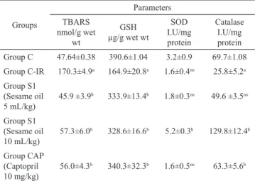
Table 1. Effect of chronic administration of Sesame oil on TBARS, GSH, SOD and catalase in rat heart homogenate.
Groups
Parameters
TBARS nmol/g wet
wt
GSH
µg/g wet wt
SOD
I.U/mg protein
Catalase I.U/mg protein
Group C 47.64±0.38 390.6±1.04 3.2±0.9 69.7±1.08
Group C-IR 170.3±4.9a 164.9±20.8a 1.6±0.4ns 25.8±5.2a Group S1
(Sesame oil 5 mL/kg)
45.9 ±3.9b 333.9±13.4b 1.8±0.3ns 49.6 ±3.5ns
Group S1
(Sesame oil 10 mL/kg)
57.3±6.0b 328.6±16.6b 5.2±0.3b 129.8±12.4b
Group CAP
(Captopril 10 mg/kg)
56.0±4.3b 340.3±32.3b 1.6±0.5ns 63.3±5.6b
All values are expressed as Mean±SEM [n=6] ap<0.001 vs Control [C] bp<0.001 vs C-IR; ns: Non signiicant.
Table 2. Effect of chronic administration of Sesame oil on LDH, CK and AST in rat heart homogenate.
Groups Parameters
LDH U/l CK U/l AST U/l
Group C 159±0.06 7.96±0.02 190±0.01
Group C-IR 72±0.03a 4.35±0.02a 82.34±0.23a
Group S1
(Sesame oil 5 mL/kg) 132±0.4
b 8.23±0.21b 112±0.32b
Group S1
(Sesame oil 10 mL/kg) 139±0.23
b 7.12±0.45b 161±0.92b
Group CAP
(Captopril 10 mg/kg) 121±0.76
b 6.98±0.5b 156±0.2b
All values are expressed as Mean±SEM [n=6] ap<0.001 vs Control [C] bp<0.001 vs C-IR [One way ANOVA].
Myocardial TBARS
There was signiicant (p<0.001) increase in
TBARS level in C-IR group when compared to the control
C. There was signiicant (p<0.001) decrease in the level of TBARS in groups S1, S2 and CAP in comparision to C-IR group.
Myocardial GSH
There was signiicant (p<0.001) decrease in
GSH level in C-IR group when compared to the control C. There was signiicant (p<0.001) increase in the level
of GSH in groups S1, S2 and CAP in comparison to C-IR
group.
Myocardial SOD
There was no signiicant change in SOD level in
C-IR group when compared to the control C. There was
no signiicant change in the level of SOD in group S1 and signiicant (p<0.001) increase in the level of SOD in
groups S2 and CAP in comparison to C-IR group.
Myocardial catalase
There was signiicant (p<0.001) decrease in catalase level in C-IR group when compared to the
control C. There was no signiicant change in the level of catalase in group S1 and signiicant (p<0.001) increase in
the level of SOD in groups S2 and CAP in comparison to
C-IR group.
Myocardial LDH, CK and AST
There was signiicant (p<0.001) decrease in
myocardial LDH, CK and AST level in C-IR group when compared to the control C. There was signiicant
(p<0.001) increase in the level of myocardial LDH, CK
and AST in groups S1, S2 and CAP in comparison to C-IR group.
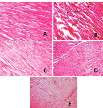
Histopathological study
Group C (Figure 1A): Light microscopy of the tissue sections of group C showed normal myoibrillar
structure with striations, branched appearance and
continuity with adjacent myoibrils.
Group C-IR (Figure 1B): Light microscopy of
the tissue sections of group C-IR showed edema, focal
haemorrhage and leukocyte iniltration. The muscle ibres
showed vascular changes with fragmentation suggestive of necrosis.
Group S1 (Figure 1C): Light microscopy of the
tissue sections of group S1 subjected to IR injury showed
minimal changes of myoibrillar structure with striations, and low level inlammation.
tissue sections of group S2 subjected to IR injury showed
normal myoibrillar structure with striations, branched appearance and continuity with adjacent myoibrils.
Group CAP (Figure 1E): Light microscopy of
the tissue sections of group CAP subjected to IR injury
showed normal architecture of myoibrillar structure with mild edema. The morphology of cardiac muscle ibres
was relatively well preserved.
Figure 1. A. Light microscopy section of group C rat myocardium showing well maintained myoibrillar structure. B. Light microscopy section of group C-IR rat myocardium showing extensive degeneration of myoibrils with leukocytic accumulation, edema and vacuolization. C. Light microscopy section of group S1 (Sesame oil 5 mL/kg) showing preserved myocardium. D. Light microscopy section of group S2 (Sesame oil 10 mL/kg) showing preserved myocardium. E. Light microscopy section of group CAP (Captopril 10 mg/kg) showing preserved myocardium.
Discussion
In-vitro myocardial ischemic reperfusion injury in rat heart model was carried out to evaluate the antioxidant and cardioprotective effect of sesame oil. In the present study ischemic reperfusion injury associated with oxidative stress, as evidenced by increased myocardial TBARS level as oxidative stress marker and depletion of myocardial endogenous antioxidant system
in C-IR group. Same indings have been observed earlier by other researchers in the same model (Krishenbaum &
Singal, 1993; Banerjee et al., 2002).
In the present study we observed that the oral chronic administration of sesame oil at two different
doses signiicantly decreases the myocardial TBARS
and concomitant increase in the levels of endogenous
antioxidants (GSH, SOD and catalase). Elevated levels
of myocardial TBARS is an indicative for increase in the oxidative stress during ischemia which might be stimulated the self protective mechanism via increasing in the level of myocardial endogenous antioxidant systems (Singal et al., 1993; Maulik et al., 1997).
Enhancement of basal endogenous antioxidant by any therapeutic alternative is presently the focus of
great scientiic interest (Maulik et al., 1997; Pathania
et al., 1998), as it is anticipated to produce better protection against oxidative stress than any exogenously administered antioxidants. In the present study, there
was a large increase in myocardial GSH, SOD and
catalase activities in group S2 (10 mL/kg-1), shows the
better activity augmented by sesame oil. It is particularly
important that both SOD and catalase were increased,
since it has been shown that an increase in cellular
SOD without a concomitant rise in catalase is more harmful by favoring the formation of H2O2 (Yim et al.,
1990; Jones et al., 1999). In this respect, the sesame oil may be a particularly useful agent, as it could enhance myocardial endogenous antioxidants, without producing any cytotoxic effects.
Another signiicant observation, made in the
present study was the protection against oxidative stress, associated with ischemic reperfusion injury in the hearts of the rats chronically treated with the sesame oil was generation of TBARS and depletion of endogenous antioxidants following ischemic reperfusion injury were
markedly inhibited in these hearts. Histopathological
evidence of myocardial injury following IR injuries in sesame oil and captopril treated rat was also absent. Much research evidence suggests that the protective role of sesame oil against oxidative stress through its
antioxidant mechanism (Kaur & Saini, 2000; Nakano et al., 2002; Hsu & Liu, 2004). It has been identiied that
presence of antioxidative principles like sesamin, sesamol and sesaminol might be a responsible components for its protective action (Suja et al., 2004). Therefore, the protection against myocardial IR injury in the treated rats is attributable to enhanced endogenous antioxidant activity.
To assess the extend of damage during ischemic reperfusion injury of myocardium we need to
analyze the speciic marker enzymes. The enzymes viz,
transaminases, creatine kinase and lactate dehydrogenase serve as sensitive indices to assess the severity of
myocardial injury (Sheela & Devi, 2000). In C-IR control rats, reduction in myocardial markers LDH, CK and AST in heart homogenate conirms the onset of myocardial injury (Ithayarasi & Devi, 1997). In the present study
chronic oral administration of sesame oil and captopril
caused signiicant change in cardiac markers LDH, CK
indicate sesame oil also shows protective action against the myocardial tissue damage during IR injury.
Further studies are required to investigate the
precise mechanisms for changes in antioxidant enzyme expression because of oxidant stress and administration
of drugs. Of particular interest will be the question of
whether these drugs operate by affecting the same or different regulatory mechanisms for the expression of an antioxidant enzyme gene.
Conclusion
Thus it is concluded that the chronic oral administration of the sesame oil in rat augments myocardial endogenous antioxidants, without causing any cellular injury. This offered protection against oxidative stress associated with myocardial ischemic reperfusion injury is dose dependent.
Acknowledgement
The irst author thanks to Management,
Annamacharya College of Pharmacy to provide the facilities to carryout the research work
References
Aebi H, Bergmeyer HU 1974. Methods of enzymatic analysis. 2nd ed. Chemic Academic Press Inc, Verlag, vol 2, p. 673-685.
Ahmad S, Khan MB, Hoda MN, Bhatia K, Haque R, Fazili IS, Jamal A, Khan JS, Katare DP 2011. Neuroprotective Effect of Sesame Seed Oil in 6-Hydroxydopamine Induced Neurotoxicity in Mice Model: Cellular, Biochemical and Neurochemical Evidence. Neurochem Res DOI: 10.1007/s11064-011-0638-4.
Banerjee SK, Dinda AK, Manchanda SC, Maulik SK 2002. Chronic garlic administration protects rat heart against oxidative stress induced by ischemic reperfusion injury. BMC Pharmacol 2: 16.
Bolli R 1998. Basic and clinical aspect of myocardial stunning. Prog Cardiovacs Dis 40: 477-516.
Borchgrevink PC, Schie R, Bergan AS, Bakoy OE, Jynge P 1989. Magnesium and reperfusion of ischemic rat heart as accessed by 31P NMR. Am J Physio 256: 11195-11204.
Bradford MM 1976. A rabid and sensitive method for the quantization of microgram quantities of protein utilizing the principle of protein-dye binding. Anal Biochem 772: 248-254.
Downey JM, Yellon DM 1992. Do free radicals contribute to myocardial cell death during ischemia-reperfusion? Myocardial protection. The pathophysiology of reperfusion and reperfusion injury. Yellon DM, Jennings RB (eds). Raven Press, New York: p. 35-57.
Ellman GL 1959. Tissue sulphydryl groups. Arch Biochem Biophy 82: 70-77.
Ferrari R, Ceconi C, Curello S, Cargnoni A, Pasini E, Visioli O 1991. The occurrence of oxidative stress during reperfusion in experimental animals and men. Cardiovasc Drug Ther 5: 277-288.
Fukuda Y 1990. Food chemical studies on the antioxidants in sesame seed. Nippon Shokuhin Kogyo Gakkaishi 37: 484-492.
Gallagher KP, Buda AJ, Pace D, Gerren RA, Shaler M 1986. Failure of superoxide dismutase and catalase to alter size of infarction in conscious dogs after 3 hours of occlusion followed by reperfusion. Circulation 73: 1065-1076
Gauthaman K, Maulik M, Kumari R, Manchanda SC, Dinda AK, Maulik SK 2001. Effect of chronic treatment with bark of Terminalia arjuna: A study on the isolated ischemic-reperfused rat heart. J Ethanopharmacol 75: 197-201.
Halliwell B 2000. The antioxidant paradox. Lancet 355: 1179-1180.
Harbone JB, Baxter HH 1993. Phytochemical Dictionary: A hand Book of Bioactive Compound from plants. Washington: Taylor and Francis; p. 237.
Hsu DZ, Liu MY 2004. Sesame oil protects against lipopolysaccharide-stimulated oxidative stress in rats. Crit Care Med 32: 227-231.
Ithayarasi PA, Devi CS 1997. Effect of α-tocopherol on isoproterenol induced changes in lipid and lipoprotein proile in rats. Ind J Pharmacol 29: 399-404.
Jones SA, McArdle F, Jack CI, Jackson MJ 1999. Effect of antioxidant supplementation on the adaptive response of human skin ibroblasts to UV-induced oxidative stress. Redox Rep 46: 291-299.
Kakkar P, Das B, Viswanathan PN 1984. A modiied spectrophotometric assay of superoxide dismutase. Ind J Biochem Biophy 21: 130-132
Kaur IP, Saini A 2000. Sesamol exhibits antimutagenic activity against oxygen species mediated mutagenicity. Mutat Res 470: 71-76.
King J 1959. A routine method for the estimation of lactic dehydrogenase activity. J Med Lab Tech 16: 265-272. Krishenbaum LA, Singal PK 1993. Increase in endogenous
antioxidant enzyme protects heart against reperfusion injury. Am J Physiol 265: H484-H493.
Kumar A, Khan SA, Parvez A, Zaheer MS, Rabbani MU, Zafar L 2011. The prevalence of hyperhomocysteinemia and its correlation with conventional risk factors in young patients with myocardial infarction in a tertiary care centre of India. Biomed Res 22: 225-229
Maulik G, Mukherjee S, Das DK 1997. Evaluation of antioxidant effectiveness of a new selected vegetable. Environ Nutr Interac 1: 287- 297.
isolated ischemic-reperfused heart. Mag Res 12: 37-42. Nakano D, Itoh C, Takaoka M, Kiso Y, Tanaka T, Matsumura
Y 2002. Antihypertensive effect of sesamin. IV. Inhibitionof vascular superoxide production by sesamin. Biol Pharmacol Bull 25: 1247-1249. Neely JR, Denton RM, England PJ, Randle PJ 1972. The effects
of increased heart work on the tricarboxylate cycle and its interactions with glycolysis in the perfused rat heart. Biochem J 1: 147-159.
Okhawa H, Oohishi N, Yagi K 1979. Assay for lipid peroxides in animal tissues by thiobarbituric acid reaction. Ann Bichem 95: 351-358.
Panwar RB, Gupta R, Gupta BK, Raja S, Vaishnav J, Khatri M, Agarwal A 2011. Atherothrombotic risk factors & premature coronary heart disease in India: A case-control study. Indian J Med Res 134: 26-32
Pathania V, Syal N, Hundal MH, Khanduja KL 1998. Geriforte stimulates antioxidant defense system. Ind J Exp Bio 36: 414-417.
Ramahi TM 2010. Cardiovascular disease in the Asia Middle East region: global trends and local implications. Asia Pac J Public Health 22: 83S-89S.
Saleem TSM, Gauthaman K 2009. Neutraceutical value of sesame oil. Phcog Rev 36: 264-269.
Saxena KK, Gupta B, Srivastava VK, Saxena RS, Singh RC, Prasad DN 1988. Creatine kinase and aspartate transaminase in experimental model to predict the size of cardiac infarct. Indian J Exp Biol 26: 235-236. Sheela SC, Devi CS 2000. Protective effect of Abana, a
polyherbal formulation, on isoproterenol induced
myocardial infarction in rats. Ind J Pharmacol 32: 198-201.
Singal PK, Dhalla AK, Hill M, Thomas TP 1993. Endogenous antioxidant changes in the myocardium in response to acute and chronic stress conditions. Mol Cell Biochem 129: 179-186.
Suja KP, Jayalekshmy A, Arumughan C 2004. Free radical scavenging behavior of antioxidant compounds of Sesame (Sesamum indicum L.) in DPPH system. J Agric Food Chem 52: 912-915.
Trease GE, Evans MC 1989. Text book of Pharmacognosy. London: Bailiere Tindall; p. 200-201, 340-348, 419-423, 626-630, 765-775.
Visweswaran P, Massin EK, Dubose TD 1997. Mannitol induced acute renal failure. J Am Soc Nephrol 8: 1028-1033 WHO 2010. World Health Statistics 2009. http://www.who.
int/whosis/whostat/2009/ en/index.html. accessed Feb 2010.
Yim MB, Chock PB, Stadtman ER 1990. Copper, Zinc super oxide Dismutase catalyzes hydroxyl radical production from hydrogen peroxide. Natl Acad Sci USA 8713: 5006-5010.
*Correspondence
T.S. Mohamed Saleem
Department of Pharmacology, Annamacharya College of Pharamacy
New Boyanapalli, Rajampet-516126, Andhrapradesh, India [email protected]