Extrace llular m atrix m o le cule s as
targe ts fo r bro wn spide r ve no m to xins
Departamentos de 1Biologia Celular, and 2Fisiologia,
Universidade Federal do Paraná, Curitiba, PR, Brasil S.S. Veiga1, V.C. Zanetti1,
A. Braz1, O .C. Mangili2
and W. Gremski1
Abstract
Loxoscelism, the term used to describe lesions and clinical manifesta-tions induced by brown spiders venom (Loxosceles genus), has attracted much attention over the last years. Brown spider bites have been reported to cause a local and acute inflammatory reaction that may evolve to dermonecrosis (a hallmark of envenomation) and hemorrhage at the bite site, besides systemic manifestations such as thrombocytopenia, disseminated intravascular coagulation, hemoly-sis, and renal failure. The molecular mechanisms by which Loxosceles venoms induce injury are currently under investigation. In this review, we focused on the latest reports describing the biological and physio-pathological aspects of loxoscelism, with reference mainly to the proteases recently described as metalloproteases and serine proteases, as well as on the proteolytic effects triggered by L. intermedia venom upon extracellular matrix constituents such as fibronectin, fibrinogen, entactin and heparan sulfate proteoglycan, besides the disruptive activity of the venom on Engelbreth-Holm-Swarm basement mem-branes. Degradation of these extracellular matrix molecules and the observed disruption of basement membranes could be related to deleterious activities of the venom such as loss of vessel and glomeru-lar integrity and spreading of the venom toxins to underlying tissues.
Co rre spo nde nce
S.S. Veiga
Departamento de Biologia Celular Universidade Federal do Paraná 81531-990 Curitiba, PR Brasil
Fax: + 55-41-266-2042 E-mail: veigass@ bio.ufpr.br
Research supported by CNPq, CAPES, Fundação Araucária-PR and FUNPAR-UFPR.
Received September 12, 2000 Accepted April 17, 2001
Ke y wo rds
·Brown spider
·Venom
·Extracellular matrix
·Proteolytic effect
Intro ductio n
Brown spiders (Loxosceles genus) have been reported to cause several clinical mani-festations. Envenomation provokes two ma-jor kinds of signals, i.e., local lesions at the bite site characterized by edema followed by vasodilatation, blood vessel degeneration, local hemorrhage and a significant cutane-ous tissue injury with gravitational spread-ing, that can exacerbate to necrotic skin ul-cers and degeneration (1-4), and systemic
effects that begin as a malaise and can be-come generalized, with hemolysis, thrombo-cytopenia, disseminated intravascular coagu-lation and renal failure. These clinical signs and toxicological effects appear to be phe-nomena similar for several Loxosceles spe-cies including the more studied L. reclusa, L. laeta, L. intermedia and L. gaucho species (4-8).
venoms cause their deleterious effects are currently under investigation, with putative explanations involving an indirect event, as is the case for endothelial cell-dependent neutrophil activation caused by the venoms and seemingly related to the dermonecrotic lesion (4,12-14). The presence of a sphingo-myelinase D-like enzyme (32-35 kDa) prob-ably associated with necrotic, hemolytic and thrombocytopenic activities triggered by the venoms has also been identified in differ-ent Loxosceles species (3,4,6,10,15,16). Other enzymes such as a hyaluronidase have been postulated to be a spreading factor during the lesions (4,17), and protease ac-tivities also appear to have some participa-tion in the noxious effects of the venoms (11,18-21). Adult plasma components ap-pear to be required for the deleterious effects of the venoms, since a purified putative der-monecrotic toxin diluted in neonate plasma or synthetic buffer did not induce platelet activation, an event that may be responsible for thrombosis, tissue ischemia and dermone-crosis (8). Indirectly, venoms also seem to cause injury by binding to cell membranes and activating the complement system of plasma (8,22,23) (see Table 1).
The key observations that some
physi-ological events closely dependent on the basement membrane and on connective or plasma extracellular matrix constituents are altered during loxoscelism, as is the case for platelet subendothelial adhesion and aggre-gation, hemostatic troubles such as dissemi-nated intravascular coagulation, hemorrhage into the dermis, renal failure and even cuta-neous tissue injury, point to the presence of molecules potentially deleterious to these structures in the venoms.
This review focuses on the specific de-grading effects triggered by Loxosceles ven-oms in extracellular matrix molecules and the biological consequences of these hydro-lytic activities related to the clinical signs induced by loxoscelism.
Pre se nce o f ge latino lytic e nzyme s in L. inte rm e d ia ve no m
Several extracellular matrix molecules have been described as targets for degrada-tion evoked by proteases present in snake venoms, which cause hemorrhage, necrosis and edema (24-28). Soluble plasma fibrino-gen is the major substrate in this family, but it is not the only one since snake venom actions on laminin, entactin, fibronectin and
Table 1. Brow n spider venom properties.
Venom effects M olecules involved References
Dermonecrotic lesion 32 kDa++, 35 kDa+, +++ 3,4,8,10,16,22
Intravascular hemolysis 32 kDa++, 35 kDa+ 4,6,16,22
Platelet aggregation and thrombocytopenia 32 kDa++, 35 kDa+ 4,8,15
Gravitational spreading 33 kDa++, 63 kDa++ 4,17
Fibrinogenolytic activity 20-28 kDa+ 19
Fibronectinolytic activity 20-28 kDa+ 19
Gelatinolytic activity 32-35 kDa+, 85 kDa+, 95 kDa+ 11,19,20
Entactinolytic activity Unknow n+ 21,57
Basement membrane-degrading effect Unknow n+ 21,57
Complement system activation 32 kDa++, 35 kDa+ 4,16,22,23
Heparan sulfate proteoglycan hydrolysis Unknow n+ 21
Dermonecrotic lesion, intravascular hemolysis, platelet aggregation and complement system activation are events dependent on the similar brow n spider toxins of 32-35 kDa devoid of proteolytic activities.
The 32-35-kDa molecule w ith gelatinolytic activity is a metalloprotease.
gelatin have been described (24,29). With respect to spider venoms and despite some similarities to snake venoms in their activi-ties, little is known about the presence of venom proteases that degrade extracellular matrix molecules. Studying L. intermedia venom (the prevalent brown spider in south-ern Brazil) we were able to detect a 32-35-kDa metalloprotease with gelatinolytic ac-tivity. The 35-kDa protease form seems to be a latent pro-enzyme molecule that under-goes cleavage, originating the 32-kDa form (19). The 32-35-kDa proteases are high-man-nose glycoproteins (20). We also detected zymogen molecules of proteolytic enzymes in the venom since trypsin activated two gelatinolytic serine proteases of 85 and 95 kDa in the venom. Other proteins such as casein, albumin, hemoglobin and laminin did not suffer any kind of cleavage. The specificity of action of these proteases should be related to spider self-protection. For ex-ample, the venom gland of L. intermedia is extremely rich in laminin, which separates muscle tissue (involved in venom secretion) from epithelial cells (involved in venom syn-thesis) (30), and the venom seems to have no lamininolytic activity (19,21). The natural substrate of the 32-35-kDa protease is un-known (since gelatin is denatured collagen and the venom does not display any activity on full length collagen), but, based on gelati-nolytic activity, we may assume that this protease has properties like vertebrate gelat-inases that appear to cleave connective com-ponents (31) and we may propose that this brown spider enzyme is functionally related to the deleterious effects of the venom. Na-tive collagen can suffer an initial effect of collagenase from polymorphonuclear neu-trophil leukocytes (which, as described above, are concentrated at the bite site and around it and seem to play a role in dermone-crosis), partially denaturating this molecule, which then can be sequentially degraded by these gelatinase-like venom proteases (see Table 1).
Effe ct o f Lo xo sce le s ve no m pro te ase s o n plasm a e xtrace llular m atrix m o le cule s
partici-pates in platelet attachment and aggregation and the proteolytic effect of the venom may disturb this event. Finally, the defective wound healing observed in some cases of envenomation may also be ascribed to the fibronectinolytic activity of the venom, since fibronectin-integrin interactions participate in an essential manner in this phenomenon (32). An identical hypothesis could be raised for the fibrinogenolytic ability of brown spi-der venom, since it has been well defined that several snake venom proteases with hem-orrhagic activities readily degrade fibrino-gen-fibrin into nonclotting fragments
(26,27,40). Disseminated intravascular co-agulation is a clinical feature detected in a few cases of brown spider envenomation. Venoms from various snakes that partially degrade fibrinogen provoke disseminated intravascular coagulation by fibrin forma-tion (25,28). As previously postulated by Bascur et al. (7) and corroborated by Feitosa et al. (19) and Veiga et al. (20), on the basis of partial fibrinogenolytic activity triggered by brown spider venoms, we may speculate that this ability is involved in disseminated intravascular coagulation, a complication that, together with other hematologic distur-bances, has been responsible for most of the deaths due to loxoscelism.
Fibrinogen is a large glycoprotein (340 kDa) formed by dimeric linking of three distinct chains, Aa, Bß and g, linked by disulfide bridges. As described above, fi-brinogen Aa chains are degraded by the venoms. It is interesting to observe that the a chains of fibrinogen have two RGD se-quences, a peptide well defined as a major binding site for several integrins (41). The cleaving of fibrinogen RGD sequences by the venoms could reduce the interaction of integrin aIIb ß3 on the platelet surface with fibrinogen, resulting in the loss of platelet adhesion and functional aggregation and contributing to the hemorrhagic effect of the venom. We may suggest this proteolysis as a conservative event for Loxosceles species, indicating some biological function related to the life cycle and envenomation of brown spiders (see Table 1).
Actio n o f bro wn spide r ve no m o n base m e nt m e m brane co nstitue nts
The basement membranes are ubiquitous specialized forms of extracellular matrices produced by several cell types such as endo-thelial, epiendo-thelial, fat, muscle and nervous cells. Molecularly they are characterized by the presence of a considerable variety of biochemically complex components arranged
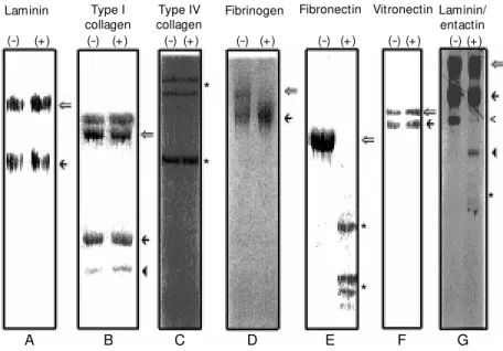
Figure 1. Proteolytic effect of Loxosceles intermedia venom on purified extracellular matrix molecules* . EHS-laminin (A), rat tail tendon type I collagen (B), human placental type IV collagen (C), human fibrinogen (D), human fibronectin (E), human vitronectin (F) and EHS-laminin/entactin dimer (G) w ere incubated w ith venom (+) or in the absence of venom, as controls for experimental stability (-) under the conditions described in References 19, 20, 21 and 57. The products obtained w ere analyzed by SDS-PAGE under reducing conditions (except for fibrinogen that w as analyzed under nonreducing conditions). In panel A the open arrow indicates the laminin a1 chain and the filled arrow indicates the ß1 and g1 laminin
chains that co-migrate. In panel B the open arrow indicates the type I collagen ß dimers, the filled arrow depicts a1 type I collagen chain and the filled arrow head indicates the a2 type I
collagen chain. In panel C the asterisks represent the major components of trypsin-extract-ed human placental type IV collagens of 100, 160 and 170 kDa. In panel D the open arrow points to an intact fibrinogen molecule and the filled arrow indicates the fibrinogen frag-ment. In panel E the open arrow show s the co-migratory fibronectin A and B chains, and asterisks indicate fibronectin fragments. In panel F the open arrow points to the 75-kDa vitronectin molecule and the filled arrow points to the 65-kDa normally processed vitronec-tin fragment. In panel G the open arrow indicates the laminin a1 chain, the filled arrow
indicates the ß1 and g1 laminin chains that co-migrate, the open arrow head indicates intact
entactin, the filled arrow head indicates the 100-kDa entactin fragment, and the asterisk show s the 50-kDa entactin fragment. * Based on results from References 19, 20, 21 and 57.
Laminin
(-) (+)
Type I collagen
(-) (+)
Type IV collagen (-) (+)
Fibrinogen
(-) (+)
Fibronectin
(-) (+)
Laminin/ entactin (-) (+) Vitronectin
(-) (+)
A
*
*
*
* *
as a network and formed by a particular set of proteins such as laminin, entactin, type IV collagen and heparan sulfate proteoglycan. Despite their tissue specificity (different iso-forms), these components are present on practically all tissue basement membrane structures. In mammals, basement mem-branes play several essential roles in angio-genesis, cell differentiation, platelet adhe-sion, neuritogenesis, and blood-urine filtra-tion through the kidney glomeruli, among several other functions (42-46). The effects of venom toxins on basement membranes have been well established for some snake venom hemorrhagic proteases that produce proteolytic degradation of basement mem-brane constituents isolated from Engelbreth-Holm-Swarm (EHS) tumor (29,40), as well as degradation of purified glomerular base-ment membrane (47). The pathogenic prop-erties of Loxosceles venoms, such as dermo-necrotic action, thrombocytopenic activity, local hemorrhage and renal disorders, are events attributable to the presence of pro-teolytic enzymes that degrade basement mem-brane molecules. By using the EHS tumor, a transplantable model which produces a char-acteristic and thick basement membrane as its capsule (48), we detected a disruptive activity of L. intermedia venom toward base-ment membrane structures. The venom ap-parently has no activity on laminin or type IV collagen (see Figure 1), the major macro-molecules of these structures, but has the ability to hydrolyze entactin (19,21), a dumb-bell-shaped molecule that links laminin, type IV collagen and heparan sulfate proteogly-can in the basement membrane organization (42,43). The hydrolytic activity of L. inter-media venom on entactin resulted in frag-ments of approximately 100 and 50 kDa when entactin was complexed to laminin and lower fragments that run out the gel when purified entactin was assayed (21). Similar results concerning this differential entactin susceptibility to the venom have been reported for other venoms such as snake
venom toxins (29,40). This difference in cleavage profile for purified entactin and for the laminin-entactin complex treated with L. intermedia venom can be explained by the fact that laminin interacting with entactin can hide domains of entactin more suscep-tible to the degrading action of the venom, as previously described for the entactinolytic effect evoked by atrolysins, metalloproteases from Crotalus atrox snake venom (40).
conse-quences of envenomation (see Table 1).
Co ncluding re m arks and future e xpe ctatio ns
The molecular mechanisms by which the brown spider venoms cause dermonecrotic injury, local and systemic hemorrhage, throm-bocytopenia, hemolysis, disseminated intra-vascular coagulation and renal failure are currently under investigation, but since the venom is composed of a mixture of several proteins, these mechanisms seem to be mo-lecularly complex and may be dependent on many different toxins.
In the present review, based on previous results obtained by our group (11,19-21,57), we introduce the idea of venom toxins acting as proteases upon molecular constituents of plasma extracellular matrix such as fibro-nectin and fibrinogen and basement mem-brane constituents such as entactin and hep-aran sulfate proteoglycan. Based on these degrading activities on plasma constituents and the disruptive effect on basement mem-brane structures triggered by venoms, we may speculate that these activities are plau-sible mechanisms for hemorrhage, delayed wound healing and renal failure and are also related to the spreading of other noxious toxins (for example, dermonecrotic protein), since disruption of the subendothelial base-ment membrane evokes blood vessel wall instability and increased permeability.
It is clear that more work is needed to elucidate the structure and function of these venom metalloproteases and to fully under-stand how they interfere with clinical signs of loxoscelism. However, it is interesting that all detected proteolytic activities and
especially the disruptive effect of L. inter-media venom on EHS-basement membranes can be inhibited by 1,10-phenanthroline, a metalloprotease inhibitor.
On the other hand, based on biodiversity, venom toxins are excellent tools for investi-gating molecular mechanisms in the cell bi-ology field, as well as for pharmaceutical applications of newly discovered medicines. In this area, venom toxins could be used as starting materials to design new drugs or directly for therapeutic use as is the case for ancrod, a defibrinogenating enzyme from Callocelasma rhodostoma venom, and ba-troxobin, a defibrinogenating enzyme from Bothrops atrox venom, toxins that have been used as defibrinogenating agents for a num-ber of clinical conditions such as deep vein thrombosis, myocardial infarction, pulmo-nary embolus, acute ischemic stroke, angina pectoris, central retinal vein occlusion and renal transplant rejection (27,58-60). With respect to the pharmaceutical applications of brown spider venom toxins, at least one possibility can emerge from our results. The fibrinogenolytic-fibrinolytic properties of venoms directly indicate that this toxin could be useful for clinical applications in cardiac and thrombotic diseases based on reduced blood fluidity. Other possibilities can di-rectly or indidi-rectly arise from brown spider venom toxins (for example, as a tool in cell biology procedures to study the inflamma-tory response). A molecular understanding of loxoscelism may greatly enhance our thera-peutic approach to this envenoming prob-lem, as well as generate potential toxins as tools for scientific protocols and medicinal applications.
Re fe re nce s
1. Atkins JA, Wingo CW, Sodeman WA & Flynn JE (1958). Necrotic arachnidism.
American Journal of Tropical M edicine and Hygiene, 7: 165-184.
2. Wasserman GS & Anderson PC (1984).
Loxoscelism and necrotic arachnidism.
Journal of Toxicology. Clinical Toxicology, 21: 451-472.
3. Babcock JL, Civello DJ & Geren CR (1981). Purification and characterization of
a toxin from brow n recluse spider ( Loxos-celes reclusa) venom gland extracts. Toxi-con, 19: 677-689.
5. Berger RS, Edw ard HA & Anderson PC (1973). Intravascular coagulation: The cause of necrotic arachnidism. Journal of Investigative Dermatology, 61: 142-150. 6. Forrester LJ, Barrett JT & Campbell BJ
(1978). Red blood cell lysis induced by the venom of the brow n recluse spider. The role of sphingomyelinase D. Archives of Biochemistry and Biophysics, 187: 355-365.
7. Bascur L, Yevenes I & Barja P (1982). Effects of Loxosceles laeta spider venom on blood coagulation. Toxicon, 20: 795-796.
8. Rees SR, Gates C, Timons S, Des-Pres RM & King Jr LE (1988). Plasma compo-nents are required for platelet activation by the toxin of Loxosceles reclusa. Toxi-con, 26: 1035-1046.
9. Norment BR, Jong Y-S & Heitz JR (1979). Separation and characterization of venom components in Loxosceles reclusa - III. Hydrolytic enzyme activity. Toxicon, 17: 539-548.
10. Barbaro KC, Cardoso JLC, Eickstedt VRD & M ota I (1992). Dermonecrotic and lethal components of Loxosceles gaucho spider venom. Toxicon, 30: 331-338.
11. Veiga SS, Da Silveira RB, Dreyfuss JL, Haoach J, Pereira AM , M angili OC & Gremski W (2000). Identification of high molecular w eight serine-proteases in Lo-xosceles intermedia (brow n spider) ven-om. Toxicon, 38: 825-839.
12. Smith WC & M icks WD (1970). The role of polymorphonuclear leukocytes in the lesion caused by the venom of the brow n spider, Loxosceles reclusa. Laboratory In-vestigation, 22: 90-93.
13. M ajeski JA, Stinnett JD, Alexander JW & Durst GG (1977). Action of venom from the brow n recluse spider (Loxosceles re-clusa) on human neutrophils. Toxicon, 15: 423-427.
14. Patel KD, M odur V, Zim m erm an GA, Prescott SM & M cIntyre TM (1994). The necrotic venom of brow n recluse spider induces dysregulated endothelial cell-de-pendent neutrophil activation. Journal of Clinical Investigation, 94: 631-642. 15. Kurpiew ski G, Forrester LJ, Barrett JT &
Campbell BJ (1981). Platelet aggregation and sphingomyelinase D activity of a puri-fied toxin from the venomof Loxosceles reclusa. Biochimica et Biophysica Acta, 678: 467-476.
16. Tambourgi DV, M agnoli FC, van den Berg CW, M organ BP, Araujo PS de, Alves EW & Dias da Silva W (1998). Sphingomyelin-ases in the venom of the spider Loxoscel-es intermedia are responsible for both
dermonecrosis and complement-depend-ent hemolysis. Biochemical and Biophysi-cal Research Communications, 251: 366-373.
17. W right RP, Elgert K, Cam pbell BJ & Barrett JT (1973). Hyaluronidase and es-terase activities of the venom of the poi-sonous brow n recluse spider. Archives of Biochemistry and Biophysics, 159: 415-426.
18. Jong Y-S, Norment BR & Heitz JR (1979). Separation and characterization of venom components in Loxosceles reclusa - II. Protease enzyme activity. Toxicon, 17: 529-537.
19. Feitosa L, Gremski W, Veiga SS, Elias M CQB, Graner E, M angili OC & Brentani RR (1998). Detection and characterization of metalloproteinases w ith gelatinolytic, fibronectinolytic and fibrinogenolytic ac-tivities in brow n spider (Loxosceles inter-media) venom. Toxicon, 36: 1039-1051. 20. Veiga SS, Gremski W, Santos VLP, Feitosa
L, M angili OC, Nader HB, Dietrich CP & Brentani RR (1999). Oligosaccharide resi-dues of Loxosceles intermedia (brow n spider) venom proteins: dependence on glycosylation for dermonecrotic activity.
Toxicon, 37: 587-607.
21. Veiga SS, Feitosa L, Santos VLP, de Souza GA, Ribeiro AS, M angili OC, Porcionatto M A, Nader HB, Dietrich CP, Brentani RR & Gremski W (2000). Effect of Loxosce-les intermedia (brow n spider) venom on basem ent m em brane structures. His-tochemical Journal, 32: 397-408. 22. Tambourgi DV, M agnoli FC, Eickstedt
VRD, Benedetti ZC, Petricevich VL & Silva WD (1995). Incorporation of a 35-kilodal-ton purified protein from Loxosceles in-termedia spider venom transforms human erythrocytes into activators of autologous complement alternative pathw ay. Journal of Immunology, 155: 4459-4466. 23. Tambourgi DV, M organ BP, de Andrade
RM G, M agnoli FC & van den Berg CW (2000). Loxosceles intermedia spider venomation induces activation of an en-dogenous metalloproteinase, resulting in cleavage of glycophorins from the eryth-rocyte surface and facilitating comple-ment-mediated lysis. Blood, 95: 683-691. 24. Bjarnason JB & Fox JW (1995). Snake venom metalloendopeptidases: reproly-sins. M ethods in Enzymology, 248: 345-368.
25. Beutler E, Lichtman M A, Coller BS & Kipps TJ (1995). Williams Hematology. 5th edn. M cGraw -Hill, New York.
26. Kamiguti AS, Hay CRM , Theakston RDG & Zuzel M (1996). Insights into the
mech-anism of hemorrhage caused by snake venom metalloproteinases. Toxicon, 34: 627-642.
27. M arkland FS (1998). Snake venoms and the hem ostatic system . Toxicon, 36: 1749-1800.
28. Lee GR, Foerster J, Lukens J, Paraskevas F, Greer JP & Rodgers GM (1999). Win-trobe’s Clinical Hematology. Williams & Wilkins, Baltimore.
29. Baramova EN, Shannon JD, Bjarnason JB & Fox JW (1989). Degradation of extracel-lular matrix proteins by haemorrhagic me-talloproteinases. Archives of Biochemis-try and Biophysics, 275: 63-71.
30. Santos VLP, Franco CRC, Viggiano RLL, da Silveira RB, Cantão M P, M angili OC, Veiga SS & Gremski W (2000). Structural and ultrastructural description of the ven-om gland of Loxosceles int erm edia
(brow n spider). Toxicon, 38: 265-285. 31. Birkedal-Hansen H, M oore WGI, Bodden
M K, W indsor LJ, Birkedal-Hansen B, DeCarlo A & Engler JA (1993). M atrix me-talloproteinases: A review . CRC Critical Review s in Oral Biology and M edicine, 4: 197-250.
32. Ruoslahti E (1988). Fibronectin and its re-ceptors. Annual Review of Biochemistry, 57: 375-413.
33. Felding-Haberm ann B & Cheresh DA (1993). Vitronectin and its receptors. Cur-rent Opinion in Cell Biology, 5: 864-868. 34. Veiga SS, Elias M CQB, Gremski W,
Por-cionatto M A, Nader HB & Brentani RR (1996). Detection of post-translational sulfation of a5ß1 integrin and its role in
integrin-fibronectin binding. Brazilian Jour-nal of M edical and Biologial Research, 29: 1235-1238.
35. Kost C, Stueber W, Ehrlich HJ, Pannekoek H & Preissner KT (1992). M apping of bind-ing sites for heparin, plasminogen activa-tor inhibiactiva-tor-1, and plasminogen to vitro-nectin’s heparin-binding region reveals a novel vitronectin-dependent feedback mechanism for the control of plasmin for-mation. Journal of Biological Chemistry, 267: 12098-12105.
36. De Boer HC, Preissner KT, Bouma BN & De Groot PG (1992). Binding of vitronec-tin-thrombin-antithrombin III complex to human endothelial cells is mediated by the heparin binding site of vitronectin.
Journal of Biological Chem istry, 267: 2264-2268.
section. Blood, 4: 929-938.
38. Ruggeri ZM (1992). Von Willebrand factor and fibrinogen. Current Opinion in Cell Biology, 5: 898-906.
39. Farrell DH, Thiagarajan P, Chung DW & David EW (1992). Role of fibrinogen a and
g chain sites in platelet aggregation. Pro-ceedings of the National Academy of Sci-ences, USA, 89: 10729-10732.
40. Fox JW & Bjarnason JB (1995). Atrolysins: metalloproteinases from Crotalus atrox
venom. M ethods in Enzymology, 248: 368-387.
41. Ruoslahti E & Pierschbater M D (1987). New perspectives in cell adhesion: RGD and integrins. Science, 238: 491-497. 42. Tim pl R, Paulsson M , Dziadek M &
Fujiw ara S (1987). Basement membranes.
M ethods in Enzymology, 145: 363-391. 43. Yurchenco P & Schittny J (1990). M
olecu-lar architecture of basement membranes.
FASEB Journal, 4: 1577-1590.
44. Farquhar M G (1991). The glomerular base-ment membrane: a selective macromo-lecular filter.In: Hay ED (Editor), Cell Biol-ogy of Extracellular M atrix.Plenum Press, New York.
45. Rohrbach DH & Timpl R (1993). M olecular and Cellular Aspects of Basement M em-branes. Academic Press, San Diego. 46. Trygvasson K (1993). The laminin family.
Current Opinion in Cell Biology, 5: 877-882.
47. Ohsaka A, Just M & Habermann E (1973). Action of snake venom hemorrhagic
prin-ciples on isolated glomerular basement m em brane. Biochim ica et Biophysica Acta, 323: 415-428.
48. Orkin RW, Gehron P, M cGoodw in EB, M artin GR, Valentine T & Sw arm R (1977). A murine tumor producing a matrix of basement membrane. Journal of Experi-mental M edicine, 145: 204-220. 49. Kjellén L & Lindahl U (1991).
Proteogly-cans: Structures and interactions. Annual Review of Biochemistry, 60: 443-475. 50. Roberts DD & M echam RP (1993).
Biol-ogy of Extracellular M atrix Series. Aca-demic Press Inc., San Diego.
51. Bernfield M , Kokenyesi R, Kato M , Hinkes M T, Spring J, Gallo RL & Lose EJ (1992). Biology of the syndecan: A family of trans-membrane heparan sulfate proteoglycan.
Annual Review of Cell Biology, 8: 365-393.
52. Veiga SS, Elias M CQB, Grem ski W , Porcionatto M A, Silva R, Nader HB & Brentani RR (1997). Post-translational modifications of a5 ß1 integrin by
gly-cosaminoglycan chains. The a5 ß1
inte-grin is a facultative proteoglycan. Journal of Biological Chem istry, 272: 12529-12535.
53. Elias M CQB, Veiga SS, Gremski W, Por-cionatto M A, Nader HB & Brentani RR (1999). Presence of a laminin-binding chondroitin sulfate proteoglycan at the cell surface of a human melanoma cell M el-85. M olecular and Cellular Biochem-istry, 197: 39-48.
54. Dietrich CP (1984). A model for cell-cell recognition and control of cell grow th mediated by sulfated glycosaminogly-cans. Brazilian Journal of M edical and Bio-logical Research, 17: 5-15.
55. Ruoslahti E (1989). Proteoglycans in cell regulation. Journal of Biological Chemis-try, 264: 13369-13372.
56. Buonassisi V & Venter JC (1976). Hor-mone and neurotransmitter receptors in an established vascular endothelial cell line. Proceedings of the National Acade-my of Sciences,USA, 73: 1612-1616. 57. de Souza GA, Ribeiro AS, Santos VLP,
Veiga SS, M angili OC & Grem ski W (1998). Proteolytic effect of Loxosceles intermedia (brow n spider) venom proteins on EHS-basement membrane structures.
Acta Biologica Paranaense, 27: 97-109. 58. Bell WR (1990). Defibrinogenating
en-zymes. In: Colman RW, Hirsh J, M arder VJ & Salzman EW (Editors), Hemostasis and Thrombosis. Lippincott, Philadelphia, 886-900.
59. Furukaw a K & Ishimaru S (1990). Use of thrombin-like snake venom enzymes in the treatment of vascular occlusive dis-eases. In: Stocker KF (Editor), M edical Use of Snake Venom Proteins. CRC Press, Boca Raton, 161-173.