Full paper published online: February 28, 2010 ISSN 1678-9199.
Identification and partial purification of an anticoagulant factor from the venom
of the Iranian snake Agkistrodon halys
Ghorbanpur M (1), Zare Mirakabadi A (2), Zokaee F (1), Zolfagarrian H (2)
(1) Chemical Engineering Department, Amirkabir University, Tehran, Iran; (2)
Department of Venomous Animals and Antivenom Production, Razi Vaccine and
Serum Research Institute, Karaj, Iran.
ABSTRACT: An anticoagulant factor was purified from the venom of the Iranian
snake Agkistrodon halys by gel filtration on Sephadex G-50 and ion-exchange
chromatography on DEAE-Sepharose. In the final stage of purification, the
percentage recovery of purified anticoagulant factor was found to be 83%. The
purified anticoagulant factor revealed a single protein band in SDS-polyacrylamide
electrophoresis under reducing conditions and its molecular weight was about 22
kDa. The purified peptide did not show any effect on casein, BApNA or plasma.
KEY WORDS: snake venom, Agkistrodon halys, anticoagulant factor,
chromatography.
CONFLICTS OF INTEREST: There is no conflict.
CORRESPONDENCE TO:
ABBAS ZARE MIRAKABADI, Department of Venomous Animals and Antivenom
Production, Razi Vaccine and Serum Research Institute, Karaj, Iran. Phone: +98 261
INTRODUCTION
Snake venoms are rich sources of pharmacologically active proteins and peptides.
They play an important role in incapacitating, immobilizing and digesting prey. Thus
toxins have evolved to specifically target various critical points in the physiological
systems of prey. Over the years, a number of toxins that affect blood circulation have
been isolated and characterized from various snake venoms (1, 2). Within each
family of snakes, the venom components seem to be fairly common and similar to
one another.
Nerve toxins are generally found in the Hydrophidae and Elapidae venoms whereas
hemorrhagic and myonecrotic toxins are generally found in the venoms of the
Viperidae and Crotalidae families of snakes (3).
Snake venom toxins affecting hemostasis have been classified by virtue of their
overall effect into procoagulant and anticoagulant ones (2-4). Snake venoms have
different types of anticoagulant proteins, some of which have enzymatic activity,
represented by phospholipase A2, metalloproteinases like α-fibrinogenase, serine
proteinases and L-Amino acid oxidase. But others (C-type lectin-related proteins and
three-finger toxins) do not show any enzymatic activity (1, 3-5). Phospholipases A2
are esterolytic enzymes that hydrolyze acyl-ester bonds of
1,2-diacyl-3-sn-phosphoglycerides and release fatty acids (3, 6, 7). Snake venoms contain a number
of serine and metalloproteinases including fibrino(geno)lytic enzymes (8, 9).
Thrombin-like enzymes and fibrinolytic enzymes, which act on fibrinogen, lead to
defibrinogenation of blood and a consequent decrease in blood viscosity. Fibrinolytic
enzymes have been reported in species of the Crotalidae and Viperidae families
including Agkistrodon contortrix contotrix and Agkistrodon acutus (10-12). L-amino
acid oxidases are flavoenzymes that catalyze the stereospecific oxidative
deamination of an L-amino acid substrate to a corresponding a-ketoacid by the
production of ammonia and hydrogen peroxide (13).
In this study, we report the purification and characterization of an anticoagulation
protein from the venom of the Iranian snake species Agkistrodon halys that exhibits
MATERIALS AND METHODS
Material
Fresh crude venom of A. halys was obtained directly from a local snake in Iran,
lyophilized and preserved at –20ºC. DEAE-Sepharose, Sephadex G-50 and C18
columns were purchased from Pharmacia Biotech Company (Sweden). Bovine
serum albumin, BApNA (N2-benzoyl-dl-arginine-p-nitroanilide), the kit of standard
protein markers and other reagents for enzymatic and biochemical assays were
purchased from Sigma (USA). All other chemicals were of analytical reagent grade.
Isolation of the Thrombin-Like Enzyme
Lyophilized crude venom of A. halys (200 mg) was dissolved in 8 mL of 50 mM
ammonium acetate buffer (pH 7.4) and centrifuged at 5,000 rpm for 15 minutes at
4ºC and was filtered by 0.45 microfilter to remove the insoluble materials.
The clear supernatant was applied on a molecular exclusion chromatographic
column of Sephadex G-50 (150 x 3 cm), previously equilibrated with the ammonium
acetate buffer (pH 7.4) and then eluted with the same buffer. Fractions of 9 mL/tube
were collected at a flow rate of 60 mL/hour at 4ºC. The obtained fractions were
denominated AH1 to AH5, indicating A. halys fractions 1 to 5.
The fraction (AH2), which showed clotting activity from the gel chromatography step,
was pooled and dialyzed overnight at 4°C against distilled water and applied on
DEAE-Sepharose CL-6B (2.5 x 20 cm) column, equilibrated with 20 mM Tris buffer,
at pH 8.2. In this step, proteins were eluted with a linear gradient of NaCl from 0.0 to
0.5 mM. The flow rate was 17 mL/hour and 5 mL fractions were collected at 4ºC. The
peaks were monitored at A280.
Blood Collection
Normal pooled plasma was obtained from ten individual healthy donors, without
history of bleeding or thrombosis. Blood was centrifuged for 20 minutes at 2,400 g,
and the plasma was fresh when used.
Determination of Molecular Weight
12% SDS-PAGE was performed and utilized a low molecular weight standard
Purity Analysis
Sample aliquots (100 μL) applied on an HPLC C18 column, were equilibrated with
solvent A (H2O, 0.1% trifluoroacetic acid), and eluted with a concentration gradient of
solvent B (acetonitrile, 0.1% trifluoroacetic acid) from 0 to 30%, at a flow rate of 0.5
mL/minute for 30 minutes.
Prothrombin Time (PT) Assay
Prothrombin time reagent (200 μL) and sample aliquots (200 μL) were pre-incubated
for ten minutes at 37ºC and mixed for five seconds and then 100 μL of plasma was
added and clotting time was recorded (13). One unit of anticoagulant activity
corresponds to an increase of 20 seconds in normal plasma coagulation.
Amidolytic Activity
Proteolytic activity in the samples was assayed using BApNA as substrate. The
substrate solution was prepared by dissolving BApNA in 5 mL of dimethyl methyl
sulfoxide (DMSO) and adding 95 mL of 0.05 M Tris-HCl buffer (pH 8.2). Proteolytic
activity was monitored as BApNA hydrolysis by mixing 50 μL of sample with 100 μL
of the substrate solution. After ten minutes of incubation at 37ºC, the absorbance at
410 nm was measured. One unit of protease activity corresponds to an increase of
A410 = 0.001/minute (9).
Coagulant Activity
Plasma (200 μL) and different amounts of purified factor were pre-incubated briefly at
37ºC, then mixed and shaken, and the clotting times were recorded (3, 8, 14).
Proteolytic Activity on Casein
Proteolytic activity was determined by addition of various amounts of the enzyme to a
buffer solution of 0.1 M Tris-HCl, pH 9.0, and the final volume was adjusted to 250
μL, followed by the addition of 750 μL of 1% (m/v) casein and incubated for 15 minutes at 37ºC. The reaction was stopped by adding 1.5 mL of 30% TCA. The
resulting proteolysis products in the supernatant solution were evaluated
spectrophotometrically at λ = 280 nm after centrifugation at 5000 rpm for ten minutes.
Protein Determination
Protein concentration was measured by the method of Lowry et al. (15), using bovine
serum albumin (BSA) as standard.
RESULTS
Selection of Snake Species
Crude venom from the Iranian snake species E. carinatus and A. halys were selected
and assayed with PT test. The E. carinatus venom coagulated plasma very rapidly,
which leads to the conclusion that E. carinatus venom contains procoagulant factors.
Crude venom of Agkistrodon halys was selected next: this venom delayed plasma
coagulation. Thus it was concluded that the A. halys snake venom contains one or
more anticoagulant factors.
Isolation of Anticoagulant Factor
In the initial Sephadex G-50 fractionation of the crude Agkistrodon halys venom, five
peaks at 280 nm were obtained (AH1 to AH5) as shown in Figure 1. When all the
fractions were tested for anticoagulation, it was found that fraction AH2 showed
anticoagulation effect. Further purification was carried out by ion exchange
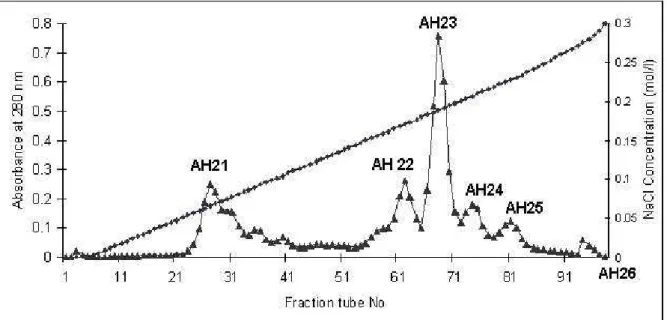
chromatography on DEAE-Sepharose resin (Figure 2). In this purification step six
fractions were obtained (AH21-AH26) out of which fraction AH21 showed
anticoagulant activity. By this purification procedure, about 16.75 mg of purified
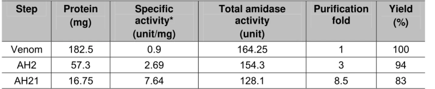
enzyme was obtained from 182.5 mg of the venom (Table 1).
Table 1. Purification of an anticoagulant from A. halys venom
Step Protein ) mg (
Specific activity* ) unit/mg (
Total amidase activity
(unit)
Purification fold
Yield (%)
Venom 182.5 0.9 164.25 1 100
AH2 57.3 2.69 154.3 3 94
AH21 16.75 7.64 128.1 8.5 83
Figure 1. Sephadex G-50 chromatography of Iranian Agkistrodon halys. Crude
venom (182.5 mg) was applied to Sephadex G-50 column (3 x 150 cm) using
ammonium acetate buffer with molarity 50 mM and pH 7.5. Flow rate was 60 mL/hour
and 9 mL fractions were collected from each tube.
Figure 2. DEAE-Sepharose chromatography of AH2 obtained from Sephadex G-50.
The pooled AH2 anticoagulant fraction was applied to DEAE-Sepharose column (2.5
x 20 cm) equilibrated with 0.05 M Tris-HCl buffer (pH 8.2). Proteins were eluted with
a linear concentration gradient of NaCl from 0.0 to 0.5 M, and 4.5 mL fractions were
collected from each tube. Series 1: NaCl concentration (mol/L) and series 2:
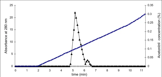
Purity and Determination of Molecular Weight
The homogeneity of purified enzyme was confirmed by SDS-PAGE and HPLC as
shown in Figures 3 and 4, respectively. Isolated anticoagulant factor (AH21) showed
high purity as analyzed by C18 reverse phase HPLC. This factor also showed a
single band in SDS-PAGE. The molecular weight of this anticoagulant factor was
estimated to be about 22 kDa under reduced conditions.
0 5 10 15 20 25
0 1 2 3 4 5 6 7 8 9 10 11
time (min) A b s o rb an c e at 2 8 0 nm 0 0.05 0.1 0.15 0.2 0.25 0.3 0.35 A c et o n it ri
l c
o nc en tr at io n (% )
Figure 3. Purity assay of anticoagulant factor by HPLC. Series 1: acetonitril
concentration (%) and series 2: absorbance at 280 nm.
A B C
Figure 4. SDS-PAGE of crude venom of (A) Agkistrodon halys, (B) AH2 and (C)
AH21, analyzed on 12.5% SDS-polyacrylamide gel in the presence of 1%
Caseinolytic Clotting Assay and Amidolytic Activity
AH21 did not exert any proteolytic activity on casein, coagulant activity on BAPNA or
clotting activity upon human plasma.
DISCUSSION
Venoms of Colubridae snakes are a rich source of novel compounds that may have
applications in medicine and biochemistry (16).This article reports a relatively simple
procedure for the isolation of an anticoagulant factor from Iranian Agkistrodon halys
venom, denominated AH22, which was isolated and purified by a combination of gel
filtration on Sephadex G-50 (Figure 1) and ion-exchange chromatography on
DEAE-Sepharose (Figure 2). By this procedure the total enzyme purification yield was about
83% (Table 1), thus enabling the conclusion that the protocol utilized was highly
efficient. Fractionation and purification of snake venom constituents have been
carried out through several chromatography methods (6-8, 16). An acidic
phospholipase A2 was purified from Agkistrodon halys pallas venom by a two-step
procedure comprised of gel filtration chromatography on Sephadex G-100 and ion
exchange chromatography on DEAE Sephadex A-50 (17). Another phospholipase A2
from B. leucurus venom was purified by a three-step procedure involving gel filtration
on Sephacryl S-200, ion exchange chromatography on Q-Sepharose and reverse
phase HPLC on Vydac C4 column (6).
By using the method described herein, about 16.75 mg of anticoagulant factor was
obtained from 182.5 mg of the venom of Agkistrodon halys (equal to 9.2% of total
venom protein). Therefore; it appears that the venom of the Iranian snake
Agkistrodon halys causes blood coagulation and, in this manner, may cause some
hemostatic disorders in the victim. The amount of this anticoagulant is quite high and
comprises four percent of the entire venom (6).
Snake venom toxins that prolong blood coagulation are proteins or glycoproteins with
molecular masses ranging from 6 to 350 kDa (1). The molecular weight of the factor
under denaturing conditions was estimated to be 22 kDa. Thus, this anticoagulant
factor should belong to the low-molecular-weight group of these factors; some
anticoagulant factors, along with their molecular weights, reported in the literature
are: l-amino acid oxidase from Agkistrodon blomhoffiiussurensis weighing 108.8 kDa
29 kDa (19); and metalloproteinase from Philodryas patagoniensis presenting 53 kDa
(5).
Venoms from snake species belonging to the genus Agkistrodon (A. contortrix
contortrix, A. c. mokasen, A. c. pictigaster, A. piscivorus, A. p. leucostoma, A. halys
halys, A. blomhoffi ussuriensis and A. bilineatus) contain protein C activators. They
are glycoproteins with a molecular mass ranging from 36 to 40 kDa. On account of its
molecular mass (22 kDa), we concluded that the anticoagulant obtained in the
present work does not belong to this low-molecular-weight group.
As described in the Introduction section, snake venom toxins that prolong blood
coagulation are proteins or glycoproteins that inhibit blood coagulation by different
mechanisms. Some of these anticoagulant proteins such as phospholipase A2,
metalloproteinases (α-fibrinogenase) and serine proteases exhibit enzymatic
activities, whereas others including C-type lectin-related proteins and three-finger
toxins do not exhibit any enzymatic activity (1, 5). We also isolated two serine
proteases, namely AH143 and AH144, from Agkistrodon halys venom (under
publishing).
This purified anticoagulant factor did not show any effect upon casein, BAPNA or
human plasma, and thus appears to be another type of anticoagulant protein rather
than protease. We also carried out a simple test on this factor by using red blood
cells in which no effect was observed (not reported), probably indicating that this
factor is not a phospholipase A2
CONCLUSION
An anticoagulant factor was purified from the venom of Agkistrodon halys. In the final
stage of purification, 83% recovery was obtained. The methods applied in the
purification were suitable and allowed a high degree of toxin recovery. The
anticoagulant factor isolated in the present work was not characterized as protease,
REFERENCES
1. Manjunatha Kini R. Anticoagulant proteins from snake venoms: structure, function
and mechanism. Biochem J. 2006;397(Pt 3):377-87.
2. Manjunatha Kini R, Rao VS, Joseph JS. Procoagulant proteins from snake
venoms. Haemostasis. 2001;31(3-6):218-24.
3. Matsui T, Fujimura Y, Titani K. Snake venom proteases affecting hemostasis and
thrombosis. Biochim Biophys Acta. 2000;1477(1-2):146-56.
4. Koh DCI, Armugam A, Jeyaseelan K. Snake venom components and their
applications in biomedicine. Cell Mol Life Sci. 2006;63(24):3030-41.
5. Kini RM. Serine proteases affecting blood coagulation and fibrinolysis from snake
venoms. Pathophysiol Haemost Thromb. 2005;34(4-5):200-4.
6. Higuchi DA, Barbosa CMW, Bincoletto C, Chagas JR, Magalhaes A,
Richardson M, et al. Purification and partial characterization of two phospholipases
A2 from Bothrops leucurus (white-tailed jararaca) snake venom. Biochimie.
2007;89(3):319-28.
7. Huang P, Mackessy SP. Biochemical characterization of phospholipase A2
(trimorphin) from the venom of the Sonoran lyre snake Trimorphodon biscutatus
lambda (family Colubridae). Toxicon. 2004;44(1):27-36.
8. Oyama E, Takahashi H. Purification and characterization of a thrombin-like
enzyme, elegaxobin II, with lys-bradykinin releasing activity from the venom of
Trimeresurus elegans (Sakishima-Habu). Toxicon. 2003;41(5):559-68.
9. Sant’Ana CD, Ticli FK, Oliveira LL, Giglio JR, Rechia CG, Fuly AL, et al.
BjussuSP-I: a new thrombin-like enzyme isolated from Bothrops jararacussu snake venom.
Comp Biochem Physiol A Mol Integr Physiol. 2008;151(3):443-54.
10. Chen HM, Guan AL, Markland Jr FS. Immunological properties of the fibrinolytic
enzyme (fibrolase) from southern copperhead (Agkistrodon contortrix contortrix)
venom and its purification by immunoaffinity chromatography. Toxicon.
1991;29(6):683-94.
11. Retzios AD, Markland Jr FS. A direct-acting fibrinolytic enzyme from the venom of
Agkistrodon contortrix contortrix: effects on various components of the human blood
coagulation and fibrinolysis systems. Thromb Res. 1988;52(6):541-2.
12. Ouyang C, Huang TF. The properties of the purified fibrinolytic principle from
13. Ande SR, Kommoju PR, Draxl S, Murkovic M, Macheroux P, Ghisla S, et al.
Mechanisms of cell death induction by L-amino acid oxidase, a major component of
ophidian venom. Apoptosis. 2006;11(8):1439-51.
14. Castro HC, Zingali RB, Albuquerque MG, Pujol-Luz M, Rodrigue CR. Snake
venom thrombin-like enzymes: from reptilase to now. CMLS. Cell Mol Life Sci.
2004;61(7-8):843-56.
15. Lowry OH, Rosebrough NJ, Farr AL, Randall RJ. Protein measurement with the
Folin phenol reagent. J Biol Chem. 1951;193(1):265-75.
16. Peichoto ME, Teibler P, Mackessy SP, Leiva L, Acosta O, Gonçalves LR, et al.
Purification and characterization of patagonfibrase, a metalloproteinase showing α
-fibrinogenolytic and hemorrhagic activities, from Philodryas patagoniensis snake
venom. Biochim Biophys Acta. 2007;1770(5):810-9.
17. Wang Y, Cui G, Zhao M, Yang J, Wang C, Giese RW, et al. Bioassay-directed
purification of an acidic phospholipase A2 from Agkistrodon halys pallas venom.
Toxicon. 2008;51(7):1131-9.
18. Wei XL, Wei JF, Li T, Qiao LY, Liu YL, Huang T, et al. Purification,
characterization and potent lung lesion activity of an L-amino acid oxidase from
Agkistrodon blomhoffii ussurensis snake venom. Toxicon. 2007;50(8):1126-39.
19. Koo BH, Sohn YD, Hwang KC, Jang Y, Kim DS, Chung KH. Characterization and
cDNA cloning of halyxin, a heterogeneous three-chain anticoagulant protein from the