7 Radiol Bras. 2013 Jan/Fev;46(1):7–14
Myocardial perfusion scintigraphy in the detection of silent
ischemia in asymptomatic diabetic patients
*
Cintilografia de perfusão miocárdica na detecção da isquemia silenciosa em pacientes diabéticos assintomáticos
Gláucia Celeste Rossatto Oki1, Elizabeth João Pavin2, Otávio Rizzi Coelho3, Maria Cândida R. Parisi2, Raitany C. Almeida4, Elba Cristina Sá de Camargo Etchebehere5, Edwaldo Eduardo Camargo6, Celso Dario Ramos7
Objective: This study was aimed to evaluate myocardial perfusion in asymptomatic patients with type 1 (DM1) and type 2 diabetes mellitus (DM2) without previous diagnoses of coronary artery disease (CAD) or cerebral infarction. Materials and Methods: Fifty-nine consecutive asymptomatic patients (16 DM1, 43 DM2) underwent myocardial perfusion scintigraphy with 99mTc-sestamibi (MPS). They were evaluated for body mass index, metabolic control of DM,
type of therapy, systemic arterial hypertension, dyslipidemia, nephropathy, retinopathy, peripheral neuropathy, smoking, and familial history of CAD. Results: MPS was abnormal in 15 patients (25.4%): 12 (20.3%) with perfusion abnormalities, and 3 with isolated left ventricular dysfunction. The strongest predictors for abnormal myocardial perfusion were: age 60 years and above (p = 0.017; odds ratio [OR] = 6.0), peripheral neuropathy (p = 0.028; OR = 6.1), nephropathy (p = 0.031; OR = 5.6), and stress ECG positive for ischemia (p = 0.049; OR = 4.08). Conclusion: Silent myocardial ischemia occurs in more than one in five asymptomatic diabetic patients. The strongest predictors of ischemia in this study were: patient age, peripheral neuropathy, nephropathy, retinopathy and a stress ECG positive for ischemia. Keywords: Diabetes mellitus; Myocardial perfusion imaging; Silent myocardial ischemia; Coronary artery disease.
Objetivo: Este estudo teve por finalidade avaliar a perfusão miocárdica de pacientes com diabetes mellitus tipo 1 (DM1) e tipo 2 (DM2) assintomáticos, sem diagnóstico prévio de doença arterial coronariana (DAC) ou acidente vas-cular cerebral. Materiais e Métodos: Cinquenta e nove pacientes consecutivos (16 DM1, 43 DM2) foram submeti-dos a cintilografia de perfusão miocárdica com sestamibi-99mTc (CPM). Foram avaliados quanto ao índice de massa
corpórea, controle metabólico do diabetes, dislipidemia, terapia para o diabetes, hipertensão arterial sistêmica, nefro-patia, retinonefro-patia, neuropatia periférica, tabagismo e história familiar de DAC. Resultados: CPM foi anormal em 25,4%: 12 (20,3%) com alterações de perfusão e 3 com disfunção ventricular esquerda isolada. Os mais fortes preditores de perfusão miocárdica anormal foram: idade igual ou maior a 60 anos (p = 0,017, odds ratio [OR] = 6,0), neuropatia periférica (p = 0,028, OR = 6,1), nefropatia (p = 0,031, OR = 5,6) e ECG de esforço positivo para isquemia (p = 0,049, OR = 4,08). Conclusão: A isquemia miocárdica silenciosa ocorre em mais de um em cada cinco diabéticos assintomáticos. Os mais fortes preditores de isquemia foram: idade avançada, neuropatia periférica, nefropatia, reti-nopatia e ECG de esforço positivo para isquemia.
Unitermos: Diabetes mellitus; Cintilografia de perfusão miocárdica; Isquemia miocárdica silenciosa; Doença arterial coronariana.
Abstract
Resumo
* Study developed at the Division of Nuclear Medicine, De-partment of Radiology, and Divisions of Endocrinology and Car-diology, Department of Internal Medicine, School of Medical Sciences, Universidade Estadual de Campinas (Unicamp), Cam-pinas, SP, Brazil.
1. Master, Physician Assistant at Serviços de Medicina Nuclear, Clínica Diagnoson and Hospital Aristides Maltez, Salvador, BA, Brazil.
2. PhDs, Physicians Assistant at Service of Endocrinology, Department of Internal Medicine, Universidade Estadual de Campinas (Unicamp), Campinas, SP, Brazil.
3. PhD, MD, Coordinator for the Service of Cardiology, Depart-ment of Internal Medicine, Universidade Estadual de Campinas (Unicamp), Campinas, SP, Brazil.
4. Master, Physician at the Service of Cardiology, Department of Internal Medicine, Universidade Estadual de Campinas (Uni-camp), Campinas, SP, Brazil.
Oki GCR, Pavin EJ, Coelho OR, Parisi MCR, Almeida RC, Etchebehere ECSC, Camargo EE, Ramos CD. Myocardial perfusion scintigra-phy in the detection of silent ischemia in asymptomatic diabetic patients. Radiol Bras. 2013 Jan/Fev;46(1):7–14.
INTRODUCTION
Coronary artery disease (CAD) is the leading cause of mortality in diabetic pa-tients. In these individuals, CAD is usually more advanced at the time of diagnosis and frequently presents an unfavorable progno-sis. In addition, patients with diabetes have a high incidence of occult CAD, observed as an increased occurrence of silent myo-cardial infarction (SMI) and not typically associated with angina, probably because
5. PhD, Physician Assistant at the Service of Nuclear Medi-cine, Department of Radiology, Universidade Estadual de Cam-pinas (Unicamp), CamCam-pinas, SP, Brazil.
6. PhD, MD, Coordinator for the Service of Nuclear Medicine, Hospital Sírio-Libanês, Campinas, SP, Brazil.
7. PhD, MD, Coordinator for the Service of Nuclear Medicine, Department of Radiology, Universidade Estadual de Campinas (Unicamp), Campinas, SP, Brazil.
Corresponding author: Dr. Celso Dario Ramos. Avenida Vital Brasil, 251, Cidade Universitária “Zeferino Vaz”, Barão Geraldo. Caixa Postal 6142, Campinas, SP, 13083-970, Brazil. E-mail: [email protected]
of diabetic autonomic neuropathy(1,2).
These factors have raised the interest in investigating CAD before its first clinical expression in diabetic patients and non-invasive tests have been advocated as an essential tool for early detection of the dis-ease(3).
In general, individuals with diabetes have at least a twofold to fourfold increased risk for cardiovascular events when com-pared with age-matched subjects without diabetes. It is estimated that SMI preva-lence in this group ranges from 10% to 20% versus from 1% to 4% estimated for non diabetic populations(4). After myocar-dial infarction, diabetic patients have a twofold to threefold greater morbidity and mortality. The high risk of CAD in these patients took the American Heart Associa-tion to not only determine diabetes as a major independent risk factor for the devel-opment of CAD as to establish that diabe-tes is a cardiovascular disease(5).
Asymptomatic diabetic patients with in-creased risk for CAD can benefit of myo-cardial perfusion scintigraphy with 99m
Tc-sestamibi (MPS) because this scintigraphic test cost-effectively measures and stratifies risk. Therefore this test is a good alterna-tive to cineangiocoronariography or tread-mill test(6). Even so, many authors have
been using cineangiocoronariography as gold standard for CAD(3,7). However, those
studies do not take into account the mi-crovascular pathophysiology of diabetes mellitus for which cineangiocoronariogra-phy and stress electrocardiogram (ECG) have a lower sensitivity(5). Indeed, there is
consistent evidence that diabetes causes al-terations in the regulation of coronary va-sodilator function in both epicardial and re-sistance coronary vessels, which are present before the appearance of obstruc-tive CAD(8). Thus, functional imaging can
more accurately identify endothelial cell dysfunction within coronary microvascu-lature among diabetic patients.
Considering that MPS is relatively ex-pensive to be used as a screening test, it is necessary to select subgroups of diabetic patients who present an increased risk for CAD, and that will effectively benefit of systematic screening for myocardial perfu-sion abnormalities. This study was aimed to evaluate myocardial perfusion in
dia-betic patients type 1 (DM1) and type 2 (DM2) without previous diagnoses or symptoms of CAD and cerebral infarction, as well as to evaluate clinical and labora-tory predictors of abnormal test results.
MATERIALS AND METHODS
The study was approved by the ethics committee of the institution and all patients signed a written consent form. The authors do not have any conflict of interest in this project.
During a 12-month period, consecutive patients who met the inclusion criteria were invited by endocrinologists during their clinical appointments to participate in the protocol. Fifty-nine patients with the diag-nosis of DM (16 type 1 and 43 type 2) were prospectively studied (44 women and 15 men; ages 26 to 74 years; mean 49.4 ± 12.2 years). Disease duration was 14.3 ± 7.2 years. Inclusion criteria were: diagnosis of type 1 diabetes or type 2 diabetes (accord-ing to American Diabetes Association cri-teria)(9); age between 18 and 75 years.
Ex-clusion criteria were: presence of clinical symptoms or ECG signs of CAD, history of cerebral infarction or myocardial infarc-tion, coronary revascularization or heart failure, and pregnancy.
Patients were clinically evaluated by the endocrinologist and the nuclear medicine physician about sex, age, type of diabetes, disease duration, treatment (insulin, insu-lin plus oral agents or oral agents), meta-bolic control of diabetes, systemic arterial hypertension (≥ 140 mmHg systolic blood pressure and/or ≥ 90 mmHg diastolic blood pressure) or treatment with an antihyperten-sive drug, smoking, body mass index (BMI) (kg/m2), regular physical activity,
familial history of CAD, dyslipidemia or hypolipemic treatment, and presence of diabetic nephropathy, retinopathy, and pe-ripheral neuropathy. The laboratory evalu-ation included: hemoglobin A1c level (good control HbA1c < 7%), total serum
cholesterol (normal value: < 200 mg/dl), LDL cholesterol (normal value: < 100 mg/ dl), HDL cholesterol (normal value: > 40 mg/dl for males and > 50 mg/dl for fe-males), triglycerides (normal value: < 150 mg/dl), albumin to creatinine ratio, as the mean of 3 nonconsecutive morning spot
urine samples (microalbuminuria: ≥ 30 to 299 mg/g creatinine, and macroalbuminuria: ≥ 300 mg/g creatinine)(10). Fundoscopy re-ports by an eye-care professional were evaluated for the presence and stage of dia-betic retinopathy. Patients were tested for peripheral neuropathy (sensation to touch by monofilament and vibration sensation by tuning fork and Achilles tendon reflex by reflex hammer). Clinical findings are summarized in Table 1.
Stress SPECT myocardial perfusion imaging
All patients underwent MPS. Patients were oriented to suspend xanthines and nitrates one day before, beta-blockers three days before, and calcium channel blockers five days before the scintigraphy. Rest and stress imaging were performed on the same day. First it was acquired resting tomo-graphic images (SPECT) after the injection of 10 mCi (370 MBq) of sestamibi-99mTc
with the following parameters: high-reso-lution collimator, matrix 64 × 64, angle range 186°, angle step 6°. Patients were then submitted to stress by the cardiologist (exercise or pharmacological stress with dipyridamole) and received an injection of 30 mCi (1110 MBq) at stress peak. Later acquisition of stress imaging followed the same gamma-camera parameters but it was also synchronized to the ECG (GATED SPECT). The left ventricle ejection fraction (LVEF) derived from these ECG-gated images. In the course of scintigraphy, elec-trocardiograms were recorded, at rest and after stress, and analyzed by the cardiolo-gist. All patients who presented perfusion defects in the inferior wall at the stress imaging repeated the acquisition in prone position to evaluate diaphragmatic arti-facts. Scintigraphies were visually ana-lyzed by two nuclear medicine physicians and, if abnormal, quantified with the 17-segment method(11).
im-ages. The sum of the differences between each of the 17 segments from these images was defined as the summed difference score, representing the amount of ischemia. Each of these variables incorporates the extent and severity of perfusion defects, which independently add prognostic infor-mation. These indices were converted to percent of the total myocardium involved with stress, ischemic, or fixed defects by di-viding the summed scores by 68, the maxi-mum potential score in the 17-segment model (4 × 17), and multiplying by 100(11).
Statistical analyses
Statistical analyses were performed with SAS software (SAS Institute Inc.; Cary, NC, USA). First, data from both groups (DM1 and DM2) were statistically analyzed together, using descriptive statis-tics (mean, standard deviation, minimum, median, and maximum) for continuous variables, and tables of frequencies for cat-egorical variables. Chi-square test and Fisher’s exact test were used to compare the categorical variables between the two groups (normal MPS and abnormal MPS) and Mann-Whitney test was used to com-pare the numerical variables due to the lack of normal distribution of the variables. To determine independent risk factors related
with ischemia and/or fixed defect a logis-tic regression analysis was performed, us-ing univariate and multiple models, with stepwise criterion for variable selection. A p value < 0.05 was considered significant. After this first evaluation, data from the DM2 group was analyzed separately, also using the descriptive statistics and the same tests.
The age cutoff according to the presence of abnormalities in MPS, in order to maxi-mize sensitivity and specificity, was deter-mined by receiver operating characteristic (ROC) curve analysis.
RESULTS
MPS was abnormal in 15/59 patients (25.4%): 12 with perfusion defects (2/16 DM1 and 10/43 DM2) and 3 with left ven-tricular dysfunction, defined by ejection fraction under 45% (3/43 DM2). For sta-tistical analyses, it was considered as ab-normal only the group with perfusion de-fects (n = 12), since isolated left ventricu-lar dysfunction could represent diabetic or hypertensive cardiomyopathy instead of true silent ischemia. Therefore in our study the term “silent ischemia” would best de-scribe patients with myocardial perfusion abnormalities.
Among patients with abnormal perfu-sion tests (n = 12), we had 2 with fixed defects and 10 with ischemia. Considering the 17-segment quantification(11) and estab-lishing a cut off of 10%, we had 7 low risk cases (quantification < 10%), and 5 high risk cases (quantification ≥ 10%) including 1 patient with TID (transient ischemic di-lation) which was also considered high risk even though it was not possible to quantify it by the same method. Still considering these subgroup, we had 3 patients with left ventricular dysfunction (LVEF < 45%) as-sociated to the perfusion defect (Table 2). In this subgroup we also observed 5 pa-tients with ischemic stress tests (1 exercise and 4 dipyridamole), and 7 patients with normal stress tests (3 exercise and 4 dipy-ridamole). On the other hand in the normal MPS group, 41 patients had normal stress tests (28 exercise and 13 dipyridamole), and 6 patients had ischemic stress tests (4 exercise and 2 dipyridamole).
The ROC curve analysis for age, using the presence of silent ischemia as the gold standard, revealed the age of 60 years as the cutoff that maximizes sensitivity and speci-ficity. Similarly, comparative analysis of age (continuous variable) between groups with and without silent ischemia in the to-tal sample showed a median of 58.0 years for the group of patients with perfusion abnormalities (p = 0.026, Mann Whitney’s test).
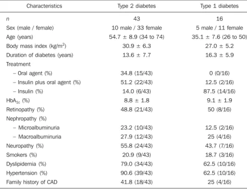
The prevalence of positive screening tests to silent ischemia varied according to type of diabetes. In the type 1 diabetes group, 2 of 16 patients (12.5%) had abnormal per-fusion tests (both women, 35 and 46 years old, and duration of diabetes of 17 and 18 years respectively). The stress ECG was normal in these 2 patients. Retinopathy, nephropathy and peripheral neuropathy were present in the first patient and were absent in the other patient. In the type 2 diabetes group, 10 of 43 patients (23.25%), including 4 men and 6 women, had posi-tive tests (2 fixed defect and 8 ischemic). The ages of patients had ranged between 46 to 74 years and the maximum time of dura-tion of disease was 28 years. The stress ECG was abnormal in half of these 10 patients. Unlikely the DM1 group, retinopathy and nephropathy were present in 80% (8/10) and neuropathy was present in 90% (9/10) Table 1 Clinical characteristics of the 59 patients.
Characteristics
n
Sex (male / female)
Age (years)
Body mass index (kg/m2)
Duration of diabetes (years)
Treatment
– Oral agent (%)
– Insulin plus oral agent (%)
– Insulin (%)
HbA1c (%)
Retinopathy (%)
Nephropathy (%)
– Microalbuminuria
– Macroalbuminuria
Neuropathy (%)
Smokers (%)
Dyslipidemia (%)
Hypertension (%)
Family history of CAD
Type 2 diabetes
43
10 male / 33 female
54.7 ± 8.9 (34 to 74)
30.9 ± 6.3
13.6 ± 7.7
34.8 (15/43)
51.2 (22/43)
14.0 (6/43)
8.8 ± 1.8
48.8 (21/43)
23.2 (10/43)
27.9 (12/43)
55.8 (24/43)
20.9 (9/43)
79.0 (34/43)
90.6 (39/43)
41.8 (18/43)
Type 1 diabetes
16
5 male / 11 female
35.1 ± 7.6 (26 to 50)
27.0 ± 5.2
16.3 ± 5.9
0 (0/16)
12.5 (2/16)
87.5 (14/16)
9.1 ± 1.9
50 (8/16)
12.5 (2/16)
25 (4/16)
43.7 (7/16)
18.7 (3/16)
62.5 (10/16)
62.5 (10/16)
25 (4/16)
of DM2 patients with perfusion abnormali-ties. However, these different rates of preva-lence between the diabetic groups (type 1 and type 2) could not be statistically ana-lyzed due to the reduced number of DM1 patients included in the study (Table 2).
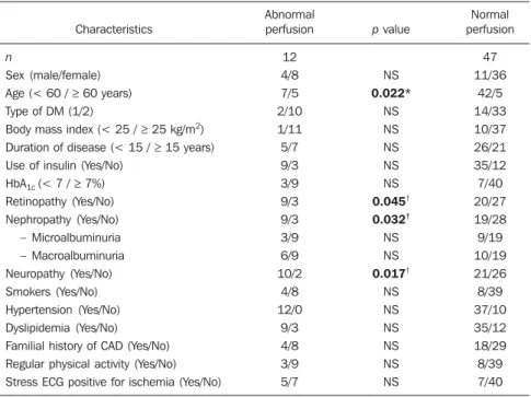
Considering the 12 patients with abnor-mal MPS (2 DM1 and 10 DM2), statisti-cal analyses of categoristatisti-cal variables using Chi-square test and Fisher’s exact test re-vealed to be statistically significant predic-tors of the presence of silent ischemia in diabetics patients: age 60 years and above (p = 0.022), retinopathy (p = 0.045), neph-ropathy (p = 0.032), and peripheral neur-opathy (p = 0.017) (Table 3).
Conversely, no difference was observed between the two groups, with and without ischemia, regarding gender, BMI, duration of diabetes, type of treatment, HbA1c level,
dyslipidemia, arterial hypertension, smok-ing, abnormal stress ECG, physical activ-ity or familial history of DAC.
Univariate logistic regression analysis showed to be statistically significant pre-dictors of silent ischemia: age 60 years and above (p = 0.017; odds ratio [OR] = 6.0), abnormal stress ECG (p = 0.049; OR = 4.08), nephropathy (p = 0.042); OR = 4.42), and peripheral neuropathy (p = 0.028; OR = 6.19). Additionally, a signifi-cant correlation was observed between si-lent ischemia and macroalbuminuric neph-ropathy (p = 0.031; OR = 5.60) (Table 4). Multiple logistic regression analysis showed that the variables which better
in-Table 2 Characteristics of 12 patients with perfusion defects.
Patient 1 2 3 4 5 6 7 8 9 10 11 12 Sex Female Female Female Female Male Female Male Female Male Female Female Male Age (years) 46 35 58 46 74 61 46 55 66 68 58 72 Type of DM 1 1 2 2 2 2 2 2 2 2 2 2 Nephropathy Macroalbuminuria No No Macroalbuminuria Microalbuminuria Microalbuminuria Macroalbuminuria Microalbuminuria Macroalbuminuria Macroalbuminuria No Macroalbuminuria Retinopathy Yes No No Yes Yes Yes Yes Yes Yes Yes Yes No Neuropathy Yes No Yes Yes Yes Yes Yes Yes Yes Yes Yes No Stress ECG Normal Normal Normal Abnormal Abnormal Abnormal Abnormal Normal Normal Normal Normal Abnormal LVEF (%) 52 56 69 43 52 56 39 55 52 47 > 70 33 MPS Ischemia Ischemia Ischemia Ischemia Ischemia Ischemia Fixed defect Ischemia Ischemia Fixed defect TID Ischemia Quantification* (%) 7 6 6 6 7 37 12 12 6 12 — 9
DM, diabetes mellitus; LVEF, left ventricular ejection fraction; MPS, myocardial perfusion scintigraphy; TID, transient ischemic dilation. * Quantification with the 17-segment method(12)
.
Table 3 Clinical characteristics of patients with or without silent coronary disease.
Characteristics
n
Sex (male/female) Age (< 60 / ≥ 60 years) Type of DM (1/2)
Body mass index (< 25 / ≥ 25 kg/m2)
Duration of disease (< 15 / ≥ 15 years) Use of insulin (Yes/No)
HbA1c (< 7 / ≥ 7%)
Retinopathy (Yes/No) Nephropathy (Yes/No) – Microalbuminuria – Macroalbuminuria Neuropathy (Yes/No) Smokers (Yes/No) Hypertension (Yes/No) Dyslipidemia (Yes/No) Familial history of CAD (Yes/No) Regular physical activity (Yes/No) Stress ECG positive for ischemia (Yes/No)
Abnormal perfusion 12 4/8 7/5 2/10 1/11 5/7 9/3 3/9 9/3 9/3 3/9 6/9 10/2 4/8 12/0 9/3 4/8 3/9 5/7
p value
NS 0.022* NS NS NS NS NS 0.045† 0.032† NS NS 0.017† NS NS NS NS NS NS Normal perfusion 47 11/36 42/5 14/33 10/37 26/21 35/12 7/40 20/27 19/28 9/19 10/19 21/26 8/39 37/10 35/12 18/29 8/39 7/40
NS, non significant; DM, diabetes mellitus; CAD, coronary arterial disease. * Fisher’s exact test (p value < 0.05); †Chi-square test (p value < 0.05).
Table 4 Univariate logistic regression analysis for ischemia (p value < 0.05).
Age (< 60 / ≥ 60 years) Stress ECG positive for ischemia (Yes/No) Nephropathy (Yes/No) – Microalbuminuria – Macroalbuminuria Neuropathy (Yes/No) MPS abnormal
n = 12
7/5 5/7 9/3 3/9 6/9 10/2 MPS normal
n = 47
42/5 7/40 19/28 9/19 10/19 21/26
p value
0.017 0.049 0.042 NS 0.031 0.028 OR 6.0 4.08 4.42 3.11 5.60 6.19
CI 95% OR
1.37–26.24 1.01–16.56 1.06–18.48 0.53–18.22 1.17–26.72 1.22–31.39
dicates concomitant silent ischemia in dia-betic patients are: age 60 years and above (p = 0.039; OR = 5.21), and peripheral neu-ropathy (p = 0.045; OR = 5.54) (Table 5). As the small number of patients with DM1 included in the study did not allow an individual statistical analysis of this sub-group, we conducted a separate analysis only the subset of patients with DM2.
The ROC curve analysis for age in DM2 patients, using the presence of silent is-chemia as the gold standard, revealed the same cutoff of 60 years. Similarly, com-parative analysis of age (continuous vari-able) between groups with and without si-lent ischemia showed a median of 59.5 years for the group of DM2 patients with perfusion abnormalities (p = 0.041; Mann Whitney’s test).
Still considering only the 10 DM2 pa-tients with abnormal scintigraphy, the sta-tistical analysis of categorical variables using Fisher’s exact test revealed to be sta-tistically significant predictors of the pres-ence of silent ischemia in DM2: age 60 years and above (p = 0.036), diabetic ret-inopathy (p = 0.034), and peripheral neur-opathy (p = 0.026). Unlike the statistical analysis of the total sample, which assessed the DM1 and DM2 together, the presence of diabetic nephropathy was not statisti-cally significant when DM2 patients were analyzed separately.
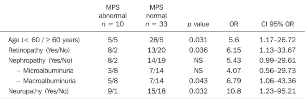
The univariate logistic regression analy-sis showed as statistically significant pre-dictors of silent ischemia in DM2 patients: age 60 years and above (p = 0.031; OR = 5.6), diabetic retinopathy (p = 0.036; OR = 6.15), peripheral neuropathy (p = 0.032; OR = 10.8), and macroalbuminuric nephr-opathy (p = 0.043; OR = 6.79) (Table 6).
Finally, multiple logistic regression analysis showed that the variable which best indicates possible silent ischemia in DM2 subjects is the presence of peripheral neuropathy (p = 0.032; OR = 10.8).
DISCUSSION
Diabetes is associated with a twofold to fourfold risk increase of developing CAD compared with age-matched subjects with-out diabetes(12–14). Many studies have been
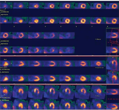
designed to evaluate the presence of silent myocardial ischemia in asymptomatic dia-Figure 2. Clinical case 2. A 68-year-old woman with diabetes type 2 for 16 years and clinical
manifes-tations of diabetic nephropathy, retinopathy, and peripheral neuropathy. Patient was asymptomatic. Phar-macological stress with dipyridamole was negative for ischemia. MPS showed fixed defect with ischemic component; 17-segment quantification = 12%.
betic patients and their prevalence rates range from 9% to 57%(2,7,15–24). The reason
for this broad range is probably due to dif-ferences in the populations studied, includ-ing age of patients, duration of disease, inclusion and exclusion criteria and defi-nition and diagnosis of SMI. Two relatively recent large retrospective studies from Ce-dars Sinai (n = 826)(25) and the Mayo Clinic
(n = 1427)(26) reported abnormal stress
SPECT images in 39% and 58% of asymp-tomatic diabetic patients, respectively. However, retrospective studies have sev-eral limitations, including selection bias of higher-risk patients.
The most important prospective study using stress SPECT imaging, the Detection of Ischemia in Asymptomatic Diabetics (DIAD) study(2), evaluated 1123 patients
with type 2 diabetes, aged 50–75 years, with no known or suspected CAD. Those patients were randomly assigned to either stress testing and 5-year clinical follow-up or to follow-up only (no imaging). A total of 113 patients (22%) had evidence of si-lent myocardial ischemia, including 83 with regional myocardial perfusion abnor-malities and 30 with normal perfusion but other abnormalities (i.e., adenosine-in-duced ST-segment depression, ventricular dilation, or rest ventricular dysfunction). Moderate or large perfusion defects were present in 33 patients. The strongest predic-tors for abnormal tests were abnormal
Val-salva, male sex, and diabetes duration. Other traditional cardiac risk factors or in-flammatory and prothrombotic markers were not predictive. The findings of the first phase of DIAD study suggest that greater than one in five asymptomatic pa-tients with type 2 diabetes, aged 50–75 years, have silent ischemia. Considering that the DIAD patient population was truly representative of asymptomatic patients with type 2 diabetes without any clinical reason to suspect CAD, the prevalence of SMI indicates that totally asymptomatic diabetic patients have at least an interme-diate probability of CAD, justifying screen-ing by noninvasive testscreen-ing such as stress myocardial perfusion imaging(2).
In our study, the prevalence of SMI was 20.3%, which is similar to DIAD results and to several other studies of the litera-ture(2,7,21). Although the prevalence of SMI
was different when we compared the DM1 and DM2 groups (12.5% versus 23.25%, respectively), this difference was not sta-tistically significant, probably due to the small number of type 1 diabetics studied.
The association between autonomic neuropathy and asymptomatic ischemia has been demonstrated by several prior stud-ies(27–29). Similarly, we did not find
signifi-cantly association among duration of dis-ease, type of diabetes and treatment, BMI, smoking, physical activity, familial history of DAC, dyslipidemia, arterial
hyperten-sion, abnormal stress ECG, HbA1c level and
presence of silent ischemia. The univariate and multiple logistic regression analyses revealed that age 60 years and above, but no male sex, was a strong predictive risk factor for myocardial ischemia. Both analy-ses also showed that peripheral neuropathy in our diabetic patients was significantly correlated to silent ischemia. Although we did not have conducted any tests for detec-tion of autonomic neuropathy, we can con-sider peripheral neuropathy as a predictor of ischemia, since it is present in most pa-tients with diabetic dysautonomia.
However we had a predominance of fe-male patients, although there are no pub-lished data that suggest a higher prevalence of diabetes in this sex. We believe that the larger number of women in our study is related to the greater availability of them to participate in research protocols. A similar number of men and women were invited to participate in the protocol, however, more women accepted the invitation and at-tended the laboratory on the day of the test. Many male patients who initially agreed to participate, did not attend the appointment. The progress made in detection and treatment of CAD allows reconsidering the screening of silent ischemia, in the hope that early CAD diagnosis leads to a more effective therapy and the decrease of car-diovascular complications and mortality. The challenge to the physician is to select the patient subgroups more likely to ben-efit from screening(30). Current guidelines
recommend screening it in asymptomatic diabetic patients selected for high cardio-vascular risk (i.e., with two or more other cardiovascular risk factors, or peripheral or carotid arterial disease, or proteinuria). ECG stress test can be recommended in first intention if maximal heart rate can be achieved. For patient with inconclusive ECG stress test, myocardial scintigraphy seems more accurate than stress echocar-diography. Coronary angiogram should be performed in case of positive stress test(31).
The second phase of the DIAD study (n = 1123) already showed that screening with MPS all asymptomatic diabetic patients is not cost-effective(32). The cumulative
car-diac event rate was 2.9% over a follow-up of 4.8 ± 0.9 years for an average of 0.6% per year. The positive predictive value of Table 5 Multiple logistic regression analysis for ischemia (p value < 0.05).
Age (< 60 / ≥ 60 years) Neuropathy (Yes/No)
MPS abnormal
n = 12
7/5 10/2
MPS normal
n = 47
42/5 21/26
p value
0.039 0.045
OR
5.21 5.54
CI 95% OR
1.09–24.96 1.04–29.58
MPS, myocardial perfusion scintigraphy; OR, odds ratio; CI, confidence interval.
Table 6 Univariate logistic regression analysis for ischemia in DM2 patients (p value < 0.05).
Age (< 60 / ≥ 60 years) Retinopathy (Yes/No) Nephropathy (Yes/No)
– Microalbuminuria – Macroalbuminuria Neuropathy (Yes/No)
MPS abnormal
n = 10
5/5 8/2 8/2 3/8 5/8 9/1
MPS normal
n = 33
28/5 13/20 14/19 7/14 7/14 15/18
p value
0.031 0.036 NS NS 0.043 0.032
OR
5.6 6.15 5.43 4.07 6.79 10.8
CI 95% OR
1.17–26.72 1.13–33.67 0.99–29.61 0.56–29.73 1.06–43.36 1.23–95.21
having moderate or large myocardial per-fusion defects was only 12%. Thus routine screening did not appear to affect overall outcome(32). However, during the course of
study there was a significant and equivalent increase in primary medical prevention in both groups and that can be an important bias. Nonprotocol stress tests were done during follow-up when clinically indicated in both groups and probably led to more stringent measures to control the DM, thus patients might have presented a better out-come than initially expected. Therefore, the goal is to identify the diabetic subgroups with the highest risk for future cardiac events to offer them more aggressive inten-sive medical therapy or coronary revascula-rization and optimum medical therapy(33). In our series of DM1 and DM2 patients, according to univariate logistic regression analysis, these subgroups would be initially determined by the patient’s age (60 years and above), presence of peripheral neur-opathy, nephrneur-opathy, retinneur-opathy, or stress ECG positive for ischemia. When the same patients were evaluated by multiple logis-tic regression analysis, the best predictors to indicate an abnormal myocardial perfu-sion scintigraphy would be age, or presence of peripheral diabetic neuropathy.
When considering the univariate logis-tic regression analysis of the subgroup of DM2 patients, individuals with an in-creased likelihood of myocardial perfusion defects are those which have age 60 years old and above, presence of retinopathy, peripheral neuropathy, or macroalbuminuric nephropathy. Attempting an even more accurate statistical selection, the multiple logistic regression analysis showed that the main predictor of silent ischemia was the presence of peripheral neuropathy, with a 10.8 times greater risk of silent ischemia in an asymptomatic diabetic patient type 2.
Interestingly, if we consider that patients with peripheral neuropathy may also present autonomic neuropathy, as both are part of the same complication, our results are in agreement with the findings of the first phase of the DIAD study(2), in which
the strongest predictor of silent ischemia was reduced heart rate response to Valsalva maneuver.
Currently there is an even greater con-cern in reducing as much as possible the
ex-posure of patients to ionizing radiation. For such, more modern equipment allow the use of a dose of 5 mCi (185 MBq) of sesta-mibi-99mTc at rest imaging and of 15 mCi
(555 MBq) in stress imaging for one-day protocol. Another alternative is to use the two-day protocol with fixed doses of 7 mCi (259 MBq) in each step. Some authors also suggest the possibility of performing first the stress imaging after the injection of 7 mCi (259 MBq), and the rest imaging only if the stress phase is abnormal(34,35). How-ever, this proposal remains controversial. Another important issue related to MPS is the definition of which patients might ben-efit from revascularization, considering myocardial viability and contractile re-serve(36,37).
Future prospective studies including only those subgroups of diabetic patients at high risk for CAD are needed to assess the real benefit of offering these patients a more aggressive therapy.
CONCLUSION
In our series, about one in five asymp-tomatic diabetic patients had silent is-chemia. Approximately 40% of DM1 and DM2 asymptomatic patients with silent is-chemia had a high risk of cardiac events, with perfusion defects of 10% and higher. The predictors of a myocardial scintig-raphy showed perfusion abnormalities were: age 60 years and above; stress ECG positive for ischemia; presence of any vas-cular complication of diabetes: retinopathy, nephropathy, or peripheral neuropathy. Peripheral neuropathy was the main predic-tor for silent ischemia in asymptomatic DM2 patients.
It is possible to clinically imply that screening of a subgroup of diabetic patients at high risk could lead to early detection and treatment of CAD, which could be useful in reducing morbidity and mortality of these patients.
Acknowledgements
This study was developed at the Divi-sion of Nuclear Medicine, Department of Radiology, and Divisions of Endocrinology and Cardiology, Department of Internal Medicine, School of Medical Sciences, University of Campinas (Unicamp),
Cam-pinas, SP, Brazil as part of a Master’s de-gree thesis and the student scholarship was granted by Coordenação de Aperfeiçoa-mento de Pessoal de Nível Superior (Capes). The project was also granted by Fundo de Apoio ao Ensino, à Pesquisa e Extensão (Faepex) grant number 193/2009.
REFERENCES
1. Nesto RW. Screening for asymptomatic coronary artery disease in diabetes. Diabetes Care. 1999; 22:1393–5.
2. Wackers FJ, Young LH, Inzucchi SE, et al. De-tection of silent myocardial ischemia in asymp-tomatic diabetic subjects: the DIAD study. Dia-betes Care. 2004;27:1954–61.
3. Kang X, Berman DS, Lewin H, et al. Compara-tive ability of myocardial perfusion single-pho-ton emission computed tomography to detect coronary artery disease in patients with and with-out diabetes mellitus. Am Heart J. 1999;137:949– 57.
4. Mieres JH, Rosman DR, Shaw LJ. The role of myocardial perfusion imaging in special popula-tions: women, diabetics, and heart failure. Semin Nucl Med. 2005;35:52–61.
5. Grundy SM, Benjamin IJ, Burke GL, et al. Dia-betes and cardiovascular disease: a statement for health professionals from the American Heart Association. Circulation. 1999;100:1134–46. 6. Hachamovitch R, Hayes SW, Friedman JD, et al.
Stress myocardial perfusion single-photon emis-sion computed tomography is clinically effective and cost effective in risk stratification of patients with a high likelihood of coronary artery disease (CAD) but no known CAD. J Am Coll Cardiol. 2004;43:200–8.
7. Janand-Delenne B, Savin B, Habib G, et al. Si-lent myocardial ischemia in patients with diabe-tes: who to screen. Diabetes Care. 1999;22:1396– 400.
8. Campisi R, Di Carli MF. Assessment of coronary flow reserve and microcirculation: a clinical per-spective. J Nucl Cardiol. 2004;11:3–11. 9. American Diabetes Association. Diagnosis and
classification of diabetes mellitus. Diabetes Care. 2010;33 Suppl 1:S62–9.
10. American Diabetes Association. Standards of medical care in diabetes – 2007. Diabetes Care. 2007;30 Suppl 1:S4–41.
11. Berman DS, Abidov A, Kang X, et al. Prognostic validation of a 17-segment score derived from a 20-segment score for myocardial perfusion SPECT interpretation. J Nucl Cardiol. 2004;11:414–23.
12. Le Feuvre C. Coronary artery disease in patients with diabetes. Presse Med. 2009;38:964–72. 13. Bax JJ, Bonow RO, Tschöpe D, et al. The
poten-tial of myocardial perfusion scintigraphy for risk stratification of asymptomatic patients with type 2 diabetes. J Am Coll Cardiol. 2006;48:754–60. 14. Zellweger MJ. Prognostic significance of silent coronary artery disease in type 2 diabetes. Herz. 2006;31:240–5.
16. Yildirimtürk O, Kiliçgedik M, Tu—cu A, et al. The relationship of microalbuminuria with left ven-tricular functions and silent myocardial ischemia in asymptomatic patients with type 2 diabetes. Turk Kardiyol Dern Ars. 2009;37:91–7. 17. Agarwal AK, Singla S, Singla S, et al. Prevalence
of coronary risk factors in type 2 diabetics with-out manifestations of overt coronary heart dis-ease. J Assoc Physicians India. 2009;57:135–42. 18. Fateh-Moghadam S, Reuter T, Htun P, et al. Stress echocardiography for risk stratification of asymp-tomatic patients with type 2 diabetes mellitus. Int J Cardiol. 2009;131:288–90.
19. Scholte AJ, Schuijf JD, Kharagjitsingh AV, et al. Different manifestations of coronary artery dis-ease by stress SPECT myocardial perfusion im-aging, coronary calcium scoring, and multislice CT coronary angiography in asymptomatic pa-tients with type 2 diabetes mellitus. J Nucl Cardiol. 2008;15:503–9.
20. Avignon A, Sultan A, Piot C, et al. Osteoprote-gerin: a novel independent marker for silent myo-cardial ischemia in asymptomatic diabetic pa-tients. Diabetes Care. 2007;30:2934–9.
21. Chico A, Tomás A, Novials A. Silent myocardial ischemia is associated with autonomic neuropa-thy and other cardiovascular risk factors in type 1 and type 2 diabetic subjects, especially in those with microalbuminuria. Endocrine. 2005;27: 213–7.
22. Mamcarz A, Chmielewski M, Braksator W, et al. Factors influencing cardiac complications in
pa-tients with type-2 diabetes mellitus and silent myocardial ischaemia: five-year follow-up. Pol Arch Med Wewn. 2004;112:1433–43. 23. Al-Attar AT, Mahussain SA, Sadanandan S.
Car-diac tests in asymptomatic type 2 diabetics. Med Princ Pract. 2002;11:171–5.
24. Langer A, Freeman MR, Josse RG, et al. Detec-tion of silent myocardial ischemia in diabetes mellitus. Am J Cardiol. 1991;67:1073–8. 25. Zellweger MJ, Hachamovitch R, Kang X, et al.
Prognostic relevance of symptoms versus objec-tive evidence of coronary artery disease in dia-betic patients. Eur Heart J. 2004;25:543–50. 26. Rajagopalan N, Miller TD, Hodge DO, et al.
Iden-tifying high-risk asymptomatic diabetic patients who are candidates for screening stress single-photon emission computed tomographic imaging. J Am Coll Cardiol. 2005;45:43–9.
27. Vinik AI, Maser RE, Mitchel BD, et al. Diabetic autonomic neuropathy. Diabetes Care. 2003;26: 1553–79.
28. Valensi P, Pariès J, Attali JR, et al. Cardiac auto-nomic neuropathy in diabetic patients: influence of diabetes duration, obesity, and the microangio-pathic complications – the French multicenter study. Metabolism. 2003;52:815–20.
29. Lee KH, Jang HJ, Kim YH, et al. Prognostic value of cardiac autonomic neuropathy independent and incremental to perfusion defects in patients with diabetes and suspected coronary artery dis-ease. Am J Cardiol. 2003;92:1458–61. 30. Doubell AF. Managing the asymptomatic diabetic
patient with silent myocardial ischaemia. Cardiovasc J S Afr. 2002;13:189–93.
31. Barthelemy O, Le Feuvre C, Timsit J. Silent myo-cardial ischemia screening in patients with dia-betes mellitus. Arq Bras Endocrinol Metabol. 2007;51:285–93.
32. Young LH, Wackers FJ, Chyun DA, et al. Cardiac outcomes after screening for asymptomatic coro-nary artery disease in patients with type 2 diabe-tes: the DIAD study: a randomized controlled trial. JAMA. 2009;301:1547–55.
33. Beller GA. Noninvasive screening for coronary atherosclerosis and silent ischemia in asymptom-atic type 2 diabetic patients: is it appropriate and cost-effective? J Am Coll Cardiol. 2007;49:1918– 23.
34. Ueyama T, Takehana K, Maeba H, et al. Prognos-tic value of normal stress-only technetium-99m myocardial perfusion imaging protocol. Compari-son with standard stress-rest protocol. Circ J. 2012;76:2386–91.
35. Chang SM, Nabi F, Xu J, et al. Normal stress-only versus standard stress/rest myocardial perfusion imaging: similar patient mortality with reduced radiation exposure. J Am Coll Cardiol. 2010; 55:221–30.
36. Lima RSL, Fonseca LMB. Evaluation of myocar-dial viability. Radiol Bras. 2010;43(5):v–vi. 37. Moraes RF, Meneghetti JC, Barroso AA.