Clinical Impact of Positron Emission Tomography by
Coincidence System with 18F-FDG on Therapeutic
Decision-Making of Patients with Ischemic
Cardiomyopathy after Myocardial Infarction
Renata Christian Martins Felix, Patrícia Lavatori Correa, Jader Cunha de Azevedo, Hans Fernando da
Rocha Dohmann, Evandro Tinoco Mesquita, Cláudio Tinoco Mesquita
Serviço de Medicina Nuclear do Hospital PróCardíaco, PROCED -Hospital Pró-Cardíaco e Universidade Federal Fluminense - Rio de Janeiro, RJ - Brazil
Mailing Address: Renata Christian Martins Felix • Rua General Polidoro, 192 – Botafogo – 22280-000 - Rio de Janeiro, RJ - Brazil E-mail: [email protected] ou [email protected] Received on 02/23/05 • Accepted on 07/25/05
O
BJECTIVETo evaluate the infl uence of the myocardium viability study by coincidence imaging using 18F-FDG in the clinical
decision-making of patients with ischemic cardiomyopathy and left ventricular dysfunction.
M
ETHODSThirty-one patients were submitted to myocardial viability study with 18F-FDG by coincidence imaging
between September 2003 and November 2004. The physician answered a questionnaire about the choice of therapeutic procedure before and after PET.
R
ESULTSTwenty-seven patients (87%) had myocardial viability. Twenty-one (68%) physicians thought that PET changed the therapeutic procedure for their patients and 27(87%) considered that PET added to the therapeutic decision. The current treatment decision (clinical or revascularization) correlated with myocardial viability (p=0.006).
C
ONCLUSIONSCoincidence imaging is a useful tool to help physicians in a difficult decision about the best treatment for patients with ischemic cardiomyopathy. Symptoms, electrocardiogram, ejection fraction and myocardial fi brosis area did not correlate with myocardium viability, so they should not be used to decide whether to perform a myocardial viability study or not.
K
EY WORDSPositron Emission Tomography (PET) is a type of exam in Nuclear Medicine Assessment recently introduced in our country. Its use in Oncology, Neurology/Psychiatry and Cardiology is well documented in the world literature 1-4. In
our country, the experience with this type of exam is still a recent one, as the production of 18fl uordesoxiglicose (18
F-FDG), a radioisotope used in PET, started in 1999 5.
As with any new technology, the introduction of a new diagnostic method can lead to two extremes: its overvalorization, with exaggerated use and lack of adequate assessment; or its underuse, due to the lack of clinical practice caused by diverse barriers, such as knowledge and trust of clinicians, the approval by health insurance companies and patients’ access to public health services. Regarding PET, the its limitations include the high cost of the exam, the low availability of radioisotope-producing centers and adequate equipment. Currently, this imaging method is available only in the states of Sao Paulo and Rio de Janeiro (in the metropolitan cities) and in relatively few Nuclear Medicine Services.
Among the clinical applications of PET, it is noteworthy the assessment of myocardial viability, which is a very important topic considering its correlation with cardiac failure, a disease of epidemic proportions with almost 5 million people with this morbidity in the United States alone, and 550,000 new cases every year 6.
Among the patients who are admitted to the hospital with a primary diagnosis of cardiac failure, coronary disease is the most important etiological factor, being present in up to 70% of the cases7,8. In a study carried
out by the Mayo Clinic from 1979 to 1994, it was observed that 36% of the patients who survived an acute myocardial infarction (AMI) developed cardiac failure9.
The success of the thromboembolytic therapy and primary angioplasty has reduced the mortality by AMI. Nevertheless, many survivors of an AMI still develop left ventricular dysfunction 7.
Up to 50% of the regions showing Q waves at the electrocardiogram (ECG) or segmented dysfunction at the echocardiogram are potentially recoverable after the revascularization of this region, as there can be viable areas with hibernating or stunned myocardium mixed with fi brotic tissue10.
A meta-analysis with 3,088 patients from 24 myocardial viability studies showed that those patients who presented myocardial viability diagnosed by stress echocardiogram (STRESS ECHO), myocardial scintillography with thallium201 or PET with 18F-FDG and
who were submitted to revascularization presented a reduction in the annual mortality of 79.6% in comparison to those who presented viability but were not submitted to revascularization. The absence of myocardial viability did not determine a survival difference in patients submitted to clinical vs. surgical treatment11,12.
When we consider that survival in individuals with
severe ventricular dysfunction treated clinically is around 50% in a fi ve-year period1, this potential possibility of
functional recovery can be signifi cant for those patients who present cardiac failure, mainly because up to 50% of the patients waiting to undergo a cardiac transplant have ischemic cardiopathy7. On the other hand, the mortality
rate within 30 days after the myocardial revascularization surgery in patients with left ventricular dysfunction can reach 20%1. Thus, it is mandatory to identify the
presence of viable myocardium in patients with ischemic cardiopathy and severe left ventricular dysfunction in order to verify which ones will indeed benefi t from the myocardial revascularization.
The medical decision regarding a procedure to be implemented must be based on some mainstays such as the patient’s clinical condition, when the risk of the clinical condition will be assessed and the cost-benefi t of certain procedure; the patient’s preferences regarding treatment; and the evidence related to the case, which include: clinical history data, physical examination and complementary test data, as well as information obtained through other scientifi c studies. The doctor’s experience is the factor that will join and assess all this information, with the objective of reaching the most adequate procedure for the patient.
Regarding the role of complementary tests, at this time of medical costs and scientifi c evidence – evidence-based cardiology – the focus has been shifted from the observation of basic operational characteristics of a complementary test such as sensitivity, specifi city and true predictive value ratio to the focus of the clinical impact, following the analysis of the test result on the medical procedure to be implemented.
There is plenty of evidence on the impact of PET on the management of patients with ventricular dysfunction of ischemic origin; however, the high cost of this technology regarding its initial investment as well as its maintenance has decreased its possibility of use, not only in our country but also in other centers around the world13.
Thus, in recent years, the conventional gamma-chamber started to be used in exams with 18F-FDG,
resulting in the so-called hybrid PET-SPECT with a coincidence detection system, using a gamma-chamber with two detectors, which are confi gured at 180o14,15.
Therefore, the primary objective of this study is to evaluate the clinical impact, i.e., the infl uence on the decision-making of Positron Emission Tomography (PET) carried out by a Coincidence System with
18fl uordesoxiglicose, to assess myocardial viability in
M
ETHODSA series of thirty-one consecutive patients of both genders, referred to the Nuclear Medicine and Molecular Imaging Service of Hospital Pró-Cardíaco were selected for undergoing a Positron Emission Tomography (PET) using a Coincidence System with 18fl uordesoxiglicose (18
F-FDG) in order to assess myocardial viability, according to the physician’s decision, regardless of the present study, from September 2003 to November 2004. The research project was approved by the Review Boards of Hospital Pró-Cardíaco.
The inclusion criteria were: previous myocardial infarction history, with or without the presence of Q wave at the ECG, but with signs of segmental fi brosis at the myocardial perfusion scintillography, such as: hypouptake of the radioisotope at rest, associated to contractility alteration and myocardial thickening at the same region; a minimum period of three weeks after the acute myocardial infarction; presence of coronary atherosclerotic disease, documented through coronary angiography or the presence of risk factors for coronary atherosclerotic disease or previous history of myocardial revascularization; presence of at least 12% of myocardial fi brosis assessed by myocardial perfusion scintillography at rest using the Emory Cardiac Toolbox software; and systolic dysfunction of the left ventricle with an ejection fraction 40% quantified by Gated SPECT (Cedars Quantitative Gated SPECT) or presence of clinical signs of cardiac failure. We excluded patients with no history of acute myocardial infarction, those with left ventricle systolic dysfunction of non-ischemic etiology, fi brosis area < 12% at scintillography at rest, younger than 18 yrs and women of reproductive age with suspected or confi rmed pregnancy.
Patients were submitted to clinical data collection. Cardiac failure symptoms were classifi ed according to the New York Heart Association (NYHA) 16 and the presence
of angina was classifi ed according to the guidelines from the Canadian Cardiovascular Society (CCS)17.
A myocardial perfusion scintillography with 99mTc – tetrofosmin (Myoview®) was carried out with the intravenous administration at rest of 740 MBq. The images were acquired between 45 and 90 min after the administration of the radioisotope by an E.cam Duet® (Siemens) gamma-chamber using two detectors with an angle of 90o between them, with a 64 X 64 matrix,
1.45 zoom and 32 projections of 20 seconds each, synchronized with the ECG (Gated SPECT), to allow the three-dimensional reconstruction and the measurement of the ejection fraction. The image processing was carried out by fi ltered backprojection with a Butterworth fi lter, with a cutoff of 0.5 and an order of 5. The images were then interpreted through the division of the left ventricle in 17 segments18; the quantifi cation of the fi brosis area
was performed using the Emory Cardiac Toolbox software and the ventricular function assessment by Gated SPECT,
using the Cedars Quantitative Gated SPECT software. Positron Emission Tomography and myocardial perfusion scintillography were carried out on different days. Patients had fasted for at least 4 hours, and the administration of 370 MBq of 18F-FDG intravenously was
carried out with a capillary glycemia < 140 mg%. Non-diabetic patients were offered an oral overload of 50-75 mg of glucose 60 min before radioisotope administration. Capillary glycemia was monitored every 15 min. If the capillary glycemia was elevated 45-60 min after the glucose overload, regular insulin was administered i.v., as described in Chart 119.
Chart 1 – Glycemia management during PET scan preparation in non-diabetic patients
130-140mg/dL 1 UI of regular insulin 140-160mg/dL 2 UI of regular insulin 160-180 mg/dL 3 UI of regular insulin 180-200 mg/dL 5 UI of regular insulin
> 200 mg/dL Re-schedule the test, consider as diabetic
When capillary glycemia is kept between 110 and 140 md/dL after two consecutive measurements, the radioisotope is administered. As for diabetic patients, two solutions are prepared: one with 100 IU of insulin in 500 ml of saline solution and another with 500 mL of 20% glucose added to 20 mL of 10% potassium chloride. Each solution is infused into an upper extremity, as 4 mU/kg/min of insulin and 6 mg/kg/min of glucose. Capillary glycemia is measured every 10 min and the infusions are adjusted according to the glycemia level, aiming at keeping it between 110 and 140 mg/dL. When capillary glycemia is maintained within this range for two consecutive measurements, the radioisotope is administered20. During
the preparation for the exam, the patient is monitored by the unit nurse and physician. Data acquisition occurred 30 to 90 minutes after the administration of the radioisotope, in an E.cam Duet® (Siemens) gamma-chamber by coincidence system, with a 1-inch thick NaI crystal doped with Tl and detectors confi gured at 180o
A clinical follow-up, ranging 2-14 months after the exam, was carried out by telephone and the patients were asked about the need to undergo revascularization, their current symptoms and use of medication.
The patients’ physician answered a questionnaire that included the following items: type of treatment proposed before PET; the clinical situation that prompted the request for PET; the treatment proposed for the patient after the PET; whether PET modifi ed or not the therapeutic strategy implemented after the exam, such as change from clinical treatment to myocardial revascularization treatment; and whether the PET result contributed for the decision-making, even in those cases where the therapeutic decision was not altered, but the physician’s initial impression was confi rmed by PET fi ndings.
The statistical analysis was carried out by the following methods: Mann-Whitney (non-parametric test) to compare quantitative data between two groups; Fisher exact test to compare ratios (qualitative data) between two groups; signifi cance level was set at 5%, i.e., there is statistical signifi cance when p value is ≤ 0.05.
R
ESULTSGeneral aspects: Clinical data of the studied population are shown in Table 1 and scintillography data are shown in Table 2. Numerical data were expressed as mean ± SD (minimum and maximal) and qualitative data as frequency and percentage.
Evaluation of the clinical impact of PET on the therapeutic decision-making – regarding PET results, we observed that 27 patients (87%) presented at least one myocardial segment with perfusion-metabolism discordance, suggesting viable myocardium. Graph I presents the division of the number of patients according to the number of viable segments.
When performing the correlation between the therapeutic decision-making and the presence of myocardial viability, we observed that none of the patients who did not present myocardial viability was referred to invasive therapy, whereas surgical or percutaneous revascularization was proposed for 21 of 27 (77.8%) of those who presented viability (p=0.006), showing that the group of patients with viable myocardium presented a signifi cantly higher revascularization indication when compared to the group with no myocardial viability.
However, we observed that the presence of viable myocardium was not signifi cant regarding the infl uence of PET on the change of therapeutic decision-making (p=0.086), i.e., the presence as well as the absence of viability was important for decision-making, although a slight tendency towards the association between viability and change of therapeutic option was noticed. Regarding the direct contribution of PET on decision-making, there was no analogous association with the presence of viability (p=0.55). From the analysis of these data, we
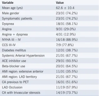
Table 1 – Clinical data of the studied population
Variable Value
Mean age (yrs) 62.6 ± 10.4 Male gender 23/31 (74.2%) Symptomatic patients 23/31 (74.2%) Dyspnea 18/31 (58.1%) Angina 9/31 (29.0%) Angina + dyspnea 4/31 (12.9%) NYHA III – IV 16/18 (88.9%) CCS III-IV 7/9 (77.8%) Diabetes mellitus 12/31 (38.7%) Systemic Arterial Hypertension 21/31 (67.7%) ACE inhibitor use 29/31 (93.5%) Beta-blocker use 20/31 (64.5%) AMI region: extensive anterior 11/31 (35.5%) AMI region: LAD territory 21/31 (67.7%) CA previous to PET 16/31 (51.6%) LAD Occlusion 11/19 (57.9%) CA with trivascular stenosis 14/19 (73.7%)
CCS III - IV = Canadian Cardiovascular Society - classes III and IV; ACE inhibitors = Angiotensin-converting enzyme inhibitors; LAD artery = Left Anterior Descending artery; CA = coronary angiography; NYHA = New York Heart Association, classes III and IV; AMI = acute myocardial infarction
Table 2 – Scintillographic data of the studied population
Variable Value
EF mean (Gated SPECT %) 29.3 ± 10.7 (11-54) Fibrosis area at SC (% de myocardium) 38.1 ± 11.2% (15-63) N of segments with fi brosis at SC 8.9 ± 2.6 (5-13) Nof patients with viability at PET 27/31 (87.0%) N of segments with viability at PET 3.52 ± 2.61 (0-9)
EF= ejection fraction; SC= scintillography
observed that both results, namely presence or absence of viability, are taken into account by the physician and infl uence therapeutic decision-making.
When we analyze the current medical strategy regarding the choice between clinical treatment and invasive procedure, taking into account the number of viable segments observed at PET, we observe that patients who underwent revascularization showed, on average, 4.29 ± 2.47 viable segments, whereas the group who received clinical treatment had 1.9 ± 2.18 viable segments (p=0.011), indicating that a higher number of viable segments was significant for the invasive treatment choice. The same happens concerning the viable myocardium area, as there was an average area of 16.1% ±13.58% for those who underwent revascularization and 7.4% ± 8.95% for those who had clinical treatment.
27
87%
4
13%
PET contribution PET did not contribute
21
68%
10
32%
PET changed the therapeutic decision
PET did not change the therapeutic decision
18 patients
67%
9 patients
33%
1-2 viable segm ents > or = 3 viable segm ents
21 presented viable myocardium; however, among the 8 symptomatic patients, 6 of them also presented viability (p=0.26).
Data on the ECG at rest of 19 patients were available, and only one of these patients presented normal ECG; the remaining 18 were distributed as follows: 10 (55.5%) showed Q waves at the ECG, whereas 8 (44.5%) presented bundle branch block (right or left). The analysis of the presence of Q wave regarding the presence of viability was impaired by the small sample size; nonetheless, the fact that 9 patients who had Q waves at ECG also showed viability is noteworthy.
When we analyze the difference between the two groups (with and without viability) regarding ejection fraction by Gated SPECT and with fi brosis area assessed by the Emory Cardiac Toolbox, we observed no statistically
Graph 1 – Number of patients with myocardial viability divided according to the number of viable segments
Graph 2 - PET infl uence on therapeutic strategy change
Graph 3 – PET contribution in decision-making
signifi cant difference between the two groups regarding these two variables, with p= 0.59 and p=0.90 for each group, respectively.
As for ejection fraction, the group that presented viability had a mean ejection fraction of 29.7%, whereas the group with no viability had 26.75%. Regarding fi brosis area, there was great similarity between both groups, with 38.04% of myocardial area with fi brosis at the scintillography at rest for the viability group and 38.75% in the group with no viability.
Follow-up – None of the 4 patients with absence of viability underwent an invasive procedure, whereas of the 27 who presented viability, 13 were submitted to myocardial revascularization (7 angioplasties and 6 surgical procedures).
treatment was not carried out due to the following reasons: in 3 cases, the coronary anatomy was diffi cult to reach; in one case, the patient died in the preoperative period, while waiting for revascularization; two patients could not undergo revascularization: one due to refractory cardiac failure and another due to structural restrictions of the hospital unit; in one case, the doctor intended to perform surgical revascularization; however, the small viable area was a negative aspect that went against the decision, when discussed with the surgical team, who considered the procedure risk/benefi t ration, and as a result, the patient is in cardiac rehabilitation. Therefore, 62% of the patients for whom there was intention to treat with myocardial revascularization really underwent the procedure. The statistical analysis of these data was not possible due to the small number of cases in one of the groups.
D
ISCUSSIONIn the real world, in addition to viability, the physician must consider other factors related to the patient when establishing the most adequate procedure; the coronary anatomy, clinical status and associated co-morbidities are included among them.
Some studies have demonstrated that between 31% and 39% of the patients undergo revascularization, regardless of the PET fi ndings21-24. Auerbach et al25
showed us that 68% of the patients with 5 or more viable myocardial segments underwent revascularization (differently from PET results in 32% of the cases); as for those with a smaller number of viable segments (1 to 4), the rate of revascularization was 52% whereas patients with no viability presented a rate of 36% of revascularization. However, these conclusions came from the observation of the patients’ clinical follow-up and not directly from inquiring doctors regarding their intention to treat.
In Oncology, the clinical impact of PET on patient management has been demonstrated by studies in which the physician answers questionnaires regarding the therapeutic decision following PET data26-33.
In Cardiology, after a search conducted by the Internet, no study was found in literature describing such study model. Our study is original regarding this type of assessment of the clinical impact on myocardial viability, using PET in a gamma-chamber by the coincidence system.
Literature shows that, concerning the research on myocardial viability, there is a signifi cant concordance between PET fi ndings with a dedicated system and PET in a gamma-chamber by the coincidence system. Tian et al34 showed that the general concordance was 93%
and that PET sensitivity by the coincidence system was 92%. Hasegawa et al35 showed that the coincidence
system presents spatial resolution, measurement
sensitivity and image quality superior to those obtained by gamma-chambers equipped with high-energy collimator; however, in order to become equivalent to dedicated PET, attenuation correction is a very important factor. One of the limitations of our study is that our equipment is not equipped with attenuation correction; nevertheless, the 1-inch crystal characteristics warrant excellent image quality.
The doctors participating in our study answered a questionnaire in which they indicated the proposed treatment before PET, what the indication for the exam was, and after it, what therapeutic approach had been adopted, and whether there had been a change of therapeutic choice due to PET results and if PET had directly infl uenced the decision-making.
We observed a signifi cant adherence on the part of these doctors regarding PET results, with concordance between myocardial viability and therapeutic proposal. Nevertheless, not only the presence but also the absence of myocardial viability were useful for doctors, and had infl uenced decision-making, regardless of the number of viable segments and area of viable myocardium. Similarly, the number of myocardial segments and extension of viable myocardium infl uence the therapeutic choice.
Among the 21 doctors who indicated myocardial revascularization, only 13 of their patients actually underwent surgery, which corresponds to 62% of the patients. This means that 38% of the patients had a fi nal therapeutic approach that was different from PET results and intention to treat, which is in accordance with the aforementioned studies25,36-39.
This means that the presence of myocardial viability is not the sole determinant that leads to myocardial revascularization, as not all patients with viable myocardium are candidates to revascularization due to a number of reasons, as previously mentioned: the complex coronary anatomy, the patient’s decision and the associated co-morbidities40.
In our country, we must add to these issues the lack of infrastructure of some hospitals to perform complex procedures such as cardiac surgery, or the prolonged waiting period that many patients have to face while waiting for a revascularization, causing many planned procedures not to be performed. We faced all these situations in our series, but the small number of cases is not enough to allow us to draw conclusions on this issue.
It is possible to infer that PET had an impact on the management of patients with ischemic cardiopathy in this population; obviously, it is necessary to increase the number of cases and doctors in order to be able to make such assertion with precision.
the presence of symptoms is not necessary to prompt the assessment of myocardial viability.
Unfortunately, we only had access to the ECG results of 19 patients, but we observed that almost all of them presented alterations (18/19 – 94.7%). Although a more accurate statistical analysis of these data was not feasible, we observed that the presence of Q wave at the ECG did not invalidate the fi nding of perfusion-metabolism discordance.
Schinkel et al40, using dobutamine stress echocardiogram
in 150 patients, found myocardial viability in 58% of the dysfunctional segments that showed Q wave at the ECG. Comparing the latter with segments that were dysfunctional but did not show Q wave at the ECG, we observed no signifi cant difference regarding viability fi nding (p=0.2). Thus, myocardial viability can be either observed or not, regardless of ECG results.
The mean ejection fraction found in our population was low (29.3%), constituting a population with more severe left ventricular dysfunction.
Studies that assessed myocardial viability analyzed patients with ejection fraction < 40% and many reported a mean ejection fraction similar to ours 23,41,42.
When comparing the ejection fraction between patients with and without myocardial viability, we did not fi nd a signifi cant difference between the two groups (p=0.59). It shows that myocardial viability can be either present or not, regardless of the degree of ventricular dysfunction. These fi ndings are in accordance with those by Di Carli et al43, who suggest that myocardial
viability assessment is useful, even in patients with severe ventricular dysfunction.
We believe that myocardial viability assessment must be carried out, even in patients with very low ejection fractions, as they can benefi t from the procedure. We mention one of our patients who presented EF at rest of 13% and the study with 18F-FDG showed a viable
myocardium area of 19%.
The study by Hurrell et al44 of 1,224 patients
showed, during a 2-year follow-up period, that patients with myocardial fi brosis area at rest > 12% of the myocardium, measured by scintillography, present a worse prognosis than those with a residual fi brosis area < 12% (p=0.003).
In our population, we found a mean fi brosis area of 38.1%. This means that the population studied presents an even higher risk for cardiac events. The possibility of fi nding myocardial viability can offer them the chance to modify the natural history of ischemic cardiomyopathy in this setting.
When we correlate the area of fi brosis with myocardial viability detection, we did not fi nd any difference between the groups with and without viability regarding fi brosis extension (p=0.9). Both groups presented a mean fi brosis area > 38%, i.e., an extensive fi brosis area. This fi nding shows that it is possible to fi nd viable myocardium even in patients with extensive fi brosis areas.
Considering our results, we can conclude that Positron Emission Tomography by Coincidence System had a clinical impact on therapeutic decision-making in this group of patients with ischemic cardiopathy after myocardial infarction. The presence of symptoms, the ECG, ejection fraction, and the myocardial fi brosis area did not correlate with the presence of PET viability in the studied population.
One of the limitations of our study is its small sample size, which does not allow conclusive assertions to be drawn from its results. The number of patients has to be increased or further studies must be carried out in order to confi rm the results obtained. The study did not assess patients’ expectations and values, which are factors that can infl uence therapeutic procedures. This work was carried out by highly specialized professionals in the Cardiology area, which might have infl uenced the results obtained.
No potential confl ict of interest relevant to this article was reported.
R
EFERENCES1. Segall G. Assessment of myocardial viability by positron emission tomography. Nucl Med Commun .2002; 23: 323-30.
2. Kostakoglu L, Agress H, Goldsmith SF. Clinical role of FDG PET in evaluation of cancer patients. Radiographics .2003; 23: 315-40.
3. Hoffman JM, Welsh-Bohmer KA, Hanson M , Gain B, Hullette C, Earl N et al. FDG PET Imaging in patients with pathologically verifi ed dementia. J Nucl Med .2000; 41: 1920-8.
4. Dohmen C, Bosche B, Graf , Staub F, Kracht L, Sobesky J et al. Prediction of malignanat course in MCA infarction by PET and microdialysis. Stroke. 2003; 34: 2152-8.
5. Cyclotron Accelerators Center.[web site on the Internet].São Paulo: IPEN;1999.[cited 2004 Nov 10].Available from:http://www.ipen. br/biblioteca/progress/cyclotron.htm
6. Ansari M, Massie BM. Heart failure: how big is the problem? Who are the patients? What does the future hold? Am Heart J .2003; 146:1-4.
7. Marwick TH. The viable myocardium: epidemiology, detection and
clinical implications. Lancet .1998; 351: 815-9.
8. Gheorghiade M, Bonow R. Chronic heart failure in the United States: a manifestation of coronary artery disease. Circulation .1998; 97: 282-9.
9. Hellerman JP, Goraya TY, Jacobsen SJ , Weston AS, Reeder GS, Gersh B et al. Incidence of heart failure after myocardial infarction: is it changing over time? Am J Epidemiol .2003; 157: 1101-07.
10. Wijins W, Vatner SF, Camici PG. Hibernating Myocardium. N Engl J Med 1998; 339: 173-81.
11. Raynes RB, Devereaux PJ, Guyatt GH. Clinical expertise in the era of evidence-based medicine and patient choice. EBM 2002; 7: 36-8.
12. Allman KC, Shaw LJ, Hachamovitch R, Udelson JE. Myocardial viability testing and impact of revascularization on prognosis in patients with coronary artery disease and left ventricular dysfunction: a meta-analysis. J Am Coll Cardiol 2002; 39: 1151-8.
Fluorine-18-deoxyglucose SPECT and coincidence imaging for myocardial viability: clinical and technologic issues. J Nucl Cardiol .2001; 8: 75-88.
14. Fitzgerrald J, Parker JA, Danias PG. F-18 fl uoro deoxyglucose SPECT for assessment of myocardial viability. J Nucl Cardiol 2000; 7: 382-7.
15. Srinivasan G, Kitsiou NA, Bacharach SL , Bartlett ML, Miller-Davis C, Dilsizian V. 18F-fl uordesoxyglucose single photon emission computed
tomography: can it replace PET and thallium SPECT for the assessment of myocardial viability? Circulation .1998; 97: 843-50.
16. The Criteria Committee of the New York Heart Association. Diseases of the heart and blood vessels: nomenclature and criteria for diagnosis. 6th ed. Boston, MA: Little Brown, 1964.
17. Campeau L. Grading of angina pectoris. Circulation .1976; 54: 522-3.
18. Cerqueira MD, Weissman NJ, Dilsizian V, Jacobs AK, Kaul S, Laskey WK et al. Standardized Myocardial Segmentation and Nomenclature for Tomographic Imaging of Heart.A statement for health care professionals from the Cardiac Imaging Committee of the Council on Clinical Cardiology of the American Heart Association. Circulation . 2002; 105: 539-42.
19. Bacharach SL, Bax JJ, Case J , Delbeke D, Kurdziel KA, Martin WH et al. PET myocardial glucose metabolism and perfusion imaging: Part 1 - Guidelines for data patient preparation and data acquisition. J Nucl Cardiol .2003; 10: 543-56.
20. Chen EQ, Maclntyre WJ, Go R, Brunken RC, Saha GB, Wong CY et al Myocardial viability studies using fl uorine-18-FDG SPECT: a comparison with fl uorine-18-FDG PET. J Nucl Med 1997; 38: 582-6.
21. Hellermann JP, Jacobsen SJ, Redfi eld MM , Reeder GS, Weston AS, Roger VL. Heart failure after myocardial infarction: clinical presentation and survival. Eur J Heart Fail .2005; 7: 119-25.
22. Mezulín J, Cerny J, Spinarova L, Toman J, Grach L, Stetka F et al. Prognosis in patients with chronic coronary artery disease and severe left ventricular dysfunction: the importance of myocardial viability. Eur J Heart Fail .2003; 5: 85-93.
23. Hass F, Haehnel CJ, Picker W , Nekolla S, Martinoff S, Meisner H et al. Preoperative positron emission tomograhic viability assessment and perioperative and postoperative risk in patients with advanced ischemic heart disease. J Am Coll Cardiol .1997; 30: 1693-700
24. Bax JJ, Visser FC, Poldermans D , Elhendy A, Boersma E, Visser CA et al.Prognostic value of perfusion-FDG mismatch in ischemic cardiomyopathy. J Nucl Cardiol .2002; 9: 675-77.
25. Auerbach MA, Schöder H, Hoh C , Gambhir SS, Yaghoubi S, Sayre JW et al. Prevalence of myocardial viability as detected by positron emission tomography in patients with ischemic cardiomyopathy. Circulation .1999; 99: 2921-6.
26. Talbot JN, Rain JD, Meignan M, Askienazaky S, Grall Y,Bok B et al. Impact of 18F-FDG-PET on medical decision making in oncology: evaluation by referring physicians during the opening year. Bull Cancer .2002; 89: 313-21.
27. Meta J, Seltzer M, Schiepers C , Silverman DH, Ariannejad M, Gamhir SS et al. Impact of 18F-FDG PET on managing patients with colorectal cancer: the referring physician´s perspective. J Nucl Med .2001; 42: 586-90.
28. Yap CS, Seltzer MA, Schiepers C ,Gambhir SS, Rao J, Phelps ME et al. Impact of whole-body 18F-FDG PET on staging and managing patients with breast cancer: the referring physician’s perspective. J Nucl Med .2001; 42: 1334-7.
29. Schoder H, Meta J, Yap C , Ariannejad M, Rao J, Phelps ME et al. Effect of whole-body 18F-FDG PET imaging on clinical staging and management of patients with malignant lymphoma. J Nucl Med .2001; 42: 1139-43.
30. Seltzer MA, Yap CS, Silverman DH, Meta J, Schiepers C, Phelps ME et al. The impact of PET on the management of lung cancer: the referring
physician’s perspective. J Nucl Med 2002; 43: 752-6.
31. Grahek D, Montravers F, Mayaud C , Regnard JF, Kerrou K, Younsi N et al. Positron emission tomography (PET) with 18F-FDG in bronchopulmonary cancer and its impact on medical decision at the time of the diagnosis, staging or recurrence evaluation. Rev Pneumol Clin .2001; 57: 393-403.
32. Kantorova I, Lipska L, Belohlvek O , Visokai V, Trubac M, Schneiderova M. Routine 18F-FDG PET preoperative staging of colorectal cancer: comparison with conventional staging and its impact on treatment decision making. J Nucl Med. 2003; 44: 1784-88.
33. Herder GJ, Van Tinteren H, Comans EF , Holkstra OS, Teule GJ, Postmus PE et al. Prospective use of serial questionnaires to evaluate the therapeutic efficacy of 18F-fluordeoxygluse (FDG) positron emission tomography (PET) in suspected lung cancer. Thorax .2003; 58: 47-51.
34. Tian M, Koyama K, Zhang H , Oriuchi N, Higuchi T, Endo K. Assessment of myocardial viability with a positron coincidence gamma camera using fl uordeoxyglucose in comparison with dedicated PET. Nucl Med Commun .2003; 24: 367-74.
35. Hasegawa S, Uehara T, Yamaguchi H, Fujino K, Kusuoka H, Hori M et al. validity of 18F-fl uorodeoxyglucose Imaging with a dual-head
coincidence gamma camera for detection of myocardial viability. J Nucl Med .1999; 40: 1884-92.
36. Lee, K, Marwick TH, Cook S , Go RT, Fix JS,James KB et al. Prognosis of patients with left ventricular dysfunction, with and without viable myocardium after myocardial infarction:relative effi cacy of medical therapy and revascularization Circulation 1994; 90: 2687-94.
37. Di Carli MF, Davidson M, Little R , Khannaady FV, Brunken RC. Value of metabolic imaging with positron emission tomography for evaluating prognosis in patients with coronary artery disease and left ventricular dysfunction. Am J Cardiol .1994; 73: 527-33.
38. Eitzman D, Al-Aouar Z, Kanter HL, Vom Dahl J, Kirsch M, Deeb GM et al. Clinical outcome of patients with advanced coronary artery disease. J Am Coll Cardiol .1992; 20: 559-65.
39. Yoshida K, Gould LK. Quantitative relation of myocardial infarction size and myocardial viability by positron emission tomography to left ventricular ejection fraction and 3-year mortality with and without revascularization. J Am Coll Cardiol 1993; 22: 984-97.
40. Schinkel AFL, Bax JJ, Boersma E , Elhendy A, Vourvouri EC, Roelandt JR et al. Assessment of residual myocardial viability in regions with chronic electrocardiographic Q-wave infarction. Am Heart J.2002; 144: 865-9.
41. Marwick TH, Zuchowskic C, Lauer MS , Secknus MA,Williams J, Lytle BW. Functional status and quality of life in patients with heart failure undergoing coronary bypass surgery after assessment of myocardial viability. J Am Coll Cardiol. 1999; 33: 750-8.
42. Beanlands RSB, Ruddy TD, deKemp RA , Iwanochko RM, Coates G, Freem,na M et al. Positron emission tomography and recovery following revascularization (PARR-1): the importance of scar and the development of a prediction rule for the degree of recovery of left ventricular function. J Am Coll Cardiol. 2002; 40: 1735-43.
43. Di Carli MF, Maddahi J, Rokhsar SBA , Schelbert HR, Bianco-Batles D, Brunken RC et al. Long-term survival of patients with coronary artery disease and left ventricular dysfunction: implications for the role of myocardial viability assessment in management decisions. J Thorac Cardiovasc Surg .1998; 116: 997-1004.